Elbow joint
Also called elbow, humeroulnar joint, humeroradial joint, proximal radioulnar joint
The hinge between the upper arm and the forearm, with two more joints in the same capsule. How it moves, what steadies it, the loads it carries, how it gets injured, and what studies report about recovery.
- Region
- Elbow
- Muscles
- biceps brachii, triceps brachii
- Updated
- October 8, 2026
- Reviewed
- October 8, 2026 · how entries are checked
Function
Three joints in one capsule
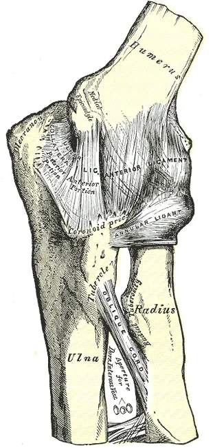
The elbow is a hinge joint formed by the humeroulnar joint1. There, the pulley-shaped trochlea of the humerus (upper arm bone) sits in the trochlear notch of the ulna1. The ulna is the forearm bone on the little-finger side, and the radius is the one on the thumb side. Two more joints sit with the hinge, inside the same joint capsule1. The humeroradial joint lies between radius and humerus, and the proximal radioulnar joint between the upper ends of radius and ulna1,6.
The radius has a disc-shaped head1. A shallow dip on top of the head meets the capitellum, a rounded knob on the humerus1. At the proximal radioulnar joint, the annular ligament holds the head against a notch on the ulna1. This is a pivot joint: the radius rotates there when the forearm turns palm up or palm down1.
The three joints work together to allow four movements6. They are flexion (bending), extension (straightening), pronation (turning the palm down) and supination (turning the palm up)6.
The back and top of the upper ulna form the olecranon, the bony tip of the elbow1. The olecranon bursa lies at that tip, under the skin1. A bursa is a small fluid-filled sac that cushions tissues where they rub.
What holds it steady
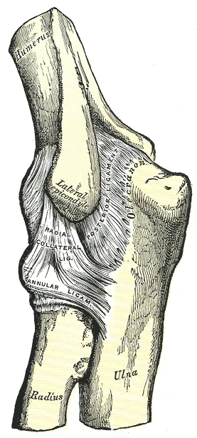
The capsule is thin at the front and back1. At the sides, strong ligaments (tough bands of tissue that hold bones together at a joint) thicken it1. They stop side-to-side movement and hyperextension (straightening a joint past its normal limit)1. The main ligaments are these:
- On the inner side, the triangular ulnar collateral ligament runs from the medial epicondyle to the inner side of the upper ulna1. The medial epicondyle is the inner bony bump at the elbow end of the upper arm bone.
- On the outer side, the radial collateral ligament runs from the lateral epicondyle, the outer bony bump1.
- The radial collateral ligament blends into the annular ligament, a ring around the radial head1.
What resists sideways bending
Valgus stress bends the forearm outward at the elbow, and varus stress bends it inward7. A preliminary study of four elbow specimens measured what resists each7. With the elbow straight, three parts shared the valgus resistance equally7. They were the collateral ligament on the inner side, the front of the capsule and the bony joint7. At 90 degrees of flexion, the inner-side ligament gave about 55% of it7.
In the same four specimens, the joint surfaces and the front of the capsule mainly resisted varus stress7. The ulnar collateral ligament page covers valgus restraint in detail.
How far it moves
In 352 healthy adults, the dominant arm reached a mean of 146 degrees of active flexion, 80 of pronation and 87 of supination8.
A study had 33 people with normal elbows do fifteen daily activities2. Most fitted within an elbow flexion arc of 100 degrees, running from 30 up to 1302. They also fitted within 100 degrees of forearm rotation, 50 degrees each of pronation and supination2.
With the elbow straight and the palm facing forward, the forearm angles outward from the upper arm’s line by 5 to 15 degrees1. This is the carrying angle1.
The loads it carries
A review of elbow biomechanics notes that the upper limb is often called non-weightbearing9. Yet it states that forces across the elbow are very large when the joint straightens from a bent position9. The reason it gives is that the triceps has a small moment arm (a muscle’s leverage) and must generate high force9. It adds that the contact force between ulna and humerus may be as much as twenty times the load on the hand and wrist9.
In nine healthy men doing the push-up, peak force on the elbow along the forearm averaged 45% of body weight10. That was with the hands in the normal position, and it was significantly lower with the hands placed further apart or further up10.
Motion analysis of 26 highly skilled, healthy adult baseball pitchers calculated an elbow varus torque (a turning force) of 64 newton-metres11. It came shortly before the arm reached its furthest outward rotation11. A video and muscle-activity study of seven healthy adult baseball pitchers reported a varus torque of 120 newton-metres, resisting valgus stress12. The ulnar collateral ligament page reports more on pitching.
Breakdown
Dislocation
A dislocation is when a bone is forced fully out of its normal place at a joint. Elbow dislocations are classed as simple, with no fracture, or complex, with a fracture13. Studies have counted how often it happens:
- A national sample of United States emergency departments from 2002 to 2006 gave an estimated rate of 5.21 dislocations in each 100,000 person-years3.
- In a study of United States emergency department data, people aged ten to nineteen had the highest rate, 6.87 per 100,000 person-years3.
- In United States emergency department data, 44.5% of dislocations in people aged ten or older happened in sports3.
- A later study of emergency department data, for 2014 to 2023 and without children under ten, estimated 3.58 per 100,000 person-years14.
Three elbow surgeons reviewed 62 online videos of elbow dislocations15. They judged that 92% happened at or near full extension15. Most showed a valgus moment (89%), a load along the arm (90%) and progressive supination (94%)15. The authors noted that this differs from earlier cadaver studies, in which elbows dislocated in flexion15. They suggested that some dislocations start with failure on the inner side under valgus force15.
Osteochondritis dissecans in young athletes
In osteochondritis dissecans, a segment of joint cartilage separates from the bone beneath it9. A review describes it as uncommon in the general population and typical of adolescent athletes who throw overhead or bear weight on their arms9. The most common site it names is the capitellum of the dominant elbow9.
A review states that the cause is still unclear9. It adds that repeated valgus forces, which compress the outer side of the joint, are thought to be the main trigger9.
One study screened the throwing elbow of 2,433 junior high and high school baseball players with ultrasound (a scan made with sound waves)4. Their mean age was 14.5 years4. It found osteochondritis dissecans of the capitellum in 82 elbows, or 3.4%4. The authors describe this figure as including latent cases4. Compared with players without a lesion, players with one:
- had begun playing baseball at an earlier age and had played competitively for longer4
- reported more present and more past elbow pain4
- did not differ in the position they played4
Stiffness after injury
A literature review names the most common causes of elbow stiffness16. They are trauma, including surgery for trauma, arthritis after trauma, and heterotopic ossification (bone forming in soft tissue, where bone does not normally grow)16. The review links the elbow’s tendency to stiffen to its closely fitting bones and its confined joint space16. It also names the tight collateral ligament complexes and the muscles lying close around the joint16.
Osteoarthritis
One study took X-rays of 318 randomly sampled residents of a Japanese town, aged over 5017. It left out anyone with a condition that could cause secondary elbow arthritis17. The X-rays showed primary elbow osteoarthritis (a long-term joint disease in which the cartilage lining the joint wears away) in 25.2%17. In 0.9%, it came with pain on movement or tenderness17.
Olecranon bursitis
In olecranon bursitis, also called student’s elbow, the bursa between the skin and the olecranon is inflamed1. Israel Defense Forces records from 2005 to 2015 put it at 29 per 100,000 person-years in soldiers overall and 97 in combat duty18.
Recovery
After a dislocation
Three follow-up studies report how people fared years after an elbow dislocation19,20,21. Two used the DASH, a questionnaire that scores disability of the arm, shoulder and hand.
- Of 110 adults reviewed a mean of 88 months after a simple dislocation, 56% reported leftover stiffness and 62% leftover pain19. Their function scores were generally good, with a mean DASH score of 6.7 points19.
- Seventy-one patients were followed for a mean of 19.3 years after a simple dislocation managed with brief splinting and early movement20. They averaged 5.22 points on the DASH, and none had later surgery for instability20.
- Of 52 patients re-examined an average of 24 years after an elbow dislocation, half had no remaining symptoms or signs21.
How motion returns after injury
In 25 people followed prospectively for a year after a traumatic elbow injury, range of motion kept improving up to one year5. At one year, 22 had a flexion-extension arc above 100 degrees5.
What studies of osteochondritis dissecans found
A retrospective study (one that looks back at past cases or records) covered young patients with early lesions of the capitellum22. It called the condition capitellar osteochondrosis and graded lesions in stages22. In it, 101 patients stopped using the elbow heavily for 6 months or more22. Among them, stage I lesions healed in 90.5% and stage II lesions in 52.9%22. Among 75 patients who did not stop, 22.7% healed22.
A 2016 review stated that the natural course of the condition in the capitellum was still undefined9. A review reports one follow-up of 31 patients, a mean of 23 years after osteochondritis dissecans of the elbow9. In it, 42% had pain or reduced range of motion, and one-third had degenerative changes on X-rays9.
Connections
The bones that form it
The humerus joins the radius and ulna at the elbow23.
The muscles and tendons that cross it
The biceps brachii, on the front of the upper arm, flexes the forearm1. The triceps brachii, on the back, extends it1. The distal biceps tendon attaches to the radial tuberosity, a bump on the radius near the elbow24. A systematic review describes the triceps as inserting through its tendon onto the tip of the olecranon25.
A review of specimen studies lists three primary and four secondary constraints of the elbow13. Two of the secondary ones are tendons: the common flexor-pronator tendon and the common extensor tendon13. The common flexor tendon and common extensor tendon pages cover overuse of each.
The ligament and nerves beside it
The ulnar collateral ligament runs down the inner side of the joint1. The ulnar collateral ligament tear page reports studies of its tears.
The ulnar nerve passes through the cubital tunnel on the inner side of the elbow26. Above the elbow crease, the median nerve gives branches to the elbow joint27.
Exercises that load it
In nine healthy men doing push-ups, valgus torque at the elbow rose significantly when the hands were placed further up10. In a dip, the exerciser lowers the body by bending the elbows and returns by straightening them28. In 21 men and 4 women, the elbow moved through a mean range of about 93 degrees in the pull-up29. Related exercise pages: push-up, dip, pull-up.
Elbow joint is formed by
Elbow joint is held by
Elbow joint is bent by
Elbow joint is straightened by
Elbow joint is crossed by
Elbow joint is passed by
Elbow joint is supplied by
Elbow joint is affected by
Elbow joint is loaded by
Sources
- Betts JG, Young KA, Wise JA, Johnson E, Poe B, Kruse DH, Korol O, Johnson JE, Womble M, DeSaix P (2022). Anatomy and Physiology 2e. OpenStax. Link
- Morrey BF, Askew LJ, Chao EY (1981). A biomechanical study of normal functional elbow motion. The Journal of bone and joint surgery. American volume. PMID 7240327
- Stoneback JW, Owens BD, Sykes J, Athwal GS, Pointer L, Wolf JM (2012). Incidence of elbow dislocations in the United States population. The Journal of bone and joint surgery. American volume. doi:10.2106/JBJS.J.01663 PMID 22298056
- Kida Y, Morihara T, Kotoura Y, Hojo T, Tachiiri H, Sukenari T, Iwata Y, Furukawa R, Oda R, Arai Y, Fujiwara H, Kubo T (2014). Prevalence and Clinical Characteristics of Osteochondritis Dissecans of the Humeral Capitellum Among Adolescent Baseball Players.. The American journal of sports medicine. doi:10.1177/0363546514536843 PMID 24944293
- Myden C, Hildebrand K (2011). Elbow joint contracture after traumatic injury. Journal of shoulder and elbow surgery. doi:10.1016/j.jse.2010.07.013 PMID 21050779
- Card RK, Lowe JB, Launico MV (2025). Anatomy, Shoulder and Upper Limb, Elbow Joint. StatPearls. PMID 30422543
- Morrey BF, An KN (1983). Articular and ligamentous contributions to the stability of the elbow joint.. The American journal of sports medicine. doi:10.1177/036354658301100506 PMID 6638246
- Zwerus EL, Willigenburg NW, Scholtes VA, Somford MP, Eygendaal D, van den Bekerom MP (2017). Normative values and affecting factors for the elbow range of motion. Shoulder & elbow. doi:10.1177/1758573217728711 PMID 31210794
- Heijink A, Vanhees M, van den Ende K, van den Bekerom MP, van Riet RP, Van Dijk CN, Eygendaal D (2016). Biomechanical considerations in the pathogenesis of osteoarthritis of the elbow. Knee surgery, sports traumatology, arthroscopy : official journal of the ESSKA. doi:10.1007/s00167-015-3518-7 PMID 25677500
- Donkers MJ, An KN, Chao EY, Morrey BF (1993). Hand position affects elbow joint load during push-up exercise. Journal of biomechanics. doi:10.1016/0021-9290(93)90026-b PMID 8514808
- Fleisig GS, Andrews JR, Dillman CJ, Escamilla RF (1995). Kinetics of baseball pitching with implications about injury mechanisms.. The American journal of sports medicine. doi:10.1177/036354659502300218 PMID 7778711
- Werner SL, Fleisig GS, Dillman CJ, Andrews JR (1993). Biomechanics of the elbow during baseball pitching. The Journal of orthopaedic and sports physical therapy. doi:10.2519/jospt.1993.17.6.274 PMID 8343786
- de Haan J, Schep NW, Eygendaal D, Kleinrensink GJ, Tuinebreijer WE, den Hartog D (2011). Stability of the elbow joint: relevant anatomy and clinical implications of in vitro biomechanical studies. The open orthopaedics journal. doi:10.2174/1874325001105010168 PMID 21633722
- Onor GI, Fellheimer H, Bagereka P, Chang M, Miltenberg B, Martinazzi B, Salako T, Burke OC, Murray J, Beredjiklian PK (2026). The epidemiology of elbow dislocation in the United States 2014-2023. Journal of shoulder and elbow surgery. doi:10.1016/j.jse.2026.04.051 PMID 42128289
- Schreiber JJ, Warren RF, Hotchkiss RN, Daluiski A (2013). An online video investigation into the mechanism of elbow dislocation. The Journal of hand surgery. doi:10.1016/j.jhsa.2012.12.017 PMID 23391358
- Siemensma MF, van der Windt AE, van Es EM, Colaris JW, Eygendaal D (2023). Management of the stiff elbow: a literature review. EFORT open reviews. doi:10.1530/EOR-23-0039 PMID 37158372
- Nakayama K, Kato H, Ikegami S, Hayashi M, Hashimoto S, Sakai N, Takahashi J, Taneichi H (2022). Prevalence and associated factors of primary elbow osteoarthritis in the Japanese general elderly population: a Japanese cohort survey randomly sampled from a basic resident registry. Journal of shoulder and elbow surgery. doi:10.1016/j.jse.2021.07.015 PMID 34454037
- Schermann H, Karakis I, Dolkart O, Maman E, Kadar A, Chechik O (2017). Olecranon Bursitis in a Military Population: Epidemiology and Evidence for Prolonged Morbidity in Combat Recruits. Military medicine. doi:10.7205/MILMED-D-16-00402 PMID 28885965
- Anakwe RE, Middleton SD, Jenkins PJ, McQueen MM, Court-Brown CM (2011). Patient-reported outcomes after simple dislocation of the elbow. The Journal of bone and joint surgery. American volume. doi:10.2106/JBJS.J.00860 PMID 21776575
- Mackinnon T, Samuel TD, Hayter E, Lee G, Huntley D, Hardman J, Anakwe RE (2023). Long-Term Follow-up (14 to 25 Years) Following Closed Reduction and Early Movement for Simple Dislocation of the Elbow. The Journal of bone and joint surgery. American volume. doi:10.2106/JBJS.23.00288 PMID 37616331
- Josefsson PO, Johnell O, Gentz CF (1984). Long-term sequelae of simple dislocation of the elbow. The Journal of bone and joint surgery. American volume. doi:10.2106/00004623-198466060-00014 PMID 6736093
- Matsuura T, Kashiwaguchi S, Iwase T, Takeda Y, Yasui N (2008). Conservative treatment for osteochondrosis of the humeral capitellum. The American journal of sports medicine. doi:10.1177/0363546507312168 PMID 18219050
- Mostafa E, Imonugo O, Varacallo MA (2023). Anatomy, Shoulder and Upper Limb, Humerus.. StatPearls. PMID 30521242
- Hsu D, Anand P, Mabrouk A, Chang KV (2023). Biceps Tendon Rupture.. StatPearls. PMID 30020607
- Shuttlewood K, Beazley J, Smith CD (2017). Distal triceps injuries (including snapping triceps): A systematic review of the literature.. World Journal of Orthopedics. doi:10.5312/wjo.v8.i6.507 PMID 28660143
- Range TL, Samra NS (2023). Anatomy, Shoulder and Upper Limb, Osborne Band.. StatPearls. PMID 30725684
- Binsaleem S (2025). Median nerve entrapment neuropathy: a review on the pronator syndrome.. JSES Reviews, Reports, and Techniques. doi:10.1016/j.xrrt.2024.10.001 PMID 39872334
- McKenzie A, Crowley-McHattan Z, Meir R, Whitting J, Volschenk W (2022). Bench, Bar, and Ring Dips: Do Kinematics and Muscle Activity Differ?. International Journal of Environmental Research and Public Health. doi:10.3390/ijerph192013211 PMID 36293792
- Youdas JW, Amundson CL, Cicero KS, Hahn JJ, Harezlak DT, Hollman JH (2010). Surface electromyographic activation patterns and elbow joint motion during a pull-up, chin-up, or perfect-pullup™ rotational exercise.. Journal of strength and conditioning research. doi:10.1519/JSC.0b013e3181f1598c PMID 21068680
Changes
- Revised to report sources added after their published corrections were read