Shoulder joint
Also called glenohumeral joint, shoulder
The ball-and-socket joint between the upper arm bone and the shoulder blade. How it moves, what holds it in place, the forces measured across it, how it dislocates or wears, and what crosses it.
- Region
- Shoulder
- Muscles
- supraspinatus, infraspinatus, teres minor, subscapularis, deltoid, triceps brachii
- Updated
- October 8, 2026
- Reviewed
- October 8, 2026 · how entries are checked
Function
A large ball on a small socket
The shoulder joint is also called the glenohumeral joint1. It is the main joint of the shoulder girdle (the shoulder blades and collarbones)5. The girdle’s smaller joints are the sternoclavicular, acromioclavicular and scapulothoracic joints5.
The rounded head of the humerus fits against the glenoid cavity as a ball-and-socket joint1,6. The glenoid cavity is the shallow socket on the scapula (shoulder blade) where the upper arm bone sits. It is small and shallow, about a third as large as the head1.
A labrum is a rim of tough tissue around the edge of a joint socket. The shoulder labrum deepens the glenoid cavity7. In 25 specimen shoulders, the labrum gave about half of the socket’s depth8.
The joint can flex, extend, abduct, adduct, circumduct and rotate9. Flexion at the shoulder is raising the arm forward, and extension is moving it backward. Abduction lifts the arm out to the side, and adduction moves it toward the midline of the body. Circumduction moves the arm so that its end traces a circle.
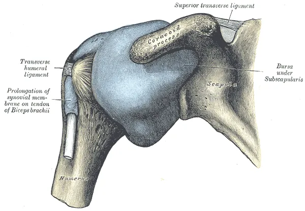
A loose sleeve with weak ligaments
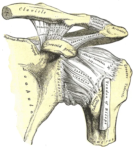
The joint capsule is the sleeve of tough tissue that encloses a joint. At the shoulder it completely encircles the joint9. It attaches around the rim of the glenoid cavity beyond the labrum, and to the humerus at a part called the anatomical neck9. The capsule is so loose that it lets the bones be drawn more than 2.5 cm apart9.
A ligament is a tough band of tissue that holds bones together at a joint. Thickenings of the capsule wall form weak ligaments: the coracohumeral ligament and three glenohumeral ligaments on the front of the capsule1. A classic anatomy text states that these ligaments limit movement and do not hold the joint surfaces together9.
| Ligament | What a review reports |
|---|---|
| Superior glenohumeral, with coracohumeral | Together, an important restraint against the head of the humerus sliding downward10 |
| Middle glenohumeral | Gives front-of-joint stability at 45 and 60 degrees of abduction10 |
| Inferior glenohumeral complex | The most important restraint against forward-and-downward dislocation, and the part of the capsule most often injured10 |
Muscles, compression and suction
A biomechanics review concludes that the shoulder’s stability rests mainly on active muscle control2. It gives the capsule, labrum and ligaments a minor role2. Muscles lie around the joint:
- above, the supraspinatus9
- in front, the subscapularis9
- behind, the infraspinatus and teres minor9
- below, the long head of the triceps9
- covering the front, back and side, the deltoid9
A tendon is a tough cord of tissue that connects a muscle to a bone. The tendons of the supraspinatus, subscapularis, infraspinatus and teres minor form the rotator cuff around the joint1.
Concavity compression is the stability gained by pressing the head of the humerus into the concave socket11. It may matter most in the middle of the range of motion, where the capsule and ligaments are slack11.
In 18 human shoulder preparations, atmospheric pressure on the sealed joint gave a mean stabilising force of 146 newtons12.
How far the arm moves
One population study set out to give norms for adults without shoulder pain or stiffness13. It measured active range of motion, how far a person can move a joint using their own muscles. In 2,404 people aged 20 to 91, average right-shoulder flexion was 161.5 degrees in men and 158.5 in women13.
An X-ray study followed twelve normal subjects raising the arm in the plane of the shoulder blade14. After about the first 30 degrees, glenohumeral movement and shoulder blade movement were in a ratio of 5 to 414. A study of 8 healthy volunteers measured with bone pins found a mean ratio of 1.7 to 115.
The roof over the joint
The acromion is the bony point at the top of the shoulder. The coracoid process is a hooked bony point at the front of the shoulder blade. Those two, with the coracoacromial ligament, form an arch that protects the joint from above9. In eight healthy men imaged with biplane fluoroscopy, the gap between acromion and humerus was smallest at roughly 90 degrees of arm elevation16. The shoulder impingement page covers pain around the acromion17.
Force across the joint
Joint contact force has been measured in patients with force-measuring shoulder implants18. One study reported these figures from four such patients:
| Task | Joint contact force, % of body weight |
|---|---|
| Passively holding a 10 kg weight at the side | 12.318 |
| Lifting the 10 kg weight to belt height | 91.518 |
| Lifting a 2 kg weight up | 98.318 |
| Lifting a 1.5 kg coffee pot with a straight arm | 10518 |
| Setting the 2 kg weight down from head height | 131.518 |
In 6 patients measured directly, raising the straight arm forward or to the side produced forces of up to 238% of body weight3.
Breakdown
Dislocations and partial slips
A review describes the glenohumeral joint as predisposed to instability by its bony anatomy20. Trauma and overuse can damage the tissues that keep it in line, leading to partial or full dislocation20. Repeated instability can follow, most often in the forward direction20.
Person-years are a count of people multiplied by the years each was followed. Three studies give rates of shoulder dislocation4,21,22:
| Where and who | Dislocations per 100,000 person-years |
|---|---|
| United States emergency departments, 2002 to 2006 | 23.94 |
| UK primary care records, 1995 to 2015: women aged 16 to 70 | 15.521 |
| UK primary care records, 1995 to 2015: men aged 16 to 70 | 40.421 |
| Oslo residents of all ages, 2009 | 56.322 |
Among 8,940 dislocations seen in United States emergency departments, 71.8% were in males, and the rate peaked at ages 20 to 294. Of those dislocations, 58.8% resulted from a fall and 48.3% occurred during sports or recreation4.
A subluxation is a partial slip of a joint out of its usual place. Among 4,141 students at the United States Military Academy followed for one academic year, 117 had a new traumatic instability event23. Of those events, 84.6% were subluxations and 15.4% were dislocations23.
In twelve patients with recurrent forward dislocation of one shoulder, a scan compared the two shoulders24. The front-lower and lower parts of the capsule were on average 19% longer on the dislocating side24. The shoulder dislocation page covers the condition and the trials in people who have it.
Stiffness and wear
A 2025 British guideline describes frozen shoulder as a painful condition that comes on gradually and typically leads to stiffness25. Two tissue studies of primary frozen shoulder describe the capsule differently26,27. A fibroblast is a cell in connective tissue that makes collagen, a strong, rope-like protein. In tissue from twelve patients, fibroblasts were actively growing and laying down thick bands of collagen, with no inflammation26. In biopsies from 22 patients, the tissue held fibroblasts and chronic inflammatory cells27.
Osteoarthritis is a long-term joint disease in which the cartilage lining the joint wears away. A review estimates that glenohumeral osteoarthritis accounts for 5% to 17% of patients with shoulder complaints28. The same review reports that X-ray findings and symptoms agree weakly, with many people having no symptoms28.
The throwing shoulder
External rotation is turning a limb outward, and internal rotation is turning it inward. A review reports that throwing athletes show more external rotation and less internal rotation in the dominant shoulder29. A 2008 review judged the evidence on the cause inconclusive29. It found a change in the shape of the upper humerus more compelling than changes in the joint’s soft tissues29.
Two pooled analyses of overhead athletes report on lost internal rotation and injury30,31. A meta-analysis is a study that pools the results of several earlier studies. One, of 17 studies and 2,195 athletes, found no shoulder motion measurement with a statistically significant link to shoulder or elbow injury30. Its non-significant results leaned toward more injury with internal rotation loss30. Another, of 819 athletes, found a weighted mean deficit of 13.8 degrees in injured athletes and 9.6 degrees in uninjured athletes31.
In the weights room
A review of resistance training injuries found up to 36% of documented injuries and disorders at the shoulder complex32. Most of the research it found was retrospective, looking back at past cases32. The authors of a dip study of 13 experienced men suggest the bench dip’s large shoulder extension may raise injury risk to the front capsule33. The study did not measure ligament strain or injuries33.
In an overhead press study of 159 healthy men, exercises needing the “high-five” position were associated with clinical characteristics of forward shoulder instability34.
Recovery
After a first dislocation
One meta-analysis pooled ten studies with 1,324 participants35. It found recurrent instability in 39% after a first traumatic forward dislocation35. The odds were higher in people aged 40 or under, in men and in people with hyperlaxity35. The same meta-analysis found that the rate of recurrent instability fell as time since the first dislocation increased35.
Twenty-five years later
A prospective Swedish study followed people aged 12 to 40 at their first forward dislocation36. X-rays of 223 shoulders 25 years later showed these findings:
| X-ray finding at 25 years | Share of 223 shoulders |
|---|---|
| Normal joint | 44%36 |
| Mild joint degeneration | 29%36 |
| Moderate degeneration | 9%36 |
| Severe degeneration | 17%36 |
Of shoulders with no further dislocation, 18% still had moderate or severe degeneration36. Of those that recurred without surgery, 39% did36.
The joint’s sense of position
Proprioception is the body’s sense of where its joints are and how they move. In 30 patients with forward shoulder instability, movement sense and joint position sense were significantly worse in the unstable shoulder than in the other37. In 20 patients who had had surgical reconstruction, the two senses did not differ significantly between the operated shoulder and the other37.
Connections
The shoulder blade joins the humerus at this joint and the clavicle (collarbone) at the acromioclavicular joint38.
The tendons of four muscles around the joint are covered on the rotator cuff page1. The long head of biceps tendon runs inside the shoulder joint but outside its lining39.
In a dissection of 15 specimens from cadavers (bodies donated for study), the axillary nerve supplied the lower part of the joint, front and back40.
Related condition pages: shoulder dislocation, frozen shoulder and shoulder impingement17,20,25.
Related exercise pages: bench press, overhead press, dip, push-up, pull-up and dead hang19,33,34,41,42,43.
Related stretch pages: sleeper stretch and doorway pec stretch44,45.
Shoulder joint is formed by
Shoulder joint is rimmed by
Shoulder joint works with
Shoulder joint is moved by
Shoulder joint is crossed by
Shoulder joint is supplied by
Shoulder joint is injured in
Shoulder joint is related to
Shoulder joint is loaded by
Shoulder joint is stretched by
Sources
- Betts JG, Young KA, Wise JA, Johnson E, Poe B, Kruse DH, Korol O, Johnson JE, Womble M, DeSaix P (2022). Anatomy and Physiology 2e. OpenStax. Link
- Veeger HE, van der Helm FC (2007). Shoulder function: the perfect compromise between mobility and stability.. Journal of biomechanics. doi:10.1016/j.jbiomech.2006.10.016 PMID 17222853
- Bergmann G, Graichen F, Bender A, Rohlmann A, Halder A, Beier A, Westerhoff P (2011). In vivo gleno-humeral joint loads during forward flexion and abduction.. Journal of biomechanics. doi:10.1016/j.jbiomech.2011.02.142 PMID 21481879
- Zacchilli MA, Owens BD (2010). Epidemiology of shoulder dislocations presenting to emergency departments in the United States.. The Journal of bone and joint surgery. American volume. doi:10.2106/JBJS.I.00450 PMID 20194311
- Chang LR, Anand P, Varacallo MA (2025). Anatomy, Shoulder and Upper Limb, Glenohumeral Joint. StatPearls. PMID 30725703
- Mostafa E, Imonugo O, Varacallo MA (2023). Anatomy, Shoulder and Upper Limb, Humerus.. StatPearls. PMID 30521242
- Almajed YA, Hall AC, Gillingwater TH, Alashkham A (2022). Anatomical, functional and biomechanical review of the glenoid labrum.. J Anat. doi:10.1111/joa.13582 PMID 34725812
- Howell SM, Galinat BJ (1989). The glenoid-labral socket. A constrained articular surface.. Clinical orthopaedics and related research. PMID 2721051
- Gray H; Lewis WH (editor) (1918). Anatomy of the Human Body. Lea & Febiger, Philadelphia (20th edition). Link
- Burkart AC, Debski RE (2002). Anatomy and function of the glenohumeral ligaments in anterior shoulder instability.. Clinical orthopaedics and related research. doi:10.1097/00003086-200207000-00005 PMID 12072743
- Lippitt S, Matsen F (1993). Mechanisms of glenohumeral joint stability.. Clinical orthopaedics and related research. PMID 8504601
- Habermeyer P, Schuller U, Wiedemann E (1992). The intra-articular pressure of the shoulder: an experimental study on the role of the glenoid labrum in stabilizing the joint.. Arthroscopy. doi:10.1016/0749-8063(92)90031-6 PMID 1637427
- Gill TK, Shanahan EM, Tucker GR, Buchbinder R, Hill CL (2020). Shoulder range of movement in the general population: age and gender stratified normative data using a community-based cohort.. BMC musculoskeletal disorders. doi:10.1186/s12891-020-03665-9 PMID 33046038
- Poppen NK, Walker PS (1976). Normal and abnormal motion of the shoulder.. The Journal of bone and joint surgery. American volume. PMID 1254624
- McClure PW, Michener LA, Sennett BJ, Karduna AR (2001). Direct 3-dimensional measurement of scapular kinematics during dynamic movements in vivo.. Journal of Shoulder and Elbow Surgery. doi:10.1067/mse.2001.112954 PMID 11408911
- Giphart JE, van der Meijden OA, Millett PJ (2012). The effects of arm elevation on the 3-dimensional acromiohumeral distance: a biplane fluoroscopy study with normative data.. Journal of shoulder and elbow surgery. doi:10.1016/j.jse.2011.11.023 PMID 22361718
- Diercks R, Bron C, Dorrestijn O, Meskers C, Naber R, de Ruiter T, Willems J, Winters J, van der Woude HJ, Dutch Orthopaedic Association (2014). Guideline for diagnosis and treatment of subacromial pain syndrome. Acta orthopaedica. doi:10.3109/17453674.2014.920991 PMID 24847788
- Westerhoff P, Graichen F, Bender A, Halder A, Beier A, Rohlmann A, Bergmann G (2009). In vivo measurement of shoulder joint loads during activities of daily living.. Journal of biomechanics. doi:10.1016/j.jbiomech.2009.05.035 PMID 19643418
- Noteboom L, Belli I, Hoozemans MJM, Seth A, Veeger HEJ, Van Der Helm FCT (2024). Effects of bench press technique variations on musculoskeletal shoulder loads and potential injury risk.. Frontiers in Physiology. doi:10.3389/fphys.2024.1393235 PMID 38974522
- Ladd LM, Crews M, Maertz NA (2021). Glenohumeral Joint Instability. Clinics in sports medicine. doi:10.1016/j.csm.2021.05.001 PMID 34509200
- Shah A, Judge A, Delmestri A, Edwards K, Arden NK, Prieto-Alhambra D, Holt TA, Pinedo-Villanueva RA, Hopewell S, Lamb SE, Rangan A, Carr AJ, Collins GS, Rees JL (2017). Incidence of shoulder dislocations in the UK, 1995-2015: a population-based cohort study.. BMJ open. doi:10.1136/bmjopen-2017-016112 PMID 29138197
- Liavaag S, Svenningsen S, Reikerås O, Enger M, Fjalestad T, Pripp AH, Brox JI (2011). The epidemiology of shoulder dislocations in Oslo.. Scandinavian journal of medicine & science in sports. doi:10.1111/j.1600-0838.2011.01300.x PMID 21507063
- Owens BD, Duffey ML, Nelson BJ, DeBerardino TM, Taylor DC, Mountcastle SB (2007). The incidence and characteristics of shoulder instability at the United States Military Academy.. The American journal of sports medicine. doi:10.1177/0363546506295179 PMID 17581976
- Urayama M, Itoi E, Sashi R, Minagawa H, Sato K (2003). Capsular elongation in shoulders with recurrent anterior dislocation. The American journal of sports medicine. doi:10.1177/03635465030310012201 PMID 12531759
- Rupani N, Gwilym SE, BESS Frozen Shoulder Working Group (2025). British Elbow and Shoulder Society patient care pathway: Frozen shoulder.. Shoulder & elbow. doi:10.1177/17585732251335955 PMID 40291049
- Bunker TD, Anthony PP (1995). The pathology of frozen shoulder. A Dupuytren-like disease.. The Journal of bone and joint surgery. British volume. PMID 7559688
- Hand GC, Athanasou NA, Matthews T, Carr AJ (2007). The pathology of frozen shoulder.. The Journal of bone and joint surgery. British volume. doi:10.1302/0301-620X.89B7.19097 PMID 17673588
- Ibounig T, Simons T, Launonen A, Paavola M (2021). Glenohumeral osteoarthritis: an overview of etiology and diagnostics.. Scandinavian journal of surgery : SJS : official organ for the Finnish Surgical Society and the Scandinavian Surgical Society. doi:10.1177/1457496920935018 PMID 32662351
- Borsa PA, Laudner KG, Sauers EL (2008). Mobility and stability adaptations in the shoulder of the overhead athlete. Sports medicine (Auckland, N.Z.). doi:10.2165/00007256-200838010-00003 PMID 18081365
- Keller RA, De Giacomo AF, Neumann JA, Limpisvasti O, Tibone JE (2018). Glenohumeral Internal Rotation Deficit and Risk of Upper Extremity Injury in Overhead Athletes: A Meta-Analysis and Systematic Review.. Sports health. doi:10.1177/1941738118756577 PMID 29381423
- Johnson JE, Fullmer JA, Nielsen CM, Johnson JK, Moorman CT (2018). Glenohumeral Internal Rotation Deficit and Injuries: A Systematic Review and Meta-analysis.. Orthopaedic journal of sports medicine. doi:10.1177/2325967118773322 PMID 29845083
- Kolber MJ, Beekhuizen KS, Cheng MS, Hellman MA (2010). Shoulder injuries attributed to resistance training: a brief review.. Journal of strength and conditioning research. doi:10.1519/JSC.0b013e3181dc4330 PMID 20508476
- McKenzie A, Crowley-McHattan Z, Meir R, Whitting J, Volschenk W (2022). Bench, Bar, and Ring Dips: Do Kinematics and Muscle Activity Differ?. International Journal of Environmental Research and Public Health. doi:10.3390/ijerph192013211 PMID 36293792
- Kolber MJ, Corrao M, Hanney WJ (2013). Characteristics of anterior shoulder instability and hyperlaxity in the weight-training population.. Journal of Strength and Conditioning Research. doi:10.1519/JSC.0b013e318269f776 PMID 22836608
- Olds M, Ellis R, Donaldson K, Parmar P, Kersten P (2015). Risk factors which predispose first-time traumatic anterior shoulder dislocations to recurrent instability in adults: a systematic review and meta-analysis.. British journal of sports medicine. doi:10.1136/bjsports-2014-094342 PMID 25900943
- Hovelius L, Saeboe M (2009). Neer Award 2008: Arthropathy after primary anterior shoulder dislocation--223 shoulders prospectively followed up for twenty-five years.. Journal of shoulder and elbow surgery. doi:10.1016/j.jse.2008.11.004 PMID 19254851
- Lephart SM, Warner JJ, Borsa PA, Fu FH (1994). Proprioception of the shoulder joint in healthy, unstable, and surgically repaired shoulders.. Journal of shoulder and elbow surgery. doi:10.1016/S1058-2746(09)80022-0 PMID 22958841
- Miniato MA, Mudreac A, Borger J (2023). Anatomy, Thorax, Scapula.. StatPearls. PMID 30855903
- Varacallo MA, Mair SD (2023). Biceps Tendon Dislocation and Instability.. StatPearls. PMID 30475566
- Tran J, Peng PWH, Agur AMR (2019). Anatomical study of the innervation of glenohumeral and acromioclavicular joint capsules: implications for image-guided intervention.. Regional anesthesia and pain medicine. doi:10.1136/rapm-2018-100152 PMID 30635516
- Polovinets O, Wolf A, Friedlander S, Wollstein R (2026). Push-Up Techniques Affect Elbows and Shoulders: How Should They Be Used?. Handchirurgie, Mikrochirurgie, plastische Chirurgie. doi:10.1055/a-2862-1658 PMID 42269686
- Summitt RJ, Cotton RA, Kays AC, Slaven EJ (2016). Shoulder Injuries in Individuals Who Participate in CrossFit Training.. Sports health. doi:10.1177/1941738116666073 PMID 27578854
- Prinold JA, Bull AM (2016). Scapula kinematics of pull-up techniques: Avoiding impingement risk with training changes.. Journal of Science and Medicine in Sport. doi:10.1016/j.jsams.2015.08.002 PMID 26383875
- Izumi T, Aoki M, Muraki T, Hidaka E, Miyamoto S (2008). Stretching positions for the posterior capsule of the glenohumeral joint. The American journal of sports medicine. doi:10.1177/0363546508318196 PMID 18567716
- Laudner KG, Wenig M, Selkow NM, Williams J, Post E (2015). Forward Shoulder Posture in Collegiate Swimmers: A Comparative Analysis of Muscle-Energy Techniques.. Journal of athletic training. doi:10.4085/1062-6050-50.11.07 PMID 26509682
Changes
- Revised to report sources added after their published corrections were read