Shoulder labrum
Also called glenoid labrum, labrum of the shoulder, glenoid ligament
The rim of fibrocartilage around the edge of the shoulder socket. What it is made of, what it adds to stability, how and in whom it tears, what scans show in shoulders without symptoms, and what studies found about healing.
- Region
- Shoulder
- Updated
- October 8, 2026
- Reviewed
- October 8, 2026 · how entries are checked
Function
A rim around the socket
The glenoid labrum is a rim of fibrocartilage attached around the whole edge of the shoulder socket1. The socket, called the glenoid fossa or glenoid cavity, is a shallow depression at the upper outer corner of the scapula (shoulder blade)1,10. The labrum has three sides1. One faces the head of the humerus (upper arm bone) and one the bone of the socket1. The third faces the joint capsule (the sleeve of tough tissue that encloses a joint)1.
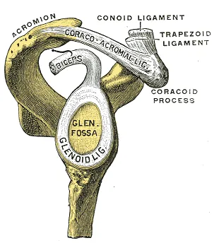
The figure labels the labrum “Glenoid lig.” and the socket “Glen. fossa”. In an anatomical study of 25 specimen shoulders, the joint surface and labrum together made a socket about 9 mm deep from top to bottom11. It was about 5 mm deep from front to back, and the labrum supplied about half of that depth11.
In an anatomical study of 23 fresh-frozen cadaver (donated body) shoulders, the top and upper-front parts of the labrum were loosely attached to the socket7. Those parts resembled the meniscus (a pad of cartilage in the knee)7. The lower part was firmly attached to the rim and looked like a fibrous, immobile extension of the joint cartilage7.
A study of 20 dry shoulder blades and 19 cadaver shoulders looked at where the long head of the biceps starts12. The biceps is the two-headed muscle on the front of the upper arm. Two thirds arose from the supraglenoid tubercle, a small bump of bone at the top of the socket12. The rest arose from the labrum12. The long head of biceps tendon page covers that origin.
Gaps and recesses under the upper labrum
In 26 fresh-frozen cadaver shoulders, 73% had a recess under the top of the labrum13. It looked like published examples of SLAP lesions on MRI13. Under the microscope in two shoulders, it had a synovial lining (the thin inner lining of a joint capsule) and no scarring to suggest injury13.
In the study of shoulder blades and cadaver shoulders, clefts and holes under the labrum were found at the upper front and nowhere else12.
What it is made of
A study of 38 cadaver shoulders from people of different ages used histology (the study of tissue under a microscope)14. The labrum was fibrocartilage in children and adults14.
Accounts of the blood supply differ. In the study of 23 fresh-frozen cadaver shoulders, blood vessels were limited to the outer edge of the labrum7. In the study of 38 cadaver shoulders, vessels were sparse throughout and became fewer with increasing age14. A dissection study of 140 shoulders from 70 cadavers, mean age 81.5, reached a different view8. Its authors conclude that the labrum has a rich blood supply, sufficient for it to reattach8.
What it adds to stability
One review describes the exact functions of the labrum in the living shoulder as not yet fully established1. It has been proposed to add stability by deepening the socket, enlarging the joint surface, centring the head of the humerus and sealing the joint1.
Concavity-compression is the holding of the head of the humerus in the concave socket by the compressive force of the surrounding muscles15. In ten fresh-frozen cadaver shoulder joints, cutting away the labrum reduced the effectiveness of compression stabilisation by about 20%16. A later study of ten cadaver shoulders put the labrum’s average contribution at about 10%, about half the value reported before17.
A biomechanics review concludes that the shoulder’s stability rests mainly on active control by muscles18. It gives the capsule, labrum and ligaments a minor role18. The shoulder joint page covers the joint as a whole.
Breakdown
The names given to tears
One review states that labral lesions can occur anywhere around the rim1. It describes the top as the most commonly affected site, as a SLAP lesion1. The Bankart lesion is a detachment of the front of the labrum along with the joint capsule, at the lower front of the socket1. The reverse Bankart lesion is at the lower back1.
SLAP lesions were first classified into four types in 19901:
- Type I. The top of the labrum is frayed, and the attachment of the biceps and labrum is intact1.
- Type II. The top of the labrum is detached, together with the long head of biceps tendon1.
- Type III. A “bucket handle” piece of the top of the labrum is displaced into the joint1.
- Type IV. A bucket-handle tear is displaced into the joint along with part of the biceps tendon1.
How often SLAP lesions are seen at arthroscopy
The counts below come from people who were already having shoulder arthroscopy2,3,19. A retrospective review of 530 arthroscopies found 32 SLAP lesions, or 6%2. A prospective (forward-following) consecutive case series of 544 arthroscopies found a SLAP lesion in 26%3. A third series found 140 injuries of the top of the labrum among 2,375 arthroscopies19. The last two report the mix of types, and they differ3,19.
| Type | Share of the 140 injuries | Share of SLAP lesions in the 544 series | Sources |
|---|---|---|---|
| I | 21% | 74% | 3,19 |
| II | 55% | 21% | 3,19 |
| III | 9% | 0.7% | 3,19 |
| IV | 10% | 4% | 3,19 |
Of the 140 injuries, 29% also had a partial-thickness tear in the rotator cuff, and 28% were isolated19. The rotator cuff is a group of four muscles that cross the shoulder joint and help hold it steady. The authors of the 544 series conclude that care is needed before blaming symptoms on a SLAP lesion when other damage is found3.
How tears happen
That review attributes labral tears to repetitive movement combined with excessive stress, or to a sudden injury such as falling on an outstretched arm1.
Dislocation. A systematic review of 22 studies, covering 1,920 shoulders, looked at front dislocations from injury4. It found a Bankart lesion in 59% after a first dislocation and 66% after repeated dislocations4. The authors urge caution because of substantial heterogeneity, meaning the studies’ results differed from one another4. Recurrence, trials of surgery and return to sport are on the shoulder dislocation page.
Throwing. Arthroscopy of 73 throwing athletes, baseball pitchers among them, found most labral tears at the upper front, near where the biceps long head starts20. In 17 athletes with unexplained shoulder pain on throwing, arthroscopy showed two rotator cuff insertions pinching against the upper-back rim of the socket21. Twelve had lesions of the upper-back labrum21. The shoulder impingement page covers that pinching.
Bracing against an opponent. A report of nine contact athletes found the back of the labrum detached from the socket at arthroscopy22. The authors hypothesise that repeated shear, a sliding force, from bracing against opponents, with the arms in front of the body, detaches the labrum22.
Changes with age
In 32 normal cadaver shoulders, mean age 57, the labrum looked no different between age groups to the naked eye23. Under the microscope, lesions in those shoulders increased significantly with age in each region23. The authors conclude that labral changes seen at arthroscopy should be judged against the changes that come with age in normal shoulders23.
Scans of shoulders without symptoms
A systematic review of imaging in adults without shoulder symptoms covered 35 studies5. It judged each of them at high risk of bias and could not pool them5. Its one population-level figure for labral abnormalities was 20%, from one study of 20 shoulders, rated very low certainty5.
Five MRI studies, each of one group, report findings in people without symptoms6,24,25,26,27.
| Who was scanned | What MRI showed | Source |
|---|---|---|
| Non-athletic adults aged 18 to 29, no history of shoulder pain | Labral abnormalities in 9% of 58 shoulders | 24 |
| 25 collegiate and professional ice hockey players | Labral abnormalities in 25% of 50 shoulders | 25 |
| 50 elite climbers aged 20 to 60, no shoulder pain | Labral pathology in 69% of shoulders, discrete tears in 56% | 26 |
| 14 professional baseball pitchers, both arms | Labrum abnormal in 79% of 28 shoulders | 27 |
| 53 adults aged 45 to 60, no history of shoulder injury or surgery | Two radiologists read a tear of the top in 55% and 72% of scans | 6 |
In the study of 53 adults, the readings did not differ by age, sex, arm dominance, physical work or a history of overhead sport6. The authors suggest that such MRI findings between 45 and 60 may be normal age-related findings6.
Recovery
Whether a torn labrum heals
Two reports looked at healing after SLAP repair9,19. In a series of 140 injuries of the top of the labrum, 18 shoulders had a repeat arthroscopy19. Four of five type II lesions fixed with an absorbable anchor had healed19. In a retrospective study of 43 patients, CT arthrography one year after arthroscopic SLAP repair showed the labrum had not healed in 32.6%9. Clinical outcomes did not differ significantly between healed and unhealed repairs9.
Sport after SLAP surgery
A systematic review and meta-analysis (a study that pools the results of several earlier studies) covered 22 studies and 617 athletes28. On limited evidence from lower-level studies, 93% of athletes returned to sport after surgery for a SLAP injury, and 72% to their previous level28.
Connections
The labrum is attached around the edge of the socket of the shoulder joint1. That socket is on the scapula, and the head of the humerus fits against it as a ball-and-socket joint10,29.
Part of the long head of biceps tendon arises from the labrum12. Among 102 type II SLAP lesions without front instability, tears of the rotator cuff were present in 31% of patients30.
The authors of an indentation study call the labrum’s stiffness in compression similar to that of knee menisci31. The hip socket has a fibrocartilage rim too, the hip labrum32. The page on cartilage covers the tissue in general.
Related exercise pages: bench press. In the report of nine contact athletes whose back labrum was detached, each had pain with bench pressing and in their sport22.
Shoulder labrum forms part of
Shoulder labrum attaches to
Shoulder labrum works with
Shoulder labrum anchors part of
Shoulder labrum is often injured with
Shoulder labrum is torn in
Shoulder labrum is related to
Shoulder labrum is compared with
Sources
- Almajed YA, Hall AC, Gillingwater TH, Alashkham A (2022). Anatomical, functional and biomechanical review of the glenoid labrum.. J Anat. doi:10.1111/joa.13582 PMID 34725812
- Handelberg F, Willems S, Shahabpour M, Huskin JP, Kuta J (1998). SLAP lesions: a retrospective multicenter study.. Arthroscopy. doi:10.1016/s0749-8063(98)70028-3 PMID 9848599
- Kim TK, Queale WS, Cosgarea AJ, McFarland EG (2003). Clinical features of the different types of SLAP lesions: an analysis of one hundred and thirty-nine cases.. J Bone Joint Surg Am. PMID 12533574
- Rutgers C, Verweij LPE, Priester-Vink S, van Deurzen DFP, Maas M, van den Bekerom MPJ (2022). Recurrence in traumatic anterior shoulder dislocations increases the prevalence of Hill-Sachs and Bankart lesions: a systematic review and meta-analysis.. Knee Surg Sports Traumatol Arthrosc. doi:10.1007/s00167-021-06847-7 PMID 34988633
- Ibounig T, Sanders S, Haas R, Jones M, Järvinen TL, Taimela S, Docking S, Rämö L, Buchbinder R (2024). Systematic Review of Shoulder Imaging Abnormalities in Asymptomatic Adult Shoulders (SCRUTINY): Abnormalities of the glenohumeral joint.. Osteoarthritis Cartilage. doi:10.1016/j.joca.2024.06.001 PMID 38876437
- Schwartzberg R, Reuss BL, Burkhart BG, Butterfield M, Wu JY, McLean KW (2016). High Prevalence of Superior Labral Tears Diagnosed by MRI in Middle-Aged Patients With Asymptomatic Shoulders.. Orthop J Sports Med. doi:10.1177/2325967115623212 PMID 26779556
- Cooper DE, Arnoczky SP, O'Brien SJ, Warren RF, DiCarlo E, Allen AA (1992). Anatomy, histology, and vascularity of the glenoid labrum. An anatomical study.. J Bone Joint Surg Am. PMID 1734013
- Alashkham A, Alraddadi A, Felts P, Soames R (2017). Blood supply and vascularity of the glenoid labrum: Its clinical implications.. J Orthop Surg (Hong Kong). doi:10.1177/2309499017731632 PMID 28920546
- Nashikkar PS, Rhee SM, Desai CV, Oh JH (2018). Is Anatomical Healing Essential for Better Clinical Outcome in Type II SLAP Repair? Clinico-Radiological Outcome after Type II SLAP Repair.. Clin Orthop Surg. doi:10.4055/cios.2018.10.3.358 PMID 30174813
- Betts JG, Young KA, Wise JA, Johnson E, Poe B, Kruse DH, Korol O, Johnson JE, Womble M, DeSaix P (2022). Anatomy and Physiology 2e. OpenStax. Link
- Howell SM, Galinat BJ (1989). The glenoid-labral socket. A constrained articular surface.. Clinical orthopaedics and related research. PMID 2721051
- Bain GI, Galley IJ, Singh C, Carter C, Eng K (2013). Anatomic study of the superior glenoid labrum.. Clin Anat. doi:10.1002/ca.22145 PMID 23001615
- Smith DK, Chopp TM, Aufdemorte TB, Witkowski EG, Jones RC (1996). Sublabral recess of the superior glenoid labrum: study of cadavers with conventional nonenhanced MR imaging, MR arthrography, anatomic dissection, and limited histologic examination.. Radiology. doi:10.1148/radiology.201.1.8816553 PMID 8816553
- Prodromos CC, Ferry JA, Schiller AL, Zarins B (1990). Histological studies of the glenoid labrum from fetal life to old age.. J Bone Joint Surg Am. PMID 2229110
- Lazarus MD, Sidles JA, Harryman DT, Matsen FA (1996). Effect of a chondral-labral defect on glenoid concavity and glenohumeral stability. A cadaveric model.. J Bone Joint Surg Am. doi:10.2106/00004623-199601000-00013 PMID 8550685
- Lippitt SB, Vanderhooft JE, Harris SL, Sidles JA, Harryman DT, Matsen FA (1993). Glenohumeral stability from concavity-compression: A quantitative analysis.. J Shoulder Elbow Surg. doi:10.1016/S1058-2746(09)80134-1 PMID 22959294
- Halder AM, Kuhl SG, Zobitz ME, Larson D, An KN (2001). Effects of the glenoid labrum and glenohumeral abduction on stability of the shoulder joint through concavity-compression : an in vitro study.. J Bone Joint Surg Am. doi:10.2106/00004623-200107000-00013 PMID 11451977
- Veeger HE, van der Helm FC (2007). Shoulder function: the perfect compromise between mobility and stability.. Journal of biomechanics. doi:10.1016/j.jbiomech.2006.10.016 PMID 17222853
- Snyder SJ, Banas MP, Karzel RP (1995). An analysis of 140 injuries to the superior glenoid labrum.. J Shoulder Elbow Surg. doi:10.1016/s1058-2746(05)80015-1 PMID 8542365
- Andrews JR, Carson WG, McLeod WD (1985). Glenoid labrum tears related to the long head of the biceps.. Am J Sports Med. doi:10.1177/036354658501300508 PMID 4051091
- Walch G, Boileau P, Noel E, Donell ST (1992). Impingement of the deep surface of the supraspinatus tendon on the posterosuperior glenoid rim: An arthroscopic study.. J Shoulder Elbow Surg. doi:10.1016/S1058-2746(09)80065-7 PMID 22959196
- Mair SD, Zarzour RH, Speer KP (1998). Posterior labral injury in contact athletes.. Am J Sports Med. doi:10.1177/03635465980260060301 PMID 9850774
- Pfahler M, Haraida S, Schulz C, Anetzberger H, Refior HJ, Bauer GS, Bigliani LU (2003). Age-related changes of the glenoid labrum in normal shoulders.. J Shoulder Elbow Surg. doi:10.1067/mse.2003.3 PMID 12610485
- Liu R, Onks C, Petscavage-Thomas J, Walker E, Berg A, Silvis M, Dhawan A (2021). Prevalence of shoulder labral abnormalities on MRI in a non-athletic asymptomatic young adult population.. Skeletal Radiol. doi:10.1007/s00256-020-03629-z PMID 33021682
- Hacken B, Onks C, Flemming D, Mosher T, Silvis M, Black K, Stuck D, Dhawan A (2019). Prevalence of MRI Shoulder Abnormalities in Asymptomatic Professional and Collegiate Ice Hockey Athletes.. Orthop J Sports Med. doi:10.1177/2325967119876865 PMID 31637270
- Cooper JD, Seiter MN, Ruzbarsky JJ, Poulton R, Dornan GJ, Fitzcharles EK, Ho CP, Hackett TR (2022). Shoulder Pathology on Magnetic Resonance Imaging in Asymptomatic Elite-Level Rock Climbers.. Orthop J Sports Med. doi:10.1177/23259671211073137 PMID 35174249
- Miniaci A, Mascia AT, Salonen DC, Becker EJ (2002). Magnetic resonance imaging of the shoulder in asymptomatic professional baseball pitchers.. Am J Sports Med. doi:10.1177/03635465020300012501 PMID 11798999
- Freijomil N, Peters S, Millay A, Sinda T, Sunset J, Reiman MP (2020). THE SUCCESS OF RETURN TO SPORT AFTER SUPERIOR LABRUM ANTERIOR TO POSTERIOR (SLAP) TEARS: A SYSTEMATIC REVIEW AND META-ANALYSIS.. Int J Sports Phys Ther. doi:10.26603/ijspt20200659 PMID 33110685
- Mostafa E, Imonugo O, Varacallo MA (2023). Anatomy, Shoulder and Upper Limb, Humerus.. StatPearls. PMID 30521242
- Morgan CD, Burkhart SS, Palmeri M, Gillespie M (1998). Type II SLAP lesions: three subtypes and their relationships to superior instability and rotator cuff tears.. Arthroscopy. doi:10.1016/s0749-8063(98)70049-0 PMID 9754471
- Carey J, Small CF, Pichora DR (2000). In situ compressive properties of the glenoid labrum.. J Biomed Mater Res. doi:10.1002/1097-4636(20000915)51:4<711::aid-jbm20>3.0.co;2-z PMID 10880120
- Groh MM, Herrera J (2009). A comprehensive review of hip labral tears.. Current Reviews in Musculoskeletal Medicine. doi:10.1007/s12178-009-9052-9 PMID 19468871
Changes
- Revised to report sources added after their published corrections were read