Cartilage
Also called cartilage tissue
The firm, rubbery tissue that lines joints, forms the knee's menisci and the spine's discs, and lets bones grow. How it responds to load, how it gets damaged, what training studies found, and how it changes with age.
- Region
- Across the body
- Updated
- September 30, 2026
- Reviewed
- September 30, 2026 · how entries are checked
What it is
Cartilage (a firm, rubbery body tissue, softer than bone) has cells called chondrocytes. They sit in small spaces called lacunae inside the cartilage matrix (the material around the cells)1. Cartilage’s distinctive appearance comes from sugar chains called chondroitin sulfates, which bind with proteins to form proteoglycans (large protein-and-sugar molecules that hold water)1. Articular cartilage has no blood vessels or nerves2. In cartilage without blood vessels, nutrients must seep through the matrix to reach the cells1.
Three kinds of cartilage
The three main kinds differ in what runs through their matrix1:
- Hyaline cartilage, the most common, has short, scattered fibres of collagen (a strong, rope-like protein) and a lot of proteoglycan. It is found in the nose and rib cage and on bone ends at movable joints1.
- Fibrocartilage is tough because thick bundles of collagen run through it. It can absorb pressure, and the knee’s menisci (pads of cartilage in the knee) and the spine’s intervertebral discs are made of it1.
- Elastic cartilage also holds elastic fibres, so it is stiff but springy. The outer ear contains it1.
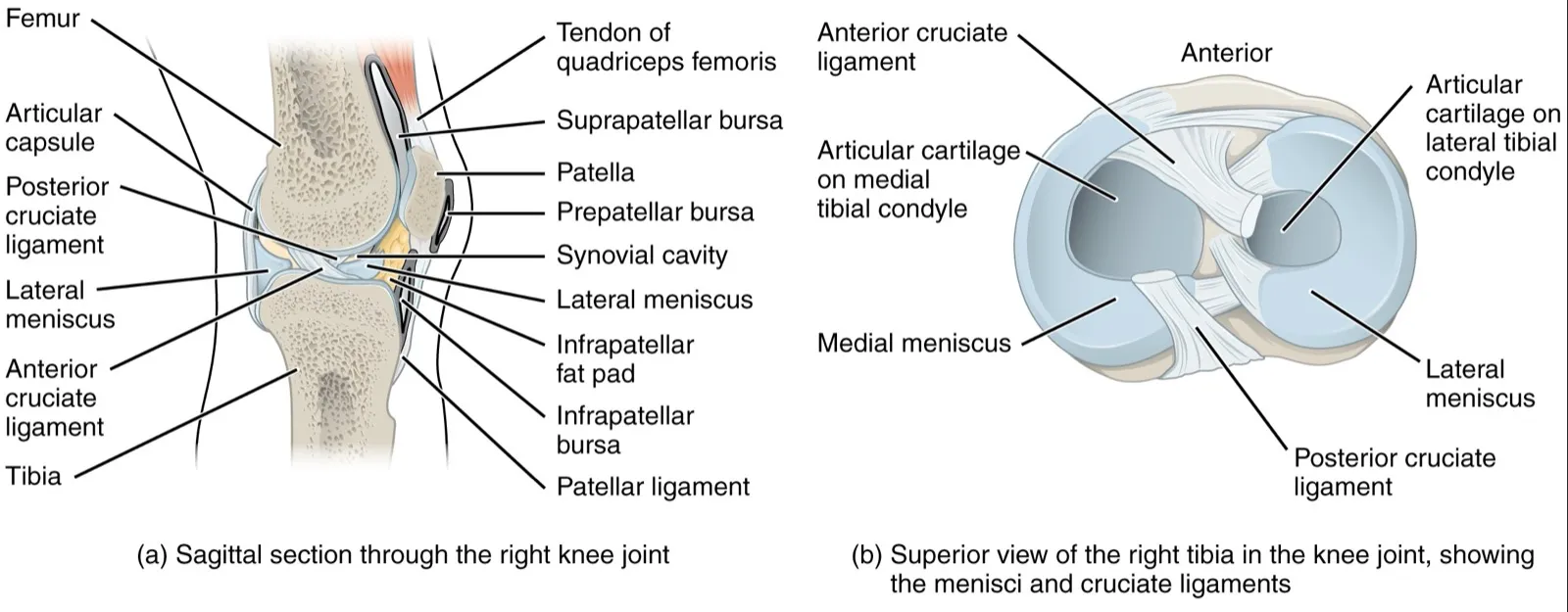
The knee holds two of these kinds. The articular cartilage (the hyaline layer on bone ends in a joint) covers the bone surfaces, and the menisci are fibrocartilage1. Each spinal disc has a tough outer ring around a soft, gel-like centre whose water resists squeezing and helps bear weight1.
Cartilage and the growing skeleton
Hyaline cartilage forms a model of the skeleton in the embryo before bone forms1. Once the fetal skeleton has formed, this cartilage model remains only at joint surfaces and at the growth plate1. The growth plate sits between a long bone’s shaft and end1. That growth plate lets bones keep growing until adulthood1.
How it responds to load
Chondrocytes renew the cartilage matrix, and mechanical forces strongly shape whether cartilage is kept up, breaks down or rebuilds7. The cells respond to growth factors, mechanical loads and fluid pressure2.
A review of ion channels (pores in a cell’s outer layer) in chondrocytes describes two paths7. Physiological (normal) loading, at 10% strain (stretch as a share of resting length), opens a channel called TRPV47. This drives a building pathway that raises production of collagen II and aggrecan (a proteoglycan)7. Injurious loading, at 50% strain, works through a channel called Piezo1 to switch on a breakdown pathway7.
Use and disuse
A review states that joint motion and load are important for keeping articular cartilage normal, and that joint inactivity leads to cartilage breakdown2. In people with complete spinal cord injury, knee cartilage was thinner than in healthy volunteers, and the gap widened over time8. At 24 months it was 23% to 25% thinner at the kneecap and inner shinbone8. In 20 people with ankle fractures who bore only partial weight for 7 weeks, knee cartilage thinned by 2.9% to 6.6%9. A review of meniscus research states that feeding the meniscus by diffusion needs repeated loading and unloading10.
What one run does
A meta-analysis of 24 MRI studies found knee cartilage thickness and volume fell right after running3. Volume fell by 3.3% in the weight-bearing thigh-bone cartilage and 4.9% behind the kneecap3. Its authors rated this evidence very low certainty and judged the changes small and apparently short-lived3. T2 (an MRI measure that changes when tissue is damaged or swollen) in the main knee joint’s cartilage was back to baseline within 91 minutes3.
A second review, of 43 MRI studies, concluded that cartilage changes after running are short-lived and likely reflect normal fluid movement11. It found moderate evidence that running does not lead to new cartilage lesions (areas of damaged tissue)11. The evidence on existing knee lesions was conflicting11.
How it gets injured
Articular cartilage injury and wear are frequent in synovial joints (joints with a fluid-filled space between the bones)12. Impact and twisting loads can injure it, and the injuries fall into three classes13:
- damage inside the cartilage under an intact surface13;
- a break limited to the cartilage13;
- a break through both the cartilage and the bone beneath13.
A review of 11 studies of 931 athletes found full-thickness cartilage defects in 36% of athletes’ knees overall14. This ranged from 2.4% to 75% between studies14. It concluded such defects are more common in athletes than in the general population14.
Why it heals poorly
Articular cartilage has a limited ability to heal and repair itself2. It cannot rebuild to its original quality12. Drilling or piercing the bone beneath prompts a repair process that forms fibrocartilage12. This fibrocartilage is weaker than normal hyaline cartilage and cannot stand up to long-term repeated use15.
Articular cartilage collagen has a calculated half-life of 117 years, against 15 years for skin collagen16.
Osteoarthritis
Osteoarthritis is the most common type of arthritis, and is associated with ageing and wear of joint cartilage1. It is linked to chondrocytes’ breakdown and building of matrix falling out of balance2. A draft definition from OARSI, a research society, describes it as starting with small and large injuries that trigger faulty repair17.
The Global Burden of Disease study estimated 595 million people had osteoarthritis in 2020, 7.6% of the world’s population6. The knee was the most common site6. High body mass index (weight for height) contributed an estimated 20.4% of all osteoarthritis, though this estimate was very uncertain6. A review of 53 studies found the odds of knee osteoarthritis about 4 times higher after an ACL (anterior cruciate ligament) injury18. After a meniscus injury, alone or with an ACL injury, the odds were about 6 times higher18.
Sport, activity and osteoarthritis
- A systematic review (a study that sums up all the research on a question) compared sport participants with non-participants19. It found knee osteoarthritis in 7.7% of 3,759 sport participants and 7.3% of 4,730 non-participants19.
- The same review found it significantly more common in soccer players, elite long-distance runners, competitive weightlifters and wrestlers19.
- A meta-analysis of 17 studies found hip and knee osteoarthritis in 3.5% of recreational runners and 13.3% of competitive runners. The figure was 10.2% in non-running controls20.
- A meta-analysis of 14 observational studies (studies that watch people without assigning groups) found 26% higher knee osteoarthritis risk with high than with moderate activity21. Neither high nor moderate activity carried lower risk than low activity21.
The sport review also looked at players with no past knee injury. Elite soccer and orienteering athletes, and non-elite soccer and American football players, still had more knee osteoarthritis than non-participants19. Its authors note it is unclear whether any added risk comes from the sport, a sport injury or something else19. The running review could not tell whether its links were causal or explained by factors such as past injury20.
Reviews of activity and knee structure on MRI also disagree. One found strong evidence that activity goes with fewer cartilage defects but more osteophytes (bone spurs), and limited evidence of more cartilage volume22. A later review of 18 studies in adults without knee osteoarthritis found no strong evidence either way23.
How training builds it
A review of 9 randomised trials (studies that assign people to groups by chance) studied people with knee osteoarthritis or at raised risk of it4. It concluded that knee-loading exercise seems not to harm cartilage, as measured on MRI4. The evidence quality was low4. In trials of people with knee osteoarthritis, six comparisons found exercise did not thicken cartilage or change its volume or defects4.
A study of athletes points the same way. Knee cartilage thickness did not differ significantly between 18 triathletes training over 10 hours a week and 18 people never regularly active5.
A review of 29 randomised trials in healthy animals found moderate daily exercise went with no change or better knee cartilage24. In 19 of 27 high-dose comparisons in these animals, cartilage measures fell24. The authors suggest the link between daily exercise dose and cartilage make-up may not be a straight line24. The authors of the 43-study running review suggest cartilage adapts to repeated running11.
The discs of the spine
A meta-analysis of 39 studies (4,152 people) linked upright loading on two legs, mostly running, with better disc health25. The certainty was very low, and no other kind of loading was linked to better or worse disc health25. In a running review, one 15-year study found no difference in disc change between runners and controls26.
Across the lifespan
Children and adolescents
In 74 children aged 9 to 18 measured about 1.6 years apart, knee cartilage volume grew at every site27. Those above the median for sport intensity gained about twice as much at the tibia (shinbone), though not at the kneecap27.
The growth plate is the cartilage layer near a growing bone’s end. A review of its injuries in young athletes notes it resists stress less than adult joint cartilage28. It also resists shear and tension less than nearby bone28. So forces that might tear a ligament in an adult can injure the growth plate instead28.
- The growth plate is involved in about 15% of children’s fractures28.
- Overuse problems of the apophyses (growth plates where major tendons attach) are common in young athletes28. One is Osgood-Schlatter disease (a painful bump just below the kneecap in growing children)28.
- In eight studies of gymnasts, 10% to 85% had X-ray changes consistent with growth plate stress in the radius (a forearm bone) at the wrist28.
A review of 24 studies looked at overuse growth plate injuries in young athletes29. It named rapid growth, age, body size, training volume and past injury as risk factors29.
Adults and older adults
With age, articular cartilage holds less water and grows stiffer when squeezed2. Chondrocytes lose some of their ability to divide and make matrix, but still release substances that drive inflammation and break down matrix30. Sugar-modified proteins build up in the matrix, and aged cartilage copes less well with stress30. MRI studies show knee cartilage thinning with age, mainly on the thigh bone and kneecap30.
Sources differ on cell numbers. One says the total number of chondrocytes stays essentially unchanged with age2. Another describes age-related thinning as consistent with falling water content and cell numbers30.
With age, the meniscus becomes stiffer and less elastic10. The disc’s centre slowly loses water, so the disc becomes thinner and less flexible1. In 3,110 people without back pain, scans showed disc degeneration (wear-type disc changes) in 37% of 20-year-olds and 96% of 80-year-olds31. The review’s authors concluded many such features are likely part of normal ageing31.
Osteoarthritis most often occurs after age 40, and becomes steeply more common with age6. In Global Burden of Disease estimates for 2020, it affected6:
| Age group | People with osteoarthritis, per 100,000 |
|---|---|
| 25 to 49 | 29846 |
| 50 to 69 | 232376 |
| 70 and over | 384196 |
Knee osteoarthritis peaked at ages 80 to 846.
Connections
The library has entries on two cartilage structures of the knee. Articular cartilage is the hyaline layer on the bone ends1. The two menisci are C-shaped pads of fibrocartilage between the thigh bone and shinbone1. Their injury has its own page, meniscus tear. The 43-study running review found no change in foot and ankle cartilage with repeated running11.
Cartilage covers the ends of bones at movable joints, and a cartilage plate lets bones grow in length1. Injury to the ACL, a ligament, was linked to higher odds of knee osteoarthritis18. Traction growth plates sit where major tendons attach28.
Cartilage has an entry on
Cartilage lines
Cartilage is injured in
Cartilage covers the ends of
Cartilage shares joints with
Cartilage meets at growth plates
Sources
- Betts JG, Young KA, Wise JA, Johnson E, Poe B, Kruse DH, Korol O, Johnson JE, Womble M, DeSaix P (2022). Anatomy and Physiology 2e. OpenStax. Link
- Sophia Fox AJ, Bedi A, Rodeo SA (2009). The basic science of articular cartilage: structure, composition, and function.. Sports health. doi:10.1177/1941738109350438 PMID 23015907
- Coburn SL, Crossley KM, Kemp JL, Warden SJ, West TJ, Bruder AM, et al. (2023). Is running good or bad for your knees? A systematic review and meta-analysis of cartilage morphology and composition changes in the tibiofemoral and patellofemoral joints.. Osteoarthritis and cartilage. doi:10.1016/j.joca.2022.09.013 PMID 36402349
- Bricca A, Juhl CB, Steultjens M, Wirth W, Roos EM (2019). Impact of exercise on articular cartilage in people at risk of, or with established, knee osteoarthritis: a systematic review of randomised controlled trials.. British journal of sports medicine. doi:10.1136/bjsports-2017-098661 PMID 29934429
- Eckstein F, Faber S, Mühlbauer R, Hohe J, Englmeier KH, Reiser M, Putz R (2002). Functional adaptation of human joints to mechanical stimuli.. Osteoarthritis and cartilage. doi:10.1053/joca.2001.0480 PMID 11795982
- GBD 2021 Osteoarthritis Collaborators (2023). Global, regional, and national burden of osteoarthritis, 1990-2020 and projections to 2050: a systematic analysis for the Global Burden of Disease Study 2021.. The Lancet. Rheumatology. doi:10.1016/S2665-9913(23)00163-7 PMID 37675071
- Gao W, Hasan H, Anderson DE, Lee W (2022). The Role of Mechanically-Activated Ion Channels Piezo1, Piezo2, and TRPV4 in Chondrocyte Mechanotransduction and Mechano-Therapeutics for Osteoarthritis.. Frontiers in cell and developmental biology. doi:10.3389/fcell.2022.885224 PMID 35602590
- Vanwanseele B, Eckstein F, Knecht H, Stüssi E, Spaepen A (2002). Knee cartilage of spinal cord-injured patients displays progressive thinning in the absence of normal joint loading and movement.. Arthritis and rheumatism. doi:10.1002/art.10462 PMID 12209511
- Hinterwimmer S, Krammer M, Krötz M, Glaser C, Baumgart R, Reiser M, Eckstein F (2004). Cartilage atrophy in the knees of patients after seven weeks of partial load bearing.. Arthritis and rheumatism. doi:10.1002/art.20378 PMID 15334465
- Fox AJ, Bedi A, Rodeo SA (2012). The Basic Science of Human Knee Menisci. Sports health. doi:10.1177/1941738111429419 PMID 23016106
- Khan MCM, O'Donovan J, Charlton JM, Roy JS, Hunt MA, Esculier JF (2022). The Influence of Running on Lower Limb Cartilage: A Systematic Review and Meta-analysis.. Sports medicine (Auckland, N.Z.). doi:10.1007/s40279-021-01533-7 PMID 34478109
- Simon TM, Jackson DW (2018). Articular Cartilage: Injury Pathways and Treatment Options.. Sports medicine and arthroscopy review. doi:10.1097/JSA.0000000000000182 PMID 29300225
- Buckwalter JA (1998). Articular cartilage: injuries and potential for healing.. The Journal of orthopaedic and sports physical therapy. doi:10.2519/jospt.1998.28.4.192 PMID 9785255
- Flanigan DC, Harris JD, Trinh TQ, Siston RA, Brophy RH (2010). Prevalence of chondral defects in athletes' knees. Medicine and science in sports and exercise. doi:10.1249/MSS.0b013e3181d9eea0 PMID 20216470
- Ghivizzani SC, Oligino TJ, Robbins PD, Evans CH (2000). Cartilage injury and repair.. Physical medicine and rehabilitation clinics of North America. PMID 10810762
- Verzijl N, DeGroot J, Thorpe SR, Bank RA, Shaw JN, Lyons TJ, Bijlsma JW, Lafeber FP, Baynes JW, TeKoppele JM (2000). Effect of collagen turnover on the accumulation of advanced glycation end products.. The Journal of biological chemistry. doi:10.1074/jbc.M006700200 PMID 10976109
- Kraus VB, Blanco FJ, Englund M, Karsdal MA, Lohmander LS (2015). Call for standardized definitions of osteoarthritis and risk stratification for clinical trials and clinical use.. Osteoarthritis and cartilage. doi:10.1016/j.joca.2015.03.036 PMID 25865392
- Poulsen E, Goncalves GH, Bricca A, Roos EM, Thorlund JB, Juhl CB (2019). Knee osteoarthritis risk is increased 4-6 fold after knee injury - a systematic review and meta-analysis.. British journal of sports medicine. doi:10.1136/bjsports-2018-100022 PMID 31072840
- Driban JB, Hootman JM, Sitler MR, Harris KP, Cattano NM (2017). Is Participation in Certain Sports Associated With Knee Osteoarthritis? A Systematic Review.. Journal of athletic training. doi:10.4085/1062-6050-50.2.08 PMID 25574790
- Alentorn-Geli E, Samuelsson K, Musahl V, Green CL, Bhandari M, Karlsson J (2017). The Association of Recreational and Competitive Running With Hip and Knee Osteoarthritis: A Systematic Review and Meta-analysis.. The Journal of orthopaedic and sports physical therapy. doi:10.2519/jospt.2017.7137 PMID 28504066
- Cui X, Xie F, Cui J, Tian Y, Bai X, Guo L, et al. (2025). Association between physical activity and knee osteoarthritis: a comprehensive systematic review and meta-analysis.. Journal of global health. doi:10.7189/jogh.15.04173 PMID 40511516
- Urquhart DM, Tobing JF, Hanna FS, Berry P, Wluka AE, Ding C, et al. (2011). What is the effect of physical activity on the knee joint? A systematic review.. Medicine and science in sports and exercise. doi:10.1249/MSS.0b013e3181ef5bf8 PMID 20631641
- Xu D, Van Middelkoop M, Bierma-Zeinstra SMA, Runhaar J (2023). Physical Activity and Features of Knee Osteoarthritis on Magnetic Resonance Imaging in Individuals Without Osteoarthritis: A Systematic Review.. Arthritis care & research. doi:10.1002/acr.25083 PMID 36576386
- Bricca A, Juhl CB, Grodzinsky AJ, Roos EM (2017). Impact of a daily exercise dose on knee joint cartilage - a systematic review and meta-analysis of randomized controlled trials in healthy animals.. Osteoarthritis and cartilage. doi:10.1016/j.joca.2017.03.009 PMID 28323138
- Samanna CL, Owen PJ, Mitchell UH, Ehrenbrusthoff K, Saueressig T, Moreira E, et al. (2026). The Impact of Exercise on Intervertebral Disc Health: A Systematic Review and Meta-Analysis.. Sports medicine (Auckland, N.Z.). doi:10.1007/s40279-025-02336-w PMID 41840183
- Shu D, Dai S, Wang J, Meng F, Zhang C, Zhao Z (2024). Impact of Running Exercise on Intervertebral Disc: A Systematic Review.. Sports health. doi:10.1177/19417381231221125 PMID 38204324
- Jones G, Ding C, Glisson M, Hynes K, Ma D, Cicuttini F (2003). Knee articular cartilage development in children: a longitudinal study of the effect of sex, growth, body composition, and physical activity.. Pediatric research. doi:10.1203/01.PDR.0000072781.93856.E6 PMID 12736391
- Caine D, DiFiori J, Maffulli N (2006). Physeal injuries in children's and youth sports: reasons for concern?. British journal of sports medicine. doi:10.1136/bjsm.2005.017822 PMID 16807307
- Arnold A, Thigpen CA, Beattie PF, Kissenberth MJ, Shanley E (2017). Overuse Physeal Injuries in Youth Athletes.. Sports health. doi:10.1177/1941738117690847 PMID 28165873
- Loeser RF (2009). Aging and osteoarthritis: the role of chondrocyte senescence and aging changes in the cartilage matrix.. Osteoarthritis and cartilage. doi:10.1016/j.joca.2009.03.002 PMID 19303469
- Brinjikji W, Luetmer PH, Comstock B, Bresnahan BW, Chen LE, Deyo RA, Halabi S, Turner JA, Avins AL, James K, Wald JT, Kallmes DF, Jarvik JG (2015). Systematic literature review of imaging features of spinal degeneration in asymptomatic populations.. AJNR American Journal of Neuroradiology. doi:10.3174/ajnr.A4173 PMID 25430861
Changes
- First published.