Wrist joint
Also called wrist, radiocarpal joint, midcarpal joint, distal radioulnar joint
Eight small bones and the joints between them and the forearm. How the wrist moves, how load crosses it, how it is hurt in falls and sport, what studies of gymnasts found, and what connects to it.
- Region
- Wrist & hand
- Muscles
- forearm flexors, forearm extensors
- Updated
- October 14, 2026
- Reviewed
- October 14, 2026 · how entries are checked
Function
Eight small bones in two rows
The wrist and the base of the hand are built from eight carpal bones (the small bones of the wrist)1. They sit in two rows of four, one next to the forearm and one next to the palm1. From the thumb side to the little-finger side, the rows are:
- next to the forearm: the scaphoid, lunate, triquetrum and pisiform1
- next to the palm: the trapezium, trapezoid, capitate and hamate1
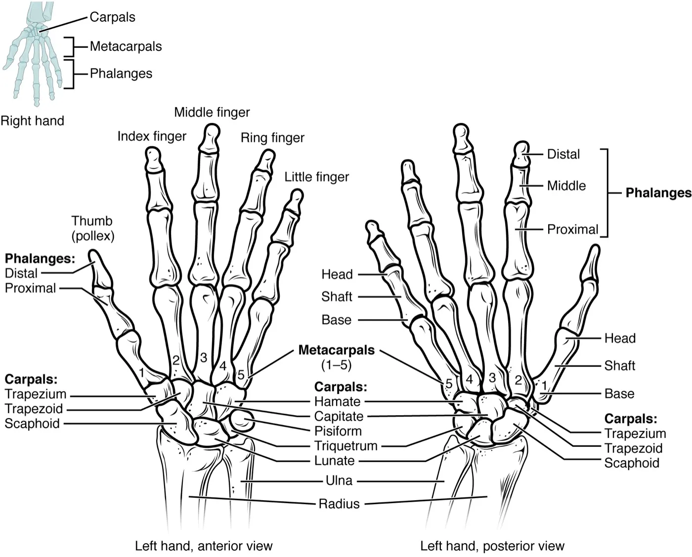
One review describes the four bones of the palm-side row as bound so tightly by ligaments that they act as one unit2. Ligaments are tough bands of tissue that hold bones together at a joint. A review of wrist biomechanics states that no muscle acting on the wrist attaches to the row of carpal bones next to the forearm2. The tendons attach further along, and that row moves through the forces of the joints around it2.
The joints between them
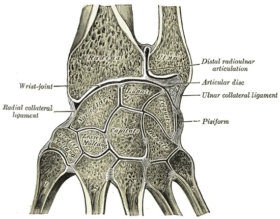
Anatomy texts describe three joints at or next to the wrist1,8:
- The radiocarpal joint. The scaphoid and lunate meet the end of the radius directly1. The triquetrum meets a pad of fibrocartilage (a tough, fibrous kind of cartilage, the firm, rubbery tissue softer than bone) instead1.
- The midcarpal joint. The two rows of carpal bones meet each other here1.
- The distal radioulnar joint. Next to the wrist, a shallow notch on the inner side of the lower radius meets the head of the ulna1.
The ulna is the forearm bone on the little-finger side. Its end does not touch any carpal bone directly1.
How it moves
The radiocarpal joint moves in two planes1,8. That gives flexion (bending the wrist forward), extension, and tilting to each side1,8. A review states that flexion and extension are shared evenly between the radiocarpal and midcarpal joints2.
Two studies measured how much wrist movement daily tasks used3,11. Radial deviation is tilting the hand toward the thumb side, and ulnar deviation is tilting it toward the little finger.
| Movement | 40 normal subjects, a whole set of daily tasks | 10 normal subjects, 52 standard tasks |
|---|---|---|
| Extension | 60 degrees3 | 30 degrees11 |
| Flexion | 54 degrees3 | 5 degrees11 |
| Ulnar deviation | 40 degrees3 | 15 degrees11 |
| Radial deviation | 17 degrees3 | 10 degrees11 |
One study of 30 healthy adults aged 23 to 30 found that wrist range differed with forearm rotation in several of its measurements12. Pronation turns the forearm palm down, and supination turns it palm up. The study’s averages, from that one small group, included:
- flexion in men: about 70 to 73 degrees palm down or in neutral, and about 62 to 63 palm up12
- extension in men: 69.5 degrees palm down and 64.6 palm up, with no significant difference in women12
- radial deviation in men: about 24 to 25 degrees palm down and about 15 to 16 palm up12
What holds it together
A review of wrist anatomy counts 33 ligaments within the wrist joint and its capsule (the sleeve of tough tissue that encloses a joint)2. They fall into two groups2:
- extrinsic ligaments, which join the carpal bones to the radius or to the metacarpals (the long bones of the hand)2
- intrinsic ligaments, which run from one carpal bone to another2
The review calls two intrinsic ligaments of the forearm-side row the most important ones2. One is the scapholunate ligament, and the other is the lunotriquetral ligament2.
The triangular fibrocartilage complex sits on the little-finger side, between the lunate, the triquetrum and the head of the ulna2. A review describes it as a load-bearing structure that passes load from the carpal bones to the ulna2.
How load crosses the wrist
The table sets a review’s figures beside a computer model built from 120 normal adult wrist X-rays2,13. The model mimicked a light grasp with the wrist in neutral13.
| Path | Review, wrist and forearm in neutral | Computer model |
|---|---|---|
| Between radius and scaphoid | about 45%2 | 55%13 |
| Between radius and lunate | 35%2 | 35%13 |
| The rest | 20%, on the ulnar side2 | 10%, through the triangular fibrocartilage13 |
A review states that load studies have consistently found the areas of higher pressure to line up well with the areas of degenerative change2.
When the wrist bears body weight
Seventeen elite male gymnasts worked on a pommel horse fitted with a load cell14. Mean peak force on the hand was 1.5 times body weight in front scissors and flairs14.
Breakdown
Falls onto the hand
Many upper limb fractures (broken bones) follow a hard fall onto an outstretched hand1. The force travels up the limb and may break the humerus, the radius or the scaphoid1. In a 2020 review, the typical scaphoid fracture patient was hurt in sport by a fall onto an outstretched hand that bent the wrist back15. A review of lower radius fractures states that many in older adults happen as a person tries to stop a fall with an outstretched hand16. The distal radius fracture and scaphoid fracture pages report the trials.
Some scapholunate ligament injuries separate the scaphoid from the lunate17. A review states that these are thought to follow, most often, a fall onto a wrist bent back and tilted toward the little finger17. The wrist sprain page reports how studies define a sprain and what they found.
In sport
Two US surveillance studies counted hand and wrist injuries together4,18. An athlete-exposure is one athlete taking part in one practice or game.
- College, 25 sports, 2004-05 to 2013-14. There were 4,851 injuries, or 41.2 per 100,000 athlete-exposures4. Wrist sprains made up 14.7%4.
- High school, 11 school years. There were 6,723 injuries, or 1.7 per 10,000 athlete-exposures18.
Wrist pain in gymnasts
Prevalence is the share of people in a group who have a condition at a given time. A 2026 meta-analysis of adolescent artistic gymnasts pooled nine studies to a wrist pain prevalence of 53%5. It rated its evidence as low to very low certainty5. The review states that a research grant from a sport science institute supported it5.
Gymnasts and the start of training
A survey and two systematic reviews report how age at the start of training relates to gymnasts’ wrist pain5,6,7. A systematic review searches for the research on a question in a set, checkable way. An odds ratio says how many times higher the odds of an outcome are in one group. The sources do not read the start of training the same way:
- A 1996 survey of 52 nonelite gymnasts. Of them, 73% had had wrist pain in the past 6 months6. Those with wrist pain had begun training later than those without, at 7.0 years against 5.16. Age at the start was one factor independently linked to wrist pain6.
- A 2015 systematic review. It found one study of risk factors7. It linked wrist pain to age 10 to 14, training intensity and an earlier start, with an odds ratio of 1.97 for the start7.
- A 2026 systematic review. It linked wrist pain to age 10 to 14, training intensity and an older age at the start5. It took its odds ratios from the 1996 survey, among them 1.97 for an older start5. Its authors suggest that a later starter may be nearer to, or within, a vulnerable growth period5.
Gymnasts’ wrists without pain
MRI (magnetic resonance imaging) is a scan that shows soft tissue in detail. A 2009 study scanned the wrists of 15 top-division college gymnasts with no history of injury or pain19. All of them showed changes in the wrist ligaments, mild in 40% and moderate in 60%19. Cartilage changes were mild in 60%, moderate in 33% and severe in 7%19. At a questionnaire 3 to 4 years later, a single gymnast reported having had a wrist injury in their career19.
Wear over time
Osteoarthritis is a long-term joint disease in which the cartilage lining the joint wears away. One centre reviewed radiographs (X-ray images) of both wrists of 1,327 patients over 4020. Advanced osteoarthritis was present in 228 of them (17%)20.
Recovery
Time away from sport
Among US high school athletes with a hand or wrist injury, 45.7% returned to play in under 7 days18. Another 12.4% of injuries kept athletes out for 3 weeks or more18.
Years later
In a cross-sectional study (one that looks at a group once) of former elite male players, some reported wrist osteoarthritis named by a physician21. They were 1.6% of 127 cricketers and 2.1% of 140 rugby union players21. In neither sport did the study find a significant association of previous hand injury with pain or osteoarthritis21.
A 2018 systematic review of wrist-loading sports in youth judged the evidence limited on two points22. One is whether their effects last, and the other is how common wrist joint degeneration is in former young athletes22.
How bone at the wrist adapts
Bone mineral density is how much mineral is packed into a given amount of bone. A meta-analysis of 17 studies compared pre-pubertal girls in gymnastics with non-gymnasts23. The gymnasts had greater bone properties, with the largest gap in bone mineral density at the lower end of the radius23.
A 2018 systematic review covered 23 studies of young people in wrist-loading sports, among them gymnastics, tennis and rowing22. Athletes had higher bone mineral density and content than controls22.
Ten cadaver wrists were scanned for bone density24. In the normal ones, the bone just under the cartilage at the end of the radius had two dense centres24. They lay in the hollows that hold the lunate and the scaphoid24. The authors state that density patterns in the wrist reflect long-term force transmission and change with loading24.
A CT scan builds X-ray images into slices or a 3D picture. In healthy adults scanned while moving the wrist through its full range, denser radius bone went with larger joint contact areas25. The authors conclude that the wrist bears load in a way similar to the knee and hip25. The distal radius page covers how that bone adapts.
Connections
Bones, ligaments and cartilage
A review calls the scapholunate ligament the most commonly injured ligament between the wrist bones26. Related pages: scaphoid, triangular fibrocartilage complex, wrist sprain, scaphoid fracture, distal radius fracture.
Muscles and tendons
A review of wrist anatomy names six forearm muscles that act only on the wrist2. According to it, the main movers in bending and straightening are:
- flexion: palmaris longus, flexor carpi ulnaris and flexor carpi radialis (see forearm flexors)2
- extension: the two radial extensors and extensor carpi ulnaris (see forearm extensors)2
A tendon is a tough cord of tissue that connects a muscle to a bone. The extensor carpi ulnaris tendon lies in a bony groove on the back of the ulna as it leaves the forearm27. The first dorsal compartment at the back of the wrist carries the tendons of two thumb muscles9.
Nerves
The median nerve crosses the wrist inside the carpal tunnel, beside several flexor tendons28. The carpal tunnel is a narrow passage at the front of the wrist. The posterior interosseous nerve, the deep branch of the radial nerve, ends in a branch that supplies the back of the wrist joint capsule29.
Exercises and stretches
Eight cadaver wrists were loaded in a knuckle (neutral) push-up position and in an extended-wrist one30. The extended position caused significantly greater peak pressure between radius and scaphoid, but not between radius and lunate30.
In 20 physically active older women, a single 30-second static stretch increased wrist range of motion31. Related pages: push-up, wrist extensor stretch, wrist flexor stretch.
Wrist joint is formed by
Wrist joint is held together by
Wrist joint is bent by
Wrist joint is extended by
Wrist joint is crossed by
Wrist joint is passed by
Wrist joint is supplied by
Wrist joint is injured in
Wrist joint is loaded by
Sources
- Betts JG, Young KA, Wise JA, Johnson E, Poe B, Kruse DH, Korol O, Johnson JE, Womble M, DeSaix P (2022). Anatomy and Physiology 2e. OpenStax. Link
- Eschweiler J, Li J, Quack V, Rath B, Baroncini A, Hildebrand F, Migliorini F (2022). Anatomy, Biomechanics, and Loads of the Wrist Joint.. Life (Basel, Switzerland). doi:10.3390/life12020188 PMID 35207475
- Ryu JY, Cooney WP, Askew LJ, An KN, Chao EY (1991). Functional ranges of motion of the wrist joint.. The Journal of hand surgery. doi:10.1016/0363-5023(91)90006-w PMID 1861019
- Chan JJ, Xiao RC, Hasija R, Huang HH, Kim JM (2023). Epidemiology of Hand and Wrist Injuries in Collegiate-Level Athletes in the United States.. The Journal of hand surgery. doi:10.1016/j.jhsa.2021.10.011 PMID 34895779
- DiLeo SDF, Noori A, Day E, Burkhart TA, Paul RA, Chan AHW (2026). Epidemiology and Risk Factors of Wrist Pain and Injury in Adolescent Artistic Gymnasts: A Systematic Review and Meta-analysis.. Orthopaedic journal of sports medicine. doi:10.1177/23259671251395329 PMID 41552624
- DiFiori JP, Puffer JC, Mandelbaum BR, Mar S (1996). Factors associated with wrist pain in the young gymnast.. The American journal of sports medicine. doi:10.1177/036354659602400103 PMID 8638761
- Kox LS, Kuijer PP, Kerkhoffs GM, Maas M, Frings-Dresen MH (2015). Prevalence, incidence and risk factors for overuse injuries of the wrist in young athletes: a systematic review.. British journal of sports medicine. doi:10.1136/bjsports-2014-094492 PMID 25872521
- Erwin J, Varacallo MA (2023). Anatomy, Shoulder and Upper Limb, Wrist Joint. StatPearls. PMID 30521200
- Gray H; Lewis WH (editor) (1918). Anatomy of the Human Body. Lea & Febiger, Philadelphia (20th edition). Link
- Agrawal A (2019). Musculoskeletal etymology: What's in a name?. Journal of clinical orthopaedics and trauma. doi:10.1016/j.jcot.2018.02.009 PMID 30828213
- Palmer AK, Werner FW, Murphy D, Glisson R (1985). Functional wrist motion: a biomechanical study.. The Journal of hand surgery. doi:10.1016/s0363-5023(85)80246-x PMID 3968403
- Fan S, Cepek J, Symonette C, Ross D, Chinchalkar S, Grant A (2019). Variation of Grip Strength and Wrist Range of Motion with Forearm Rotation in Healthy Young Volunteers Aged 23 to 30.. Journal of hand and microsurgery. doi:10.1055/s-0038-1676134 PMID 31413492
- Schuind F, Cooney WP, Linscheid RL, An KN, Chao EY (1995). Force and pressure transmission through the normal wrist. A theoretical two-dimensional study in the posteroanterior plane.. Journal of biomechanics. doi:10.1016/0021-9290(94)00093-j PMID 7775494
- Markolf KL, Shapiro MS, Mandelbaum BR, Teurlings L (1990). Wrist loading patterns during pommel horse exercises.. Journal of biomechanics. doi:10.1016/0021-9290(90)90315-t PMID 2229083
- Clementson M, Björkman A, Thomsen NOB (2020). Acute scaphoid fractures: guidelines for diagnosis and treatment.. EFORT open reviews. doi:10.1302/2058-5241.5.190025 PMID 32175096
- Nellans KW, Kowalski E, Chung KC (2012). The epidemiology of distal radius fractures.. Hand clinics. doi:10.1016/j.hcl.2012.02.001 PMID 22554654
- Konopka G, Chim H (2018). Optimal management of scapholunate ligament injuries.. Orthopedic research and reviews. doi:10.2147/ORR.S129620 PMID 30774459
- Johnson BK, Brou L, Fields SK, Erkenbeck AN, Comstock RD (2017). Hand and Wrist Injuries Among US High School Athletes: 2005/06-2015/16.. Pediatrics. doi:10.1542/peds.2017-1255 PMID 29162658
- Fredericson M, Ho C, Waite B, Jennings F, Peterson J, Williams C, Mathesonn GO (2009). Magnetic resonance imaging abnormalities in the shoulder and wrist joints of asymptomatic elite athletes.. PM R. doi:10.1016/j.pmrj.2008.09.004 PMID 19627884
- Cornette B, Hollevoet N (2025). Patterns of osteoarthritis of the wrist: a single-centre observational cohort study.. The Journal of hand surgery, European volume. doi:10.1177/17531934241275450 PMID 39752285
- Jones ME, Davies MAM, Shah K, Kemp S, Peirce N, Leyland KM, Stokes KA, Judge AD, Newton JL, Furniss D, Arden NK (2019). The prevalence of hand and wrist osteoarthritis in elite former cricket and rugby union players.. Journal of science and medicine in sport. doi:10.1016/j.jsams.2019.03.004 PMID 30940442
- Kox LS, Kuijer PPFM, Thijssen DAJ, Kerkhoffs GMMJ, van Rijn RR, Frings-Dresen MHW, Maas M (2018). Health Effects of Wrist-Loading Sports During Youth: A Systematic Literature Review.. Journal of physical activity & health. doi:10.1123/jpah.2017-0328 PMID 29741447
- Burt LA, Greene DA, Ducher G, Naughton GA (2013). Skeletal adaptations associated with pre-pubertal gymnastics participation as determined by DXA and pQCT: a systematic review and meta-analysis.. Journal of science and medicine in sport. doi:10.1016/j.jsams.2012.07.006 PMID 22951266
- Hoogbergen MM, Niessen WJ, Schuurman AH, Spauwen PH, Kauer JM (2002). Subchondral bone mineral density patterns representing the loading history of the wrist joint.. Journal of hand surgery (Edinburgh, Scotland). doi:10.1054/jhsb.2001.0714 PMID 12027489
- Straatman L, Norman E, Knowles N, Walton D, Suh N, Lalone E (2023). Use it or lose it: The relationship between two image-based biomarkers in better understanding osteoarthritis progression in the wrist.. Journal of biomechanics. doi:10.1016/j.jbiomech.2023.111849 PMID 37931557
- Andersson JK (2017). Treatment of scapholunate ligament injury: Current concepts.. EFORT open reviews. doi:10.1302/2058-5241.2.170016 PMID 29071123
- Campbell D, Campbell R, O'Connor P, Hawkes R (2013). Sports-related extensor carpi ulnaris pathology: a review of functional anatomy, sports injury and management.. British journal of sports medicine. doi:10.1136/bjsports-2013-092835 PMID 24096897
- Henry BM, Zwinczewska H, Roy J, Vikse J, Ramakrishnan PK, Walocha JA, Tomaszewski KA (2015). The Prevalence of Anatomical Variations of the Median Nerve in the Carpal Tunnel: A Systematic Review and Meta-Analysis.. PLoS One. doi:10.1371/journal.pone.0136477 PMID 26305098
- Wheeler R, DeCastro A (2023). Posterior Interosseous Nerve Syndrome.. StatPearls. PMID 31082090
- Daly BT, Werner FW, Cavallaro SM, Tucci ER, Harley BJ (2018). Effect of Push-Up Position on Wrist Joint Pressures in the Intact Wrist and Following Scapholunate Interosseous Ligament Sectioning.. The Journal of hand surgery. doi:10.1016/j.jhsa.2017.10.019 PMID 29157783
- Palmer TB, Palmer BM (2025). Acute Effects of Static Stretching on Handgrip Strength and Wrist Joint Flexibility in Physically Active Older Women.. Journal of aging and physical activity. doi:10.1123/japa.2024-0344 PMID 40015283
Changes
- First published.