Forearm flexors
Also called forearm flexor muscles, wrist flexors, finger flexors, flexor-pronator muscles
The eight muscles on the front of the forearm that bend the wrist, fingers and thumb and turn the palm down. Where they attach, how they are injured in climbers and motorcycle racers, how they respond to training, and what they work with.
- Region
- Forearm
- Starts on (pronator teres)
- just above the upper arm bone's inner elbow bump, from the common flexor tendon, and a slip from the ulna1,6
- Starts on (flexor carpi radialis)
- the upper arm bone's inner elbow bump by the common flexor tendon, plus the forearm's fascia and partitions between muscles1,6
- Starts on (palmaris longus)
- the upper arm bone's inner elbow bump by the common flexor tendon, plus the forearm's fascia and partitions between muscles1,6
- Starts on (flexor carpi ulnaris)
- the upper arm bone's inner elbow bump by the common flexor tendon, and the ulna's elbow point and back border1,6
- Starts on (flexor digitorum superficialis)
- three heads, from the upper arm bone's inner elbow bump and a ligament there, the ulna, and the radius (Common flexor tendon)1,6
- Starts on (flexor digitorum profundus)
- the upper three-quarters of the front and inner surfaces of the ulna, and the membrane between radius and ulna1
- Starts on (flexor pollicis longus)
- the grooved front of the radius's shaft, and the nearby part of the membrane between radius and ulna1
- Starts on (pronator quadratus)
- a ridge and the front of the lower quarter of the ulna1
- Ends on (pronator teres)
- a rough area at the middle of the outer surface of the radius, the forearm bone on the thumb side1,33
- Ends on (flexor carpi radialis)
- the base of the second metacarpal, a long bone of the hand, with a slip to the third1
- Ends on (palmaris longus)
- the middle of a wrist ligament (transverse carpal ligament) and the palm's flat tendon sheet (palmar aponeurosis)1
- Ends on (flexor carpi ulnaris)
- the pisiform, a small wrist bone, and by ligaments onward to the hamate, another wrist bone, and the fifth metacarpal1,33
- Ends on (flexor digitorum superficialis)
- the sides of the middle bone (phalanx) of each finger, about halfway along, after each tendon splits1
- Ends on (flexor digitorum profundus)
- the base of the end bone (last phalanx) of each finger, by four tendons1
- Ends on (flexor pollicis longus)
- the base of the end bone (distal phalanx) of the thumb1
- Ends on (pronator quadratus)
- the front and outer border of the lower quarter of the radius's shaft1
- Tendons
- Common flexor tendon
- Updated
- October 14, 2026
- Reviewed
- October 14, 2026 · how entries are checked
Function
Eight muscles in two layers
Gray’s Anatomy (1918) divides the muscles on the front of the forearm into a superficial group and a deep group1. The superficial group has five muscles and the deep group has three1.
The five superficial muscles share one starting point6. It is the medial epicondyle, the inner bony bump at the elbow end of the humerus (upper arm bone)6. They attach there by a shared tendon (a tough cord joining muscle to bone), the common flexor tendon1. Near the elbow they also take fibres from the forearm’s fascia, a sheet of tough connective tissue, and from the partitions between the muscles1.
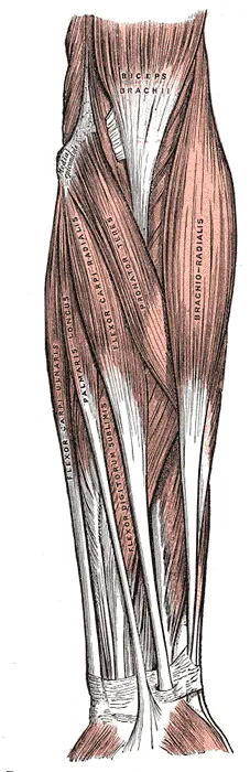
Some of the five have more than one head, or starting part1. Flexor digitorum superficialis is the largest of the superficial group and has three heads1. They start on the humerus and on the two forearm bones, the ulna and the radius1. Flexor carpi ulnaris has two heads joined by an arch of tendon, and the ulnar nerve passes under that arch1.
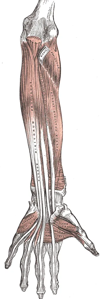
Flexor digitorum profundus lies directly under the superficial flexors and starts on the upper three-quarters of the ulna1. Flexor pollicis longus (the long thumb flexor) lies in the same layer and starts on the front of the radius1. Pronator quadratus is a small, flat, four-sided muscle across the front of the lower radius and ulna1.
What each muscle does
The first five rows of the table are the superficial group1. A phalanx is a finger bone, and a metacarpal is one of the long bones of the hand.
| Muscle | Where it ends | What it does | Nerve |
|---|---|---|---|
| Pronator teres | The middle of the outer surface of the radius1 | Turns the palm down1 | Median6 |
| Flexor carpi radialis | The base of the second metacarpal, with a slip to the third1 | Bends the wrist and tilts the hand toward the thumb side1 | Median7 |
| Palmaris longus | A ligament at the wrist (the transverse carpal ligament) and the palmar aponeurosis, a flat sheet of tendon1 | Bends the wrist1 | Median7 |
| Flexor carpi ulnaris | The pisiform bone1 | Bends the wrist and tilts the hand toward the little-finger side1 | Ulnar1 |
| Flexor digitorum superficialis | The sides of the middle phalanx of each finger1 | Bends the middle phalanx of each finger, then the proximal (nearer) one1 | Median7 |
| Flexor digitorum profundus | The base of the last phalanx of each finger1 | Bends the end phalanx of each finger1 | Ulnar for its ulnar half1. Anterior interosseous for the index-finger part8 |
| Flexor pollicis longus | The base of the end phalanx of the thumb1 | Bends the phalanges of the thumb1 | Anterior interosseous8 |
| Pronator quadratus | The lower quarter of the radius1 | Turns the palm down1 | Anterior interosseous8 |
The median nerve supplies most of the forearm flexors2. The anterior interosseous nerve is a branch of the median nerve to deep muscles of the forearm. One source describes flexor carpi ulnaris as the most powerful wrist flexor9.
How the finger tendons reach the fingers
Flexor digitorum superficialis forms four tendons1. These four, the four deep finger tendons and the long thumb tendon pass through the carpal tunnel with the median nerve10. The carpal tunnel is a narrow passage at the front of the wrist.
Opposite the base of the first phalanx, each superficial tendon splits into two slips1. The matching deep tendon passes through the gap1. It runs on to the base of the last phalanx1.
A review of climbing injuries describes a string of ligaments (tough bands of tissue) that hold these tendons against the finger bones4. There are five annular pulleys, named A1 to A5, and three cruciate pulleys, named C1 to C34. The pulleys stop the tendons from bowstringing, which means pulling away from the finger bones as the finger bends4.
How strong and how large they are
Two studies measured wrist torque, or turning force, in both directions11,12. In 20 people making isometric efforts, the greatest flexion torque was about 32 Nm, against 17 Nm for extension11. Isometric means the muscle works but the joint does not move. In 40 healthy arms, flexion torque was also the higher of the two12. One model, built from measurements in cadaver (donated body) forearms, predicted near-maximal flexor torque over the whole range of wrist motion13.
In 86 young adults, thicker front-of-forearm muscles over the ulna went with a stronger handgrip, in both men and women14. That thickness was measured by ultrasound, a scan made with sound waves14.
Breakdown
Finger pulley injuries in climbers
One prospective study, which follows people forward in time, recorded 911 climbing injuries in 836 patients over four years3. Finger injuries made up 52% of them, and pulley injuries were the most frequent finger injury3.
A review of pulley injuries in rock climbers describes A2 and A4 as the strongest pulleys, and the two most often injured in climbing4. The review reports forces on the A2 pulley of up to 380 N in climbing4. It reports up to 450 N under a sudden shock load, such as a foot slipping4.
The same review compares two climbing grips4. It describes the open-hand grip as giving very low loads on the pulleys4. It describes the full crimp as setting the conditions for the highest loads on A2 and A44. Pulley injuries can follow one high-intensity overload, or continued overuse4.
Pressure that builds inside the forearm
The forearm is usually described as having four compartments, two of them on the volar (palm) side16. Chronic exertional compartment syndrome is compartment syndrome that comes on with exercise and eases with rest. In the forearm, pressure inside the compartments rises during repeated activities17. It shows as forearm pain, stiffness, weaker muscles and tingling or numbness, which go when the exercise stops16.
The condition is rare in the general population, but can be seen in motorcycle racers, climbers and rowers16. A systematic review, a study that searches for all the research on a question in a set way, covered 12 studies16. Its 336 patients were 98.5% male, with a mean age of 25.1 years16. Motorcycle racers were 93.7% of them16.
A questionnaire went to 20,641 licensed French motorcycle racers, and 1,311 answered18. Of those, 8.7% reported the condition in the forearm18.
The systematic review’s authors suggest the racers’ continuous two-handed grip on the handlebars could explain their share16. The cause is still debated16. The most accepted theory is that a stiff fascia stops the compartment adapting as blood flow and muscle volume rise in exercise16.
A torn muscle belly
One case report, a report on a single patient, describes a rupture of the flexor digitorum superficialis muscle belly during isometric weight training19. The report states that rupture of the belly of the forearm flexors is rare19. Its authors note that such a rupture can form a haematoma, a pocket of blood in the tissue19. That can raise the pressure inside the forearm compartment19.
Where the muscles start and where nerves pass
A review describes medial epicondylitis, often called golfer’s elbow, as degeneration of the tendon where these muscles start20. The common flexor tendon page covers it.
Two nerves pass between heads of these muscles1,7. The median nerve passes between the two heads of pronator teres7. One review names that gap as the most common place above the wrist for the nerve to be trapped7. The ulnar nerve passes into the forearm between flexor carpi ulnaris’s two heads1. A review names the arcade joining those heads as one of two main places where that nerve is compressed at the elbow21.
Recovery
What training studies found
In one study, fourteen men did wrist-flexion training three times a week: 5 sets of 8 at 70 to 80% of their one-repetition maximum5. That is the heaviest load a person can lift once. After six weeks, wrist-flexor thickness and local muscle endurance had increased5.
A meta-analysis pooled seven trials from four studies of active climbers22. Training on a fingerboard, a board with small edges to grip, improved finger strength more than continuing climbing training as usual22.
A crossover study, in which each person tries every option in turn, compared two kinds of fingerboard training in thirteen intermediate climbers23. One used 30% of maximum with blood flow restriction, a cuff that partly limits blood flow in the limb23. The other used 60% of maximum without it, and each ran for five weeks23. Neither period changed maximal finger flexor strength significantly, and both maintained strength and endurance23. A university grant supported the study, and its authors state the funding did not influence it23.
After hard lowering work
In 25 healthy adults, eccentric wrist contractions lowered wrist flexion and extension force at 24 hours24. Eccentric means muscle work done while the muscle lengthens. Soreness and forearm circumference rose at the same point24. All were back to normal by seven days24.
After surgery for compartment syndrome
A systematic review gathered reports of fasciotomy, surgery that cuts through fascia, for the forearm condition25. It found 15 case series and 23 case reports covering 500 patients, 88.0% of them motorcycle racers25. Of the athletes, 94.2% returned to sport at any level and 86.8% at their previous level or higher25. The mean time to return was 5.1 weeks, and 2.4% had a recurrence25.
Connections
The inner side of the elbow
A clinical reference describes the common flexor tendon as lying next to and in front of the ulnar collateral ligament6. This is the ligament on the inner side of the elbow. Two cadaver studies disagree on which muscle helps the ligament most26,27. In eight elbow specimens loaded one muscle at a time, flexor carpi ulnaris relieved the most strain on the ligament26. In a second study of eight specimens, the authors found flexor digitorum superficialis to be the biggest contributor27.
The wrist bones
The flexor carpi radialis tendon uses a bump on the scaphoid, a wrist bone, as a pulley on its way to the second metacarpal28. The muscle has been suggested to help steady the scaphoid28. In ten cadaver wrists, loading the muscle rotated the scaphoid and another bone, the triquetrum, in opposite directions28. The authors suggest this is likely to relax the back part of the scapholunate ligament28.
The muscles on the back of the forearm
One study recorded the extensor digitorum, a forearm muscle that straightens the fingers, in 20 people making isometric wrist efforts11. At 50% and 75% of maximum, it co-contracted more during wrist flexion than flexor carpi ulnaris did during wrist extension11. Co-contraction means opposing muscles working at the same time. The forearm extensors page describes the muscles on the back of the forearm.
Exercises and stretches that load them
Related exercise pages: wrist curl, dead hang, deadlift, farmer’s carry and wrist flexor stretch12,29,30,31,32. Several of the studies used electromyography (EMG), which measures a muscle’s activity from its electrical signals.
| Exercise | What studies found |
|---|---|
| Wrist curl | In 40 healthy arms holding a wrist-flexion effort at 75% of maximum, EMG showed flexor carpi radialis working harder with the forearm palm-up12 |
| Dead hang | In 25 climbers doing maximal finger hangs, one grip, the sloper, gave more flexor digitorum superficialis and flexor carpi radialis activity than two others29 |
| Deadlift | In 29 trained athletes, the mixed grip gave the least flexor carpi ulnaris activity of three grips, whatever the load30 |
| Farmer’s carry | The authors of a systematic review of strongman exercise suggest that grip strength may limit performance in the farmer’s walk31 |
| Wrist flexor stretch | In 30 participants, a bout of static stretching of the finger flexors on one side lowered handgrip peak force in the stretched hand only32 |
Forearm flexors starts on
Forearm flexors is supplied by branches of
Forearm flexors works against
Forearm flexors lies next to
Forearm flexors has a tendon that bends around
Forearm flexors is loaded by
Forearm flexors is stretched by
Sources
- Gray H; Lewis WH (editor) (1918). Anatomy of the Human Body. Lea & Febiger, Philadelphia (20th edition). Link
- Murphy KA, Ryan N, Fakoya AO (2025). Anatomy, Shoulder and Upper Limb, Median Nerve.. StatPearls. PMID 28846302
- Schöffl V, Popp D, Küpper T, Schöffl I (2015). Injury trends in rock climbers: evaluation of a case series of 911 injuries between 2009 and 2012. Wilderness & Environmental Medicine. doi:10.1016/j.wem.2014.08.013 PMID 25712297
- Larsson R, Nordeman L, Blomdahl C (2022). To tape or not to tape: annular ligament (pulley) injuries in rock climbers-a systematic review. BMC Sports Science, Medicine and Rehabilitation. doi:10.1186/s13102-022-00539-6 PMID 35915476
- Yamane M, Ohnishi N, Matsumoto T (2015). Does Regular Post-exercise Cold Application Attenuate Trained Muscle Adaptation?. International journal of sports medicine. doi:10.1055/s-0034-1398652 PMID 25760154
- Li D, Hammad A, Kaiser K (2026). Medial Epicondylitis (Golfer's Elbow).. StatPearls. PMID 30085542
- Binsaleem S (2025). Median nerve entrapment neuropathy: a review on the pronator syndrome.. JSES Reviews, Reports, and Techniques. doi:10.1016/j.xrrt.2024.10.001 PMID 39872334
- Löppönen P, Hulkkonen S, Ryhänen J (2022). Proximal Median Nerve Compression in the Differential Diagnosis of Carpal Tunnel Syndrome.. Journal of Clinical Medicine. doi:10.3390/jcm11143988 PMID 35887752
- Lung BE, Siwiec RM (2024). Anatomy, Shoulder and Upper Limb, Forearm Flexor Carpi Ulnaris Muscle.. StatPearls. PMID 30252307
- Henry BM, Zwinczewska H, Roy J, Vikse J, Ramakrishnan PK, Walocha JA, Tomaszewski KA (2015). The Prevalence of Anatomical Variations of the Median Nerve in the Carpal Tunnel: A Systematic Review and Meta-Analysis.. PLoS One. doi:10.1371/journal.pone.0136477 PMID 26305098
- Salonikidis K, Amiridis IG, Oxyzoglou N, Giagazoglou P, Akrivopoulou G (2011). Wrist flexors are steadier than extensors.. International journal of sports medicine. doi:10.1055/s-0031-1280777 PMID 21913160
- Ikeda K, Kaneoka K, Matsunaga N, Ikumi A, Yamazaki M, Yoshii Y (2025). Effects of forearm rotation on wrist flexor and extensor muscle activities.. Journal of orthopaedic surgery and research. doi:10.1186/s13018-024-05363-x PMID 39819577
- Loren GJ, Shoemaker SD, Burkholder TJ, Jacobson MD, Fridén J, Lieber RL (1996). Human wrist motors: biomechanical design and application to tendon transfers. Journal of Biomechanics. doi:10.1016/0021-9290(95)00055-0 PMID 8850639
- Abe T, Counts BR, Barnett BE, Dankel SJ, Lee K, Loenneke JP (2015). Associations between Handgrip Strength and Ultrasound-Measured Muscle Thickness of the Hand and Forearm in Young Men and Women. Ultrasound in Medicine & Biology. doi:10.1016/j.ultrasmedbio.2015.04.004 PMID 25959055
- Yammine K (2013). Clinical prevalence of palmaris longus agenesis: a systematic review and meta-analysis. Clinical Anatomy. doi:10.1002/ca.22289 PMID 23825029
- Smeraglia F, Tamborini F, Garutti L, Minini A, Basso MA, Cherubino M (2021). Chronic exertional compartment syndrome of the forearm: a systematic review. EFORT Open Reviews. doi:10.1302/2058-5241.6.200107 PMID 33828852
- Hirardot T, Pomares G, Menu P, Grondin J, Dauty M, Fouasson-Chailloux A (2025). Diagnostic criteria of forearm Chronic Exertional Compartment Syndrome: A systematic review. Orthopaedics & Traumatology: Surgery & Research. doi:10.1016/j.otsr.2024.104001 PMID 39313023
- Regas I, Pluvy I, Sakek F, Tuphe P, Ortega P, Guinchard B, Obert L, Lepage D (2021). Epidemiology of upper limb chronic exertional compartment syndrome (CECS) in the French Motorcycle Federation racers: Results of a national questionnaire-based study. Hand Surgery & Rehabilitation. doi:10.1016/j.hansur.2020.12.017 PMID 33667652
- Chow W, Dickson J, Khan U (2013). Rupture of the flexor digitorum superficialis muscle belly during isometric weight training. BMJ Case Reports. doi:10.1136/bcr-2013-201257 PMID 24177460
- Amin NH, Kumar NS, Schickendantz MS (2015). Medial epicondylitis. The Journal of the American Academy of Orthopaedic Surgeons. doi:10.5435/JAAOS-D-14-00145 PMID 26001427
- Mezian K, Jačisko J, Kaiser R, Machač S, Steyerová P, Sobotová K, Angerová Y, Naňka O (2021). Ulnar Neuropathy at the Elbow: From Ultrasound Scanning to Treatment.. Frontiers in neurology. doi:10.3389/fneur.2021.661441 PMID 34054704
- Stien N, Riiser A, Shaw MP, Saeterbakken AH, Andersen V (2023). Effects of climbing- and resistance-training on climbing-specific performance: a systematic review and meta-analysis.. Biol Sport. doi:10.5114/biolsport.2023.113295 PMID 36636194
- Javorský T, Saeterbakken AH, Andersen V, Baláš J (2023). Comparing low volume of blood flow restricted to high-intensity resistance training of the finger flexors to maintain climbing-specific strength and endurance: a crossover study. Frontiers in Sports and Active Living. doi:10.3389/fspor.2023.1256136 PMID 37841889
- Tiidus PM, Brown L, Brant A, Enns D, Bryden PJ (2008). Physiological, sensory, and functional measures in a model of wrist muscle injury and recovery.. Physiotherapy Canada. Physiotherapie Canada. doi:10.3138/physio/60/1/30 PMID 20145740
- Gawel RJ, Wang Y, Kemler BR, Coladonato C, Tjoumakaris FP, Freedman KB (2024). Return to Sport After Fasciotomy for Chronic Exertional Compartment Syndrome of the Forearm: A Systematic Review. The American Journal of Sports Medicine. doi:10.1177/03635465231216368 PMID 38343382
- Lin F, Kohli N, Perlmutter S, Lim D, Nuber GW, Makhsous M (2007). Muscle contribution to elbow joint valgus stability.. Journal of shoulder and elbow surgery. doi:10.1016/j.jse.2007.03.024 PMID 17936028
- Udall JH, Fitzpatrick MJ, McGarry MH, Leba TB, Lee TQ (2009). Effects of flexor-pronator muscle loading on valgus stability of the elbow with an intact, stretched, and resected medial ulnar collateral ligament.. Journal of shoulder and elbow surgery. doi:10.1016/j.jse.2009.03.008 PMID 19487136
- Salvà-Coll G, Garcia-Elias M, Llusá-Pérez M, Rodríguez-Baeza A (2011). The role of the flexor carpi radialis muscle in scapholunate instability. The Journal of Hand Surgery. doi:10.1016/j.jhsa.2010.09.023 PMID 21093178
- Ferrer-Uris B, Arias D, Torrado P, Marina M, Busquets A (2023). Exploring forearm muscle coordination and training applications of various grip positions during maximal isometric finger dead-hangs in rock climbers.. PeerJ. doi:10.7717/peerj.15464 PMID 37304875
- Pratt J, Hoffman A, Grainger A, Ditroilo M (2020). Forearm electromyographic activity during the deadlift exercise is affected by grip type and sex. Journal of Electromyography and Kinesiology. doi:10.1016/j.jelekin.2020.102428 PMID 32446132
- Hindle BR, Lorimer A, Winwood P, Keogh JWL (2019). The Biomechanics and Applications of Strongman Exercises: a Systematic Review.. Sports Medicine - Open. doi:10.1186/s40798-019-0222-z PMID 31820223
- Jelmini JD, Cornwell A, Khodiguian N, Thayer J, Araujo AJ (2018). Acute effects of unilateral static stretching on handgrip strength of the stretched and non-stretched limb.. European journal of applied physiology. doi:10.1007/s00421-018-3810-6 PMID 29453756
- Betts JG, Young KA, Wise JA, Johnson E, Poe B, Kruse DH, Korol O, Johnson JE, Womble M, DeSaix P (2022). Anatomy and Physiology 2e. OpenStax. Link
Changes
- First published.