Forearm extensors
Also called wrist extensors, finger extensors, forearm extensor muscles, extensor compartment of the forearm, posterior compartment of the forearm
The muscles on the back of the forearm that bend the wrist back and straighten the fingers and thumb. Where they run, what they do in a grip, how they tire and get hurt, and how they change with use.
- Region
- Forearm
- Starts on (brachioradialis)
- the upper two-thirds of the upper arm bone's ridge above the outer elbow, and a nearby partition between muscles2
- Starts on (extensor carpi radialis longus)
- the upper arm bone's ridge above the outer elbow (lower third), and a few fibres from the common extensor tendon2
- Starts on (extensor carpi radialis brevis)
- the upper arm bone's outer elbow bump, by the common extensor tendon it shares with other wrist and finger extensors9,26
- Starts on (extensor digitorum)
- the upper arm bone's outer elbow bump by the common extensor tendon, plus nearby partitions and the forearm's fascia2,26
- Starts on (extensor digiti minimi)
- the upper arm bone's outer elbow bump by a thin slip of the common extensor tendon, and nearby partitions2,26,30
- Starts on (extensor carpi ulnaris)
- the outer elbow bump's lower back, by the common extensor tendon in some accounts, and the ulna's back border (Humerus)2,7,9,26
- Starts on (anconeus)
- the back of the upper arm bone's outer elbow bump, by its own tendon2
- Starts on (supinator)
- the upper arm bone's outer elbow bump, two ligaments of the elbow, and a ridge on the ulna2
- Starts on (abductor pollicis longus)
- the back of the ulna's shaft, and the middle third of the back of the radius's shaft2
- Starts on (extensor pollicis brevis)
- the back of the radius's shaft, below abductor pollicis longus, and the membrane between radius and ulna2
- Starts on (extensor pollicis longus)
- the middle third of the back of the ulna's shaft2
- Starts on (extensor indicis)
- the back of the ulna's shaft, below extensor pollicis longus, and the membrane between radius and ulna2
- Ends on (brachioradialis)
- the outer base of the styloid process, the bony point on the thumb side of the lower radius2,31,32,33
- Ends on (extensor carpi radialis longus)
- the back of the base of the second metacarpal, a long bone of the hand, on its thumb side2
- Ends on (extensor carpi radialis brevis)
- the back of the base of the third metacarpal, on its thumb side9
- Ends on (extensor digitorum)
- the middle and end bones of the fingers, by four tendons fanning out over the back of the hand2
- Ends on (extensor digiti minimi)
- the back of the first bone of the little finger, where it joins the extensor digitorum's tendon sheet2
- Ends on (extensor carpi ulnaris)
- the base of the fifth metacarpal, after running in a groove on the ulna through its own wrist compartment (Extensor carpi ulnaris tendon)2,7,34
- Ends on (anconeus)
- the side of the elbow's bony point (the olecranon) and the upper fourth of the back of the ulna2
- Ends on (supinator)
- the upper radius, which its surface fibres wrap round, as far down as the pronator teres attachment2
- Ends on (abductor pollicis longus)
- the thumb side of the base of the first metacarpal, the bone at the root of the thumb (First dorsal compartment tendons)2,35,36,37
- Ends on (extensor pollicis brevis)
- the base of the first bone (proximal phalanx) of the thumb (First dorsal compartment tendons)2,36,37
- Ends on (extensor pollicis longus)
- the base of the end bone of the thumb, after crossing the tendons of the two thumb-side wrist extensors2
- Ends on (extensor indicis)
- the extensor digitorum tendon to the index finger, which it joins opposite the head of the second metacarpal2
- Tendons
- Common extensor tendon, Extensor carpi ulnaris tendon, First dorsal compartment tendons
- Updated
- October 14, 2026
- Reviewed
- October 14, 2026 · how entries are checked
Function
Two groups on the back of the forearm
The forearm has a front (flexor) compartment and a back (extensor) compartment6. Together they hold twenty muscles that act on the elbow, the wrist and the joints of the hand6. The back compartment’s muscles mainly extend the wrist, fingers and thumb1. Wrist extension means bending the wrist back. Gray’s Anatomy calls these muscles the direct opponents of the flexor muscles and of the muscles that turn the palm down2.
Gray describes a superficial group and a deep group2. Sources count the superficial group in different ways:
- One anatomy reference counts seven superficial muscles, the seven in the table below7.
- Another sets three apart as the “mobile wad”: the brachioradialis and the extensor carpi radialis longus and brevis, the two radial wrist extensors1. It lists the other four as the superficial group1.
Many of the wrist extensors share a starting point at or near the lateral epicondyle4. This is the outer bony bump at the elbow end of the upper arm bone.
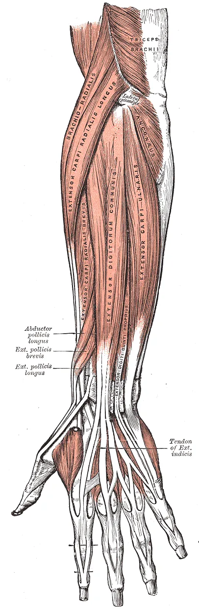
| Muscle | Starts on | Ends on | What it does |
|---|---|---|---|
| Brachioradialis | The upper two-thirds of a ridge above the outer elbow, on the humerus (upper arm bone)2 | The outer side of the radius (the forearm bone on the thumb side), at the base of its styloid process2 | Bends the elbow and turns the forearm both palm-down and palm-up8 |
| Extensor carpi radialis longus (ECRL) | The lower third of the same ridge2 | The back of the base of the second metacarpal, one of the long bones of the hand2 | Bends the wrist back and tilts the hand toward the thumb2 |
| Extensor carpi radialis brevis (ECRB) | The lateral epicondyle, by a tendon shared with three other muscles9 | The back of the base of the third metacarpal9 | Bends the wrist back and may tilt the hand slightly toward the thumb2 |
| Extensor digitorum | The lateral epicondyle, by the common extensor tendon2 | The middle and end bones of the fingers, by four tendons2 | In Gray’s account, straightens the fingers, acting mainly on the first finger bones2 |
| Extensor digiti minimi | The common extensor tendon, by a thin slip2 | The back of the first bone of the little finger, where it joins the extensor digitorum’s tendon sheet2 | Straightens the little finger and helps bend the wrist back2 |
| Extensor carpi ulnaris | The lateral epicondyle, by the common extensor tendon, and the back border of the ulna2,7 | The base of the fifth metacarpal2,7 | Bends the wrist back and tilts the hand toward the little finger2,7 |
| Anconeus | The back of the lateral epicondyle, by its own tendon2 | The side of the olecranon (the bony point of the elbow) and the upper fourth of the back of the ulna2 | Called a weak elbow extensor by the authors of a study of ten healthy volunteers10 |
The ulna is the forearm bone on the little-finger side. A styloid process is a slender, pointed part of a bone. The brachioradialis is counted as an extensor-compartment muscle although it works as a flexor8.
The deep group
The deep group has five muscles1.
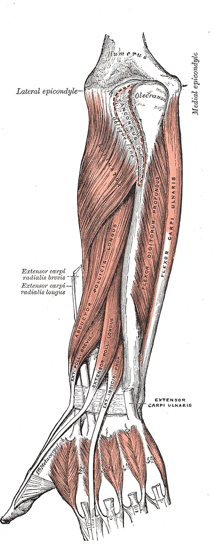
| Muscle | Starts on | Ends on | What it does |
|---|---|---|---|
| Supinator | The lateral epicondyle, two ligaments of the elbow and a ridge on the ulna2 | The upper radius, which its superficial fibres wrap round2 | Helps the biceps turn the hand palm-up2 |
| Abductor pollicis longus | The back of the shaft of the ulna and of the radius2 | The base of the first metacarpal, on its thumb side2 | In Gray’s account, mainly carries the thumb sideways away from the palm2 |
| Extensor pollicis brevis | The back of the radius, and the interosseous membrane (a sheet of tough tissue joining two side-by-side bones)2 | The base of the first bone of the thumb2 | Straightens the first bone of the thumb2 |
| Extensor pollicis longus | The middle third of the back of the ulna2 | The base of the end bone of the thumb2 | Straightens the end bone of the thumb2 |
| Extensor indicis | The back of the ulna, and the interosseous membrane2 | The extensor digitorum tendon to the index finger, which it joins2 | Straightens the index finger and helps bend the wrist back2 |
How the tendons cross the wrist
A strong fibrous band, the extensor retinaculum, lies across the back of the wrist2. Between the band and the bones are six compartments for tendons, each with its own lubricating sheath2. Each compartment carries set tendons:
- First: two thumb tendons, the abductor pollicis longus with the extensor pollicis brevis, covered on the first dorsal compartment page2.
- Second: the two radial wrist extensors2.
- Third: the extensor pollicis longus2.
- Fourth: the extensor digitorum and extensor indicis2.
- Fifth: the extensor digiti minimi2.
- Sixth: the extensor carpi ulnaris tendon2.
Their nerve
A review states that the radial nerve supplies all the muscles of the back of the arm and forearm3. In Gray’s account, the nerve itself supplies the brachioradialis, the two radial wrist extensors and the anconeus2. Its deep branch supplies the rest2. The review instead places the ECRB on the deep branch3.
The deep branch lies between the two layers of the supinator2. It emerges as the posterior interosseous nerve, which supplies the muscles of the back of the forearm11.
What they do in a grip
The wrist extensors add to stability during gripping by holding the wrist slightly extended4. This lets the flexors produce their best force4. In 20 healthy subjects making a free maximal grip, the wrist settled at 35 degrees of extension12. Grip strength was significantly lower in any wrist position moved away from the one they chose12.
One study used surface electromyography (EMG), which measures a muscle’s activity by picking up its electrical signals13. It recorded three wrist flexors and three wrist extensors during combined grip and wrist forces of 15% to 60% of maximum13. Across the grip and wrist tasks, flexor activity changed significantly from task to task, while extensor activity stayed consistently high13.
Breakdown
How they tire in grip work
In nine women doing on-and-off gripping at 25% of maximum, EMG recorded three extensor muscles14. At every work-rest pattern tested, at least two of them showed significant signs of fatigue14. The two flexor muscles showed them at one pattern only: 30 seconds of work to 10 of rest14.
At the outer elbow
Lateral epicondylitis, pain where the forearm extensor tendons attach at the outer elbow, is commonly called tennis elbow15. A clinical reference describes it as an overuse injury of the common extensor tendon, particularly where the ECRB starts15. Studies of work disagree on what brings it on16,17. A systematic review of five worker studies found no significant link with gripping, wrist extension or repeated movements at work16. In a Finnish sample aged 30 to 64, arm work both repetitive and forceful carried 5.6 times the odds, compared with doing neither17.
Where the thumb muscles cross the wrist extensors
The ECRL tendon runs under two thumb muscles, the extensor pollicis brevis with the abductor pollicis longus, before it reaches the wrist band2. A 2017 review describes intersection syndrome as a rare sports overuse injury18. In the review’s account, it arises from friction where the first and second compartments cross18. The review lists rowing, canoeing, horseback riding, skiing, weight training and racket sports as sports in which it is reported, each with repeated wrist extension18.
The review reports an open debate about its nature18. One side holds it is a chronic tendon degeneration without inflammation, the other an inflammatory process of the tendon sheaths18.
A case series, a report on a group of patients with no comparison group, came from a national rowing squad19. About 5% of the squad had intersection syndrome over three years19. One study compared patients with intersection syndrome and patients with De Quervain’s tenosynovitis, painful swelling around two thumb tendons at the wrist20. Compared with De Quervain’s tenosynovitis, intersection syndrome affected men and the dominant hand more often20.
Pressure inside the forearm
Compartment syndrome is a rise in pressure inside a closed space of muscle wrapped by tough tissue and bone. In the chronic exertional form, pressure inside the forearm compartments rises during repeated activities21. It is rare in the general population but can be seen in motorcycle racers, climbers and rowers22.
When the nerve is hurt
A review describes “wrist drop” as the loss of wrist straightening after radial nerve injury3. It reads this as a sign of weakness of the two radial wrist extensors3.
A review describes “finger drop” when the posterior interosseous nerve is impaired3. The knuckle joints of the fingers and thumb do not straighten properly, while skin feeling stays normal3. Posterior interosseous nerve syndrome means trouble with that deep branch. A clinical reference states that wrist straightening is then expected to be kept, because the radial nerve itself supplies the ECRL11.
Recovery
Growth with use
One MRI study measured muscle volume in 7 male tennis players of about 11, before puberty5. The forearm extensors of the playing arm were 25% larger than those of the other arm5. The side-to-side difference was significantly greater in the players than in 7 non-active controls5. The authors worded their conclusion as an association5. The funders, a Real Madrid and Madrid university research chair and the Spanish science ministry, had no role in it, the study states5.
What training studies found
In 13 healthy subjects, 8 weeks of isometric wrist extension training at 70% of maximum raised wrist extension strength23. Isometric means the muscle works but the joint does not move. Grip strength on the trained side rose too23.
A four-week trial randomly assigned experienced climbers to finger flexion, finger extension or alternating training on a hang board24. A hang board is a board with small edges to grip. Only the extension-based training affected finger extension strength24. One author declared a role in developing a training product, and the paper states that the firm took no part in the research24.
As the nerve comes back
One study followed 14 arms of seven healthy men as a radial nerve block wore off25. A nerve block is an injection that numbs a nerve for a short time. Grip strength was very strongly correlated with wrist extension strength25. Making a fist stopped pulling the wrist into flexion (bending forward) once wrist extension strength was back to a mean of 51% of normal25.
Connections
The forearm flexors
The extensors and the forearm flexors also work at the same time, which is called co-contraction13. In the surface EMG study of grip and wrist forces, co-contraction at the wrist was significantly higher when the wrist extensors were the opposing muscles13.
The tendon they share
Sources differ on which muscles share the common extensor tendon9,26. One account has the ECRB sharing it with three muscles: the extensor carpi ulnaris, the extensor digitorum and the extensor digiti minimi9. An MRI and anatomical study of 10 cadaver elbows found a shared origin for the ECRB, extensor digitorum and extensor digiti minimi26. In that study, the tendon of the extensor carpi ulnaris came from the lower back part of the lateral epicondyle26.
Exercises and stretches that load them
Related exercise pages: wrist extension and dead hang27,28. Studies recorded the following:
| Movement | What studies found |
|---|---|
| Wrist extension | In 40 healthy arms holding a wrist-extension effort at 75% of maximum, surface EMG showed the ECRB working harder with the forearm turned palm-down27 |
| Dead hang | In 11 male climbers hanging from a 20 mm edge until failure, the authors classed the finger extensor as a motor muscle28 |
The wrist extensor stretch page covers stretching29. A 2015 anatomical paper describes the stretch used in lateral epicondylitis as bending the wrist forward with the elbow straight and the palm down29.
Forearm extensors is loaded by
Forearm extensors is stretched by
Forearm extensors starts on
Forearm extensors pulls through
Forearm extensors is supplied by
Forearm extensors works against
Sources
- Fazal ZL, Hafeez AM, Sinkler MA, Arain A (2023). Anatomy, Shoulder and Upper Limb, Forearm Compartments.. StatPearls. PMID 30969606
- Gray H; Lewis WH (editor) (1918). Anatomy of the Human Body. Lea & Febiger, Philadelphia (20th edition). Link
- Węgiel A, Karauda P, Zielinska N, Tubbs RS, Olewnik Ł (2023). Radial nerve compression: anatomical perspective and clinical consequences.. Neurosurgical review. doi:10.1007/s10143-023-01944-2 PMID 36781706
- Reeves N, Crow T, Varacallo MA (2026). Anatomy, Shoulder and Upper Limb, Wrist Extensor Muscles.. StatPearls. PMID 30521226
- Sanchis-Moysi J, Idoate F, Serrano-Sanchez JA, Dorado C, Calbet JA (2012). Muscle hypertrophy in prepubescent tennis players: a segmentation MRI study.. PloS one. doi:10.1371/journal.pone.0033622 PMID 22428074
- Mitchell B, Whited L (2023). Anatomy, Shoulder and Upper Limb, Forearm Muscles.. StatPearls. PMID 30725660
- Sawyer E, Sajjad H, Tadi P (2023). Anatomy, Shoulder and Upper Limb, Forearm Extensor Carpi Ulnaris Muscle.. StatPearls. PMID 30969582
- Lung BE, Ekblad J, Bisogno M (2024). Anatomy, Shoulder and Upper Limb, Forearm Brachioradialis Muscle.. StatPearls. PMID 30252366
- Walkowski AD, Goldman EM (2023). Anatomy, Shoulder and Upper Limb, Forearm Extensor Carpi Radialis Brevis Muscle.. StatPearls. PMID 30969541
- Miguel-Andres I, Alonso-Rasgado T, Walmsley A, Watts AC (2017). Effect of Anconeus Muscle Blocking on Elbow Kinematics: Electromyographic, Inertial Sensors and Finite Element Study.. Annals of biomedical engineering. doi:10.1007/s10439-016-1715-2 PMID 27573695
- Wheeler R, DeCastro A (2023). Posterior Interosseous Nerve Syndrome.. StatPearls. PMID 31082090
- O'Driscoll SW, Horii E, Ness R, Cahalan TD, Richards RR, An KN (1992). The relationship between wrist position, grasp size, and grip strength.. The Journal of hand surgery. doi:10.1016/0363-5023(92)90136-d PMID 1538102
- Forman DA, Forman GN, Holmes MWR (2021). Wrist extensor muscle activity is less task-dependent than wrist flexor muscle activity while simultaneously performing moderate-to-high handgrip and wrist forces.. Ergonomics. doi:10.1080/00140139.2021.1934564 PMID 34024262
- Hägg GM, Milerad E (1997). Forearm extensor and flexor muscle exertion during simulated gripping work -- an electromyographic study.. Clinical biomechanics (Bristol, Avon). doi:10.1016/s0268-0033(96)00049-6 PMID 11415670
- Buchanan BK, Varacallo MA (2023). Lateral Epicondylitis (Tennis Elbow).. StatPearls. PMID 28613744
- Bretschneider SF, Los FS, Eygendaal D, Kuijer PPFM, van der Molen HF (2022). Work-relatedness of lateral epicondylitis: Systematic review including meta‐analysis and GRADE work-relatedness of lateral epicondylitis. American journal of industrial medicine. doi:10.1002/ajim.23303 PMID 34674287
- Shiri R, Viikari-Juntura E, Varonen H, Heliövaara M (2006). Prevalence and determinants of lateral and medial epicondylitis: a population study.. American journal of epidemiology. doi:10.1093/aje/kwj325 PMID 16968862
- Balakatounis K, Angoules AG, Angoules NA, Panagiotopoulou K (2017). Synthesis of evidence for the treatment of intersection syndrome.. World journal of orthopedics. doi:10.5312/wjo.v8.i8.619 PMID 28875127
- Hoy G, Trease L, Braybon W (2019). Intersection syndrome: an acute surgical disease in elite rowers.. BMJ open sport & exercise medicine. doi:10.1136/bmjsem-2019-000535 PMID 31258929
- Sato J, Ishii Y, Noguchi H (2016). Clinical and ultrasound features in patients with intersection syndrome or de Quervain's disease.. The Journal of hand surgery, European volume. doi:10.1177/1753193415614267 PMID 26546605
- Hirardot T, Pomares G, Menu P, Grondin J, Dauty M, Fouasson-Chailloux A (2025). Diagnostic criteria of forearm Chronic Exertional Compartment Syndrome: A systematic review. Orthopaedics & Traumatology: Surgery & Research. doi:10.1016/j.otsr.2024.104001 PMID 39313023
- Smeraglia F, Tamborini F, Garutti L, Minini A, Basso MA, Cherubino M (2021). Chronic exertional compartment syndrome of the forearm: a systematic review. EFORT Open Reviews. doi:10.1302/2058-5241.6.200107 PMID 33828852
- Shimose R, Matsunaga A, Muro M (2011). Effect of submaximal isometric wrist extension training on grip strength.. European journal of applied physiology. doi:10.1007/s00421-010-1675-4 PMID 20931218
- Devise M, Pasek L, Goislard De Monsabert B, Vigouroux L (2023). Finger flexion to extension ratio in healthy climbers: a proposal for evaluation and rebalance.. Frontiers in sports and active living. doi:10.3389/fspor.2023.1243354 PMID 38077281
- Suzuki T, Kunishi T, Kakizaki J, Iwakura N, Takahashi J, Kuniyoshi K (2012). Wrist extension strength required for power grip: a study using a radial nerve block model.. The Journal of hand surgery, European volume. doi:10.1177/1753193411427831 PMID 22095402
- Zoner CS, Buck FM, Cardoso FN, Gheno R, Trudell DJ, Randall TD, Resnick D (2010). Detailed MRI-anatomic study of the lateral epicondyle of the elbow and its tendinous and ligamentous attachments in cadavers.. AJR. American journal of roentgenology. doi:10.2214/AJR.09.3173 PMID 20729438
- Ikeda K, Kaneoka K, Matsunaga N, Ikumi A, Yamazaki M, Yoshii Y (2025). Effects of forearm rotation on wrist flexor and extensor muscle activities.. Journal of orthopaedic surgery and research. doi:10.1186/s13018-024-05363-x PMID 39819577
- Exel J, Kaufmann P, Froschauer O, Baca A, Kainz H, Mochizuki L (2026). Upper-Body Neuromechanical Coordination Strategies During Fatiguing Sustained Dead Hangs in Climbers.. European Journal of Sport Science. doi:10.1002/ejsc.70197 PMID 42168114
- Shirato R, Wada T, Aoki M, Iba K, Kanaya K, Fujimiya M, Yamashita T (2015). Effect of simultaneous stretching of the wrist and finger extensors for lateral epicondylitis: a gross anatomical study of the tendinous origins of the extensor carpi radialis brevis and extensor digitorum communis.. Journal of orthopaedic science : official journal of the Japanese Orthopaedic Association. doi:10.1007/s00776-015-0758-9 PMID 26260257
- Betts JG, Young KA, Wise JA, Johnson E, Poe B, Kruse DH, Korol O, Johnson JE, Womble M, DeSaix P (2022). Anatomy and Physiology 2e. OpenStax. Link
- Summers K, Mabrouk A, Fowles SM (2023). Colles Fracture. StatPearls. PMID 31971712
- Satteson E, Tannan SC (2023). De Quervain Tenosynovitis.. StatPearls. PMID 28723034
- Gupta A, Mirzaee F, Farahanchi Baradaran A, Aslani MA, Aslani H (2021). Biomechanical study of the abductor pollicis longus & extensor pollicis brevis after release and reconstruction of the first dorsal compartment of the wrist.. Annals of medicine and surgery (2012). doi:10.1016/j.amsu.2021.102863 PMID 34584688
- Campbell D, Campbell R, O'Connor P, Hawkes R (2013). Sports-related extensor carpi ulnaris pathology: a review of functional anatomy, sports injury and management.. British journal of sports medicine. doi:10.1136/bjsports-2013-092835 PMID 24096897
- El-Beshbishy RA, Abdel-Hamid GA (2013). Variations of the abductor pollicis longus tendon: an anatomic study.. Folia morphologica. doi:10.5603/fm.2013.0027 PMID 23740505
- Beutel BG, Taylor K, Michols NJ, Taqi M (2025). Intersection Syndrome.. StatPearls. PMID 28613649
- Bonczar M, Walocha J, Pasternak A, Depukat P, Dziedzic M, Ostrowski P, Bonczar T, Warchoł Ł, Koziej M (2023). Anatomical variations in the first dorsal compartment of the wrist: meta-analysis.. Folia morphologica. doi:10.5603/FM.a2022.0081 PMID 36165900
Changes
- First published.