Humerus
Also called upper arm bone
The upper arm bone, from the shoulder to the elbow. How it is shaped, how it breaks, how it heals and thickens with throwing and racquet sports, and what attaches to it.
- Region
- Upper arm
- Muscles
- Brachialis, Deltoid, Latissimus dorsi, Pectoralis major, Triceps brachii, Supraspinatus, Infraspinatus, Teres minor, Subscapularis, Forearm flexors, Forearm extensors
- Updated
- September 30, 2026
- Reviewed
- September 30, 2026 · how entries are checked
Function
Where the bone sits
Among the bones of the upper limb, the humerus is the largest, and it forms the skeleton of the upper arm1,2. Above, it meets the scapula (shoulder blade) at the glenohumeral joint, the main shoulder joint1. Below, it meets the radius and ulna, the two forearm bones, at the elbow1.
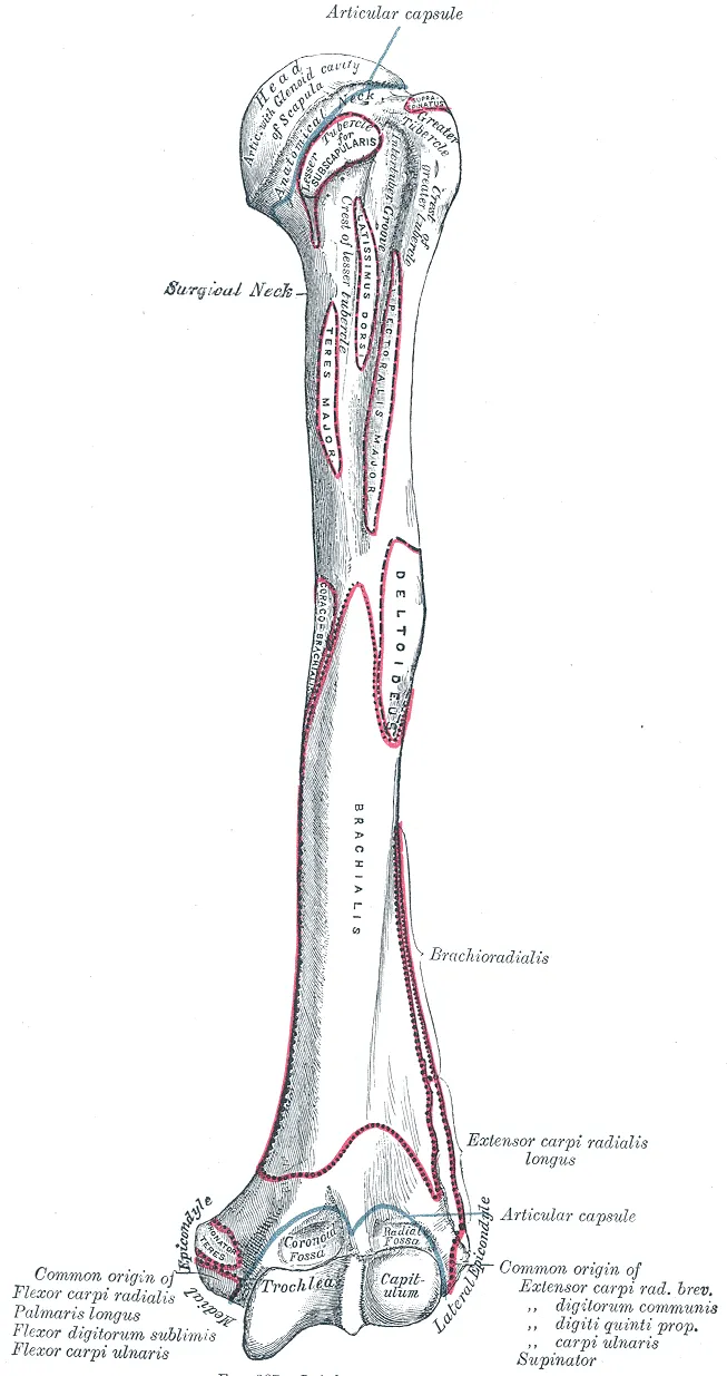
The upper end: a ball and two bumps
The head of the humerus is a large, smooth, rounded surface that faces inward1,2. It sits against the glenoid cavity (the shoulder socket) of the shoulder blade, and the two form a ball-and-socket joint1,2.
Two tubercles (bony bumps) sit beside the head: the greater tubercle on the outer side and the smaller lesser tubercle at the front2. Both are anchor points for muscles that work across the shoulder2. The anatomical neck is the rim around the smooth head, marking it off from the two tubercles1,2. Just below the tubercles, the surgical neck is where the wide upper end narrows into the shaft1,2. Between the tubercles runs the bicipital groove, which carries a tendon of the biceps brachii2.
The shaft and its groove
The shaft of the humerus is shaped like a cylinder1. It has the deltoid tuberosity on its outer side and the radial groove, also called the spiral groove, on its back1. The deltoid tuberosity is a rough, V-shaped patch halfway down the shaft where the deltoid (the rounded muscle that caps the shoulder) attaches2. The radial groove runs along the back and outer side of the middle third of the bone6. The radial nerve and the profunda brachii artery (the deep artery of the upper arm) run in it6.
The lower end: the elbow surfaces
Toward the elbow, the bone flattens and has two epicondyles (bony bumps where muscles attach) and a ridge above the outer one2. These are anchor points for muscles that move the forearm, wrist and hand2. The medial epicondyle (the inner bony bump) is the larger of the two2. The strong gripping muscles of the front of the forearm start there2. The weaker muscles of the back of the forearm start from the lateral epicondyle, the bump on the outer side2.
The lower end has two joint surfaces2. The trochlea is shaped like a pulley and meets the ulna2. The capitulum is a small knob just outside it that meets the radius2.
Where it grows in length
The growth plate (the layer of cartilage where a bone grows longer) at the top of the humerus supplies about 80% of its length growth7. A systematic review gives up to 80% and says the plate typically closes between 16 and 20 years of age8.
How throwing and lifting load it
A review of arm stress fractures (small cracks in a bone from repeated loading) describes lifts like the bench press and overhead press9. In these, the deltoid and rotator cuff (four muscles that steady the shoulder joint) counter the pull of the pectoralis major, the large chest muscle9. This balance moves some stress off the humerus, which the review calls stress shielding9.
Breakdown
Breaks at the upper end
Breaks of the upper humerus are common, and older people have them most3. Osteoporosis is a disease in which bones become thin and weak. Among fractures linked to it in older people, these breaks rank third, after the wrist and the hip10.
One study counted upper-humerus fractures in a Finnish town from 2006 to 201010. It found 678 people with these breaks, and 73% of them were women10. The breaks became more common with age10. Of 692 breaks, 62% were two-part fractures (the bone split into two main pieces), and 78% were managed in a sling without surgery10.
| Group in the Finnish study | New upper-humerus fractures per 100,000 people a year |
|---|---|
| Everyone | 8210 |
| Women | 11410 |
| Men | 4710 |
Breaks of the shaft
Breaks of the shaft make up 1% to 3% of fractures overall, and happen in both young and older people11.
Nerves close to the bone
The radial nerve runs in the groove on the back of the shaft6. Two systematic reviews counted radial nerve palsy with shaft fractures4,12:
- One found radial nerve palsy in 11.8% of shaft fractures, or 532 of 4,5174.
- An updated review of studies from 1964 to 2017 put it at 12.3%, or 890 of 7,26212.
Stress fractures in sport and lifting
The review of stress fractures in the arm describes Little League shoulder as one type9. It calls it a twisting stress fracture through the upper growth plate of the humerus9. It reports it in roughly 5% of Little League players aged 9 to 129. A systematic review of 23 studies covered 266 young athletes with Little League shoulder, with an average age of 12.88. Widening of the upper growth plate of the throwing arm was reported in 99% of them8.
Frequent weightlifting rarely leads to humeral stress fractures, the review reports, and the bench press is the lift most often involved9. It describes muscle fatigue as weakening stress shielding and raising the bending load on the bone9.
Recovery
How a broken bone mends
An anatomy textbook describes how a broken bone heals in stages2:
- First, a blood clot called a fracture hematoma forms2.
- Next, calluses (new tissue that bridges the break as it heals) of cartilage and bone form2.
- Later, spongy bone (light bone made of a honeycomb of thin struts) replaces the cartilage in the callus2.
- Remodelling can then remove the outward signs of the break2.
A review of fracture healing reports that repair usually restores bone’s structure and function, but about 10% of fractures do not heal normally13.
A Cochrane review (a systematic review done to the Cochrane Collaboration’s published standards) calls upper-humerus breaks a serious injury, frequent in older people3. Getting back the use of the arm can take several months3. Some loss of movement and some pain are common long-term problems3.
What studies of surgery found
The same review looked at randomised trials in upper-humerus fractures3. Its 47 trials were mostly in women, mainly aged 60 or over, and 10 trials of 717 people looked at displaced breaks3. It found high-certainty evidence that surgery gave no clinically important gain in shoulder function at one and two years over non-surgical care3. It also found low-certainty evidence of a higher risk of further surgery after surgical care3.
Two reviews compared surgery with non-surgical care for breaks of the shaft11,14:
- One pooled 12 studies of 1,406 adults. It found fewer nonunions (broken bones that fail to join and heal) after surgery, but more deep infections11.
- A meta-analysis (a study that pools the results of several earlier studies) of 17 studies reported trends that favoured surgery over splint or brace care14. The trends were toward faster return of arm function and fewer nonunions and repeat procedures14.
For radial nerve palsy with shaft fractures, one systematic review found that 70.7% of nerves recovered on their own in people managed without surgery4. An updated review of palsy with humeral fractures found recovery on its own in 77.2% managed without surgery12. It found recovery in 89.8% of those who had surgical exploration and fracture repair within 3 weeks of injury12.
For Little League shoulder managed without surgery, the review of arm stress fractures reports that symptoms fully cleared within two to eight months9. Of those imaged again, 84.6% showed healing9. In the systematic review, 94% returned to sport and 92.5% to their pre-injury level, while 18.7% had symptoms come back8.
How the bone builds up with use
A review of bone strength reports that bone adapts to the loads it usually bears, shaping its structure to their size, rate and frequency15. An anatomy textbook adds that bone changes its structure as mechanical stress rises or falls, so its size and weight suit its activity2. Without stress, bone loses mineral and collagen (a strong, rope-like protein), and so loses strength2. The review of bone strength likewise says strain below a minimum effective level leads to bone loss15.
Comparing the two arms of 103 professional baseball players with arm differences in 94 controls, throwing loaded the shaft heavily and nearly doubled its strength5.
In professional baseball players, the upper humerus of the throwing arm differed from the other arm in these ways16:
| Measure in the throwing arm | How much more than the other arm |
|---|---|
| Bone mass of the upper humerus | 28%16 |
| Cross-sectional area at the surgical neck | 12%16 |
| Thickness of the cortex (dense outer layer) at the surgical neck | 31%16 |
| Estimated resistance to twisting at the surgical neck | 57%16 |
When the gains are made and how long they last
In 90 male throwing athletes and 51 controls, the gap between arms at mid-humerus was larger in each more mature group17. In professional baseball players, the extra bone mass and cortex thickness built in youth faded after throwing stopped5. Yet about half the extra bone size and a third of the extra strength lasted for life5. Players who kept throwing as they aged kept more of the strength, because their marrow cavity (the hollow centre of the bone) widened less5.
One study looked at 105 female national-level tennis and squash players18. The playing arm’s bone benefit was about twice as large in those who started at or before their first period18. In 64 female tennis and squash players, the playing arm’s shaft had about 19% more bone mineral in early starters and 9% in later starters19. This came from a larger outer cortex, while cortex density matched the other arm19.
A meta-analysis covered 761 males aged 10 to 2620. In it, tennis raised bone mineral in the playing arm but not in the other arm20.
A twist in throwers’ arms
A systematic review of 14 studies covered 1,152 overhead throwing athletes21. Each study that measured bone shape found more backward twist (retroversion) in the throwing arm, by average differences of 9.6 to 25.8 degrees21. These studies also found less internal rotation, meaning less inward turning, in that arm21.
A review of 32 baseball studies found humeral torsion (twist along the bone) did not differ between players with and without shoulder or elbow injuries22.
Connections
Muscles and tendons attached to it
Apart from the pectoralis major and latissimus dorsi, the muscles that cross the shoulder and move the humerus start on the shoulder blade2.
| Structure | Where it meets the humerus |
|---|---|
| Deltoid | Deltoid tuberosity, on the outer side2,23,24 |
| Pectoralis major | Outer lip of the bicipital groove25 |
| Latissimus dorsi | Floor of the bicipital groove26,27 |
| Rotator cuff tendons | Connect the shoulder blade to the humerus2 |
| Triceps brachii | Its lateral and medial heads start on the humerus28 |
| Long head of the biceps tendon | Runs in the bicipital groove2,29 |
| Superficial back-of-forearm muscles | Start on the humerus2 |
| Common extensor tendon | Shared by several of these muscles, starting on the lateral epicondyle30 |
The shoulder blade and the upper arm
The scapula joins the humerus at the glenohumeral joint31. The upper arm has two compartments around the humerus6:
- The front holds three muscles: the biceps brachii, brachialis and coracobrachialis6. Brachialis bends the elbow from under the biceps; coracobrachialis is a small muscle at the front of the arm. The front also holds the brachial artery, the main artery of the upper arm6. Three nerves run there too: the musculocutaneous, median and ulnar nerves6. The musculocutaneous nerve works the front muscles; the ulnar nerve runs past the inner elbow to the little finger.
- The back holds the three heads of the triceps and the radial nerve6.
Humerus meets at the shoulder joint
Humerus is attached to by
Humerus carries in its groove
Humerus shares the upper arm with
Humerus is loaded by
Sources
- Mostafa E, Imonugo O, Varacallo MA (2023). Anatomy, Shoulder and Upper Limb, Humerus.. StatPearls. PMID 30521242
- Betts JG, Young KA, Wise JA, Johnson E, Poe B, Kruse DH, Korol O, Johnson JE, Womble M, DeSaix P (2022). Anatomy and Physiology 2e. OpenStax. Link
- Handoll HH, Elliott J, Thillemann TM, Aluko P, Brorson S (2022). Interventions for treating proximal humeral fractures in adults.. Cochrane Database of Systematic Reviews. doi:10.1002/14651858.CD000434.pub5 PMID 35727196
- Shao YC, Harwood P, Grotz MR, Limb D, Giannoudis PV (2005). Radial nerve palsy associated with fractures of the shaft of the humerus: a systematic review.. Journal of Bone and Joint Surgery. British Volume. doi:10.1302/0301-620X.87B12.16132 PMID 16326879
- Warden SJ, Mantila Roosa SM, Kersh ME, Hurd AL, Fleisig GS, Pandy MG, Fuchs RK (2014). Physical activity when young provides lifelong benefits to cortical bone size and strength in men.. Proceedings of the National Academy of Sciences of the United States of America. doi:10.1073/pnas.1321605111 PMID 24706816
- Bounds EJ, Frane N, Jajou L, Weishuhn LJ, Kok SJ (2023). Humeral Shaft Fractures.. StatPearls. PMID 28846218
- Collins AC, May T (2026). Proximal Humeral Epiphysiolysis.. StatPearls. PMID 30485006
- Bednar ED, Kay J, Memon M, Simunovic N, Purcell L, Ayeni OR (2021). Diagnosis and Management of Little League Shoulder: A Systematic Review.. Orthopaedic Journal of Sports Medicine. doi:10.1177/23259671211017563 PMID 34377716
- Koslosky EJ, Heath DM, Atkison CL, Dutta A, Brady CI (2024). Upper Extremity Stress Fractures.. Sports Medicine - Open. doi:10.1186/s40798-024-00769-7 PMID 39327396
- Launonen AP, Lepola V, Saranko A, Flinkkilä T, Laitinen M, Mattila VM (2015). Epidemiology of proximal humerus fractures.. Archives of Osteoporosis. doi:10.1007/s11657-015-0209-4 PMID 25675881
- Lode I, Nordviste V, Erichsen JL, Schmal H, Viberg B (2020). Operative versus nonoperative treatment of humeral shaft fractures: a systematic review and meta-analysis.. Journal of Shoulder and Elbow Surgery. doi:10.1016/j.jse.2020.05.030 PMID 32553853
- Ilyas AM, Mangan JJ, Graham J (2020). Radial Nerve Palsy Recovery With Fractures of the Humerus: An Updated Systematic Review.. Journal of the American Academy of Orthopaedic Surgeons. doi:10.5435/JAAOS-D-18-00142 PMID 31714418
- Einhorn TA, Gerstenfeld LC (2015). Fracture healing: mechanisms and interventions.. Nature reviews. Rheumatology. doi:10.1038/nrrheum.2014.164 PMID 25266456
- Li Y, Luo Y, Peng J, Fan J, Long XT (2024). Clinical effect of operative vs nonoperative treatment on humeral shaft fractures: Systematic review and meta-analysis of clinical trials.. World Journal of Orthopedics. doi:10.5312/wjo.v15.i8.783 PMID 39165869
- Hart NH, Nimphius S, Rantalainen T, Ireland A, Siafarikas A, Newton RU (2017). Mechanical basis of bone strength: influence of bone material, bone structure and muscle action.. Journal of musculoskeletal & neuronal interactions. PMID 28860414
- Warden SJ, Carballido-Gamio J, Avin KG, Kersh ME, Fuchs RK, Krug R, Bice RJ (2019). Adaptation of the proximal humerus to physical activity: A within-subject controlled study in baseball players.. Bone. doi:10.1016/j.bone.2019.01.008 PMID 30634064
- Warden SJ, Weatherholt AM, Gudeman AS, Mitchell DC, Thompson WR, Fuchs RK (2017). Progressive skeletal benefits of physical activity when young as assessed at the midshaft humerus in male baseball players.. Osteoporosis International. doi:10.1007/s00198-017-4029-9 PMID 28396902
- Kannus P, Haapasalo H, Sankelo M, Sievänen H, Pasanen M, Heinonen A, Oja P, Vuori I (1995). Effect of starting age of physical activity on bone mass in the dominant arm of tennis and squash players.. Annals of Internal Medicine. doi:10.7326/0003-4819-123-1-199507010-00003 PMID 7762910
- Kontulainen S, Sievänen H, Kannus P, Pasanen M, Vuori I (2003). Effect of long-term impact-loading on mass, size, and estimated strength of humerus and radius of female racquet-sports players: a peripheral quantitative computed tomography study between young and old starters and controls.. Journal of Bone and Mineral Research. doi:10.1359/jbmr.2003.18.2.352 PMID 12568413
- Huiwen H, Jun H, Dong F, Yan F, Tao W (2025). The effect of tennis on male bone mineral density: A meta-analysis.. PLOS ONE. doi:10.1371/journal.pone.0328636 PMID 40853978
- Kay J, Kirsch JM, Bakshi N, Ekhtiari S, Horner N, Gichuru M, Alolabi B, Khan M, Bedi A (2018). Humeral Retroversion and Capsule Thickening in the Overhead Throwing Athlete: A Systematic Review.. Arthroscopy. doi:10.1016/j.arthro.2017.10.049 PMID 29373297
- Helmkamp JK, Bullock GS, Rao A, Shanley E, Thigpen C, Garrigues GE (2020). The Relationship Between Humeral Torsion and Arm Injury in Baseball Players: A Systematic Review and Meta-analysis.. Sports Health. doi:10.1177/1941738119900799 PMID 32027223
- Stokey PJ, Kaur S, Lee A, Behrens K, Ebraheim N (2024). Anatomy and Deficiency of the Deltoid Muscle: A Review of Literature.. Orthopedic Reviews. doi:10.52965/001c.115352 PMID 38562147
- Sanderson B, Bogard M, Jazayeri R (2021). Traumatic rupture of the posterior deltoid tendon during weight lifting: A case report and review of literature.. Clinical Case Reports. doi:10.1002/ccr3.3710 PMID 34466228
- Solari F, Burns B (2023). Anatomy, Thorax, Pectoralis Major Major.. StatPearls. PMID 30252247
- Lehane KM, Faust T, Romeo AA, Erickson BJ (2025). Approach to diagnosing and treating tears of the latissimus dorsi and teres major.. JSES Reviews, Reports, and Techniques. doi:10.1016/j.xrrt.2025.05.015 PMID 41179423
- Hovsepian JM, Cartaya MA, Glaser J, Oehler N, Solovyov I, Abi Mansour C, Avila JC, Vogt S (2026). Surgical anatomy, biomechanics and clinical outcomes of latissimus dorsi transfer for irreparable rotator cuff tears: a review.. Journal of Orthopaedic Surgery and Research. doi:10.1186/s13018-026-07200-9 PMID 42661214
- Tiwana MS, Sinkler MA, Bordoni B (2023). Anatomy, Shoulder and Upper Limb, Triceps Muscle.. StatPearls. PMID 30725681
- Varacallo MA, Mair SD (2023). Proximal Biceps Tendinitis and Tendinopathy.. StatPearls. PMID 30422594
- Walkowski AD, Goldman EM (2023). Anatomy, Shoulder and Upper Limb, Forearm Extensor Carpi Radialis Brevis Muscle.. StatPearls. PMID 30969541
- Miniato MA, Mudreac A, Borger J (2023). Anatomy, Thorax, Scapula.. StatPearls. PMID 30855903
Changes
- First published.