Deltoid
The thick, three-part muscle that rounds the shoulder and lifts the arm. How it moves the arm, how it loses strength or tears, how it grows with training, and what it works with.
- Region
- Shoulder
- Starts on (front part)
- the outer third of the collarbone2
- Starts on (middle part)
- the acromion, the bony tip of the shoulder at the outer end of the ridge across the shoulder blade2,4
- Starts on (back part)
- the ridge across the back of the shoulder blade2
- Ends on
- the deltoid tuberosity, a V-shaped rough patch halfway down the outer side of the upper arm bone2,3,4
- Updated
- September 29, 2026
- Reviewed
- September 29, 2026 · how entries are checked
Function
How three parts make one muscle
The deltoid is the thick muscle that gives the shoulder its rounded outline4. It is described as having three parts: front, middle and back1. The parts are told apart by where each one starts on the bones of the shoulder2. The fibres of the three parts come together in a short tendon, a tough cord of tissue joining muscle to bone2,3,4.
That tendon attaches to the humerus (upper arm bone) at the deltoid tuberosity2,3,4. This is a rough, V-shaped area on the outer side of the bone, halfway down its shaft4.
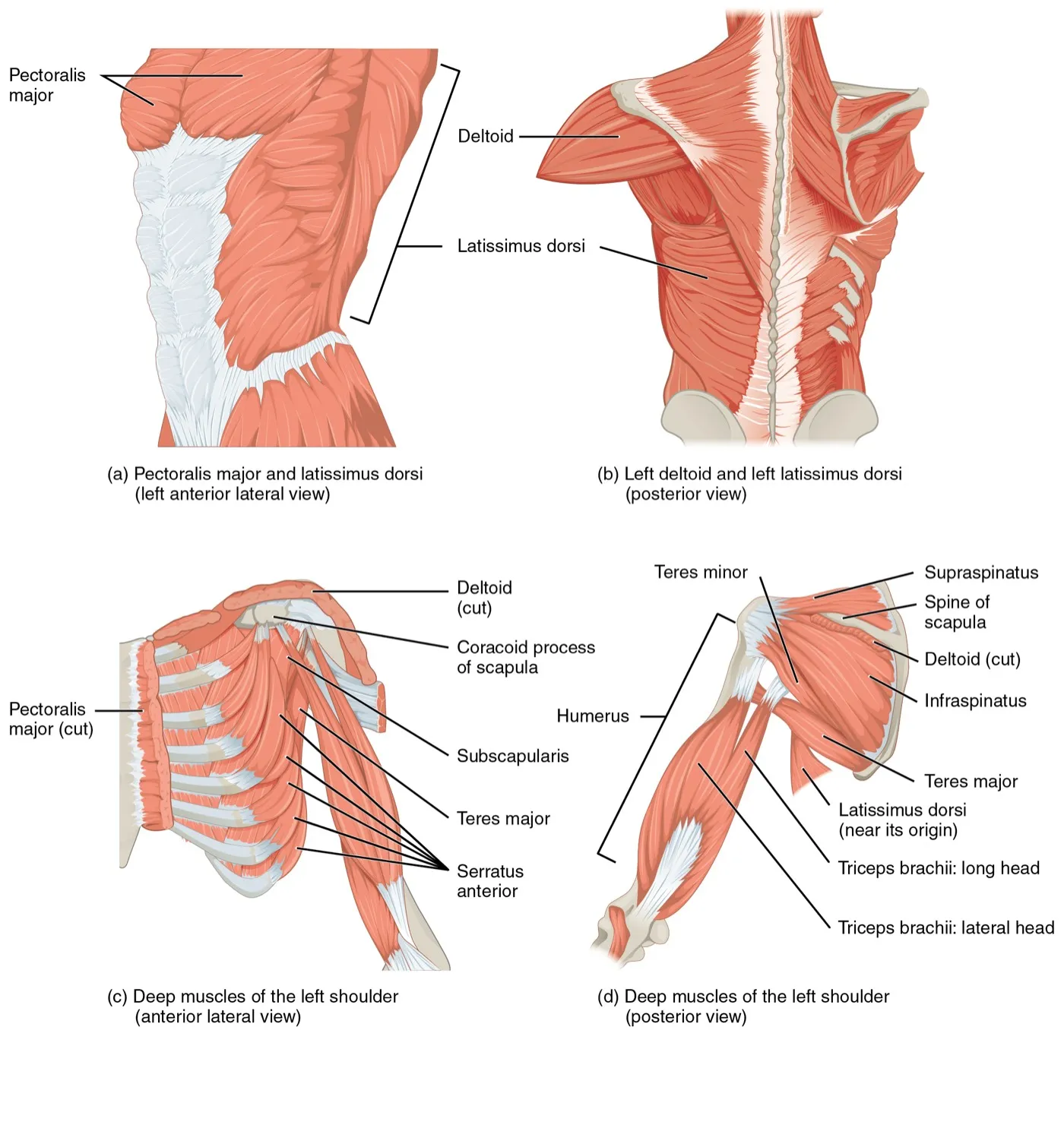
What each part does
The deltoid is the major muscle for abduction, which means lifting the arm out to the side4. It also helps swing the arm forward and back, and turn it inward and outward4. Each part has its own main job2:
| Part | Where it starts | What it does |
|---|---|---|
| Front (anterior) | Outer third of the clavicle (collarbone)2 | Lifts the arm forward and turns it inward2 |
| Middle | The acromion, the bony point on top of the scapula (shoulder blade)2 | Lifts the arm out to the side2 |
| Back (posterior) | The spine of the scapula, the ridge across the back of the shoulder blade2 | Draws the arm back and turns it outward2 |
In its introduction, one review describes the deltoid as the shoulder’s main source of force, giving up to 75% of its greatest strength5. It adds that, for this reason, a torn deltoid can cost a lot of shoulder function5.
Its nerve and blood supply
The axillary nerve carries the signals that make the deltoid contract2. It also carries feeling from the skin over the shoulder2. The nerve comes from the brachial plexus, the network of nerves that serves the arm2,9.
Two reviews place the nerve, or its front branch, roughly 6 to 7 cm below the acromion as it wraps around the upper arm2,9. One of them describes it running from back to front2,9. Its front branch reaches the front part of the deltoid and its back branch reaches the back part9. The middle part gets branches from both9.
The deltoid gets its blood from two arteries2. A branch of one feeds the front part, and the other artery feeds the back part2.
Breakdown
Three ways the deltoid loses strength
A review of the research lists three causes of a deltoid that works less well2:
- Injury to the axillary nerve2.
- A rupture, or tear, of the muscle itself2.
- Damage to the muscle during a medical procedure2.
The review says a deltoid that is not working shows up as a weaker lift of the arm to the side2.
How often the deltoid tears
A systematic review, which gathers and sums up the research on a question, looked at how often the deltoid tears5. Reported rates ranged from 0.3% to 7%5:
| Group studied | Share with a deltoid tear |
|---|---|
| 8,562 shoulders scanned by MRI (a detailed body scan); the 24 patients with a tear averaged 73 years old | 0.3%5 |
| 271 shoulders with rotator cuff tears, average age 65 | 7%5 |
Most people with a torn deltoid also had a torn rotator cuff5. The shoulder joint gets most of its support from the muscles crossing it, especially the cuff’s four4. The review found large tears that go right through the rotator cuff to be a significant risk factor, and describes how this may happen5. Without a working cuff, the deltoid can pull the top of the arm bone upward, so the muscle’s starting point rubs on the acromion5. Over time this can wear down the fibres and may end in a tear where the muscle starts5.
Tears from injury and without injury
One review says injuries at sport or work can tear the deltoid2.
Some tears happen without a fall, blow or strain5. Both axillary nerve injury and the loss of muscle strength that comes with age have been found to lead to such tears5.
When the nerve is hurt
The axillary nerve is often injured when the shoulder is broken or dislocated2. A systematic review of 13 studies, covering 17,087 patients, looked at nerve injuries after a shoulder dislocation6. The share of patients whose nerves were hurt ranged from 0.4% to 65.5%6. The axillary nerve was the nerve most often hurt6. Such an injury may show as a weak sideways lift, lost feeling over the outer shoulder, and a shrinking deltoid2.
The nerve can also be squeezed9,10. One review reports squeezing of this nerve alone mostly in adults under 40, and particularly in people who do a lot of overhead movement9. It may weaken the deltoid and the teres minor, another muscle this nerve supplies9.
Quadrilateral space syndrome is rare10. In it, the nerve or an artery is trapped in the quadrilateral space (a gap between muscles at the back of the shoulder)10. It typically affects adults aged 20 to 35, especially athletes in overhead sports10.
Recovery
How the deltoid grows with training
In two studies, parts of the deltoid grew after eight to ten weeks of lifting7,8. In one study, 13 healthy men did the bench press three times a week for 10 weeks, and 11 did not train7. The front deltoid’s cross-sectional area, the size of a slice across the muscle, grew more in the trained men7. The chest and back-of-arm muscles measured grew more too7. The middle deltoid grew less than each of the other muscles measured7.
In another study, 24 trained men and women did five sets of lateral raises twice a week for eight weeks8. Each set went until they could not lift again8. The middle deltoid got 3.3% to 4.6% thicker8.
Recovery after nerve injury
One review says nerve recovery can be checked with nerve tests about three months after the injury2. Suppose the arm still does not lift to the side after 4 to 6 months, despite therapy2. The review reads this as a sign2. It suggests the rotator cuff has not made up for the deltoid’s lost pull2.
Connections
The rotator cuff
The deltoid works with the rotator cuff to lift the arm out to the side2. Muscles crossing the joint, especially the cuff’s four, give the shoulder most of its support4. When the deltoid contracts, it tends to slide the top of the arm bone upward11. Three other shoulder muscles, the subscapularis, infraspinatus and teres minor, pull downward to hold it in place11. The deltoid also makes up for weak neighbours in people with cuff disease2.
The muscles that move the shoulder blade
Muscles that hold the shoulder blade in place give the deltoid a steady base to move the arm from4. These include the serratus anterior (a muscle on the side of the chest) and the trapezius4. The trapezius is the large muscle across the upper back and neck that moves the shoulder blade. As the arm rises, the upper and lower trapezius and the serratus anterior work as a pair11. They turn the shoulder blade upward, and control it as the arm comes down11.
One review says that when the deltoid is not working, the arm may still rise past 90 degrees2. This holds if it can first be raised to the side that far with help2. That higher range comes from shoulder blade movement2.
The pectoralis major
The pectoralis major (the large muscle at the front of the chest) meets the deltoid near their tendons12. In an ultrasound study of 46 people, one of its two tendons emerged between the deltoid and the chest muscle’s upper fibres12. That tendon merged with the front tendon inside the deltoid12. One study measured leverage (moment arms) in 8 arms from body donors13. The front deltoid and the upper chest fibres had among the best leverage for swinging the arm forward13.
Exercises that load it
Research and studies that record the electrical activity of working muscles describe how common exercises use the deltoid14,15,16:
| Exercise | What studies found |
|---|---|
| Overhead press | Described as suited to working the deltoids, upper trapezius and triceps (the muscle on the back of the upper arm that straightens the elbow)14 |
| Overhead press | Standing with dumbbells gave the highest deltoid activity but the lowest one-lift maximum17 |
| Overhead press | Standing, front deltoid activity was about 15% lower with a barbell than with dumbbells17 |
| Overhead press | Back deltoid activity was about 25% lower seated than standing with a barbell17 |
| Overhead press | Seated barbell presses: in 8 bodybuilders, pressing behind the neck worked the middle and back parts more, and partly the front14. In 14 trained men, pressing in front gave more front deltoid activity on the way up18 |
| Bench press | The front deltoid is one of the traditional main movers, but is less active than the chest and triceps15 |
| Bench press | Of five bench angles from flat to 60 degrees, front deltoid activity was highest at 60 degrees, the steepest tested19 |
| Bench press | Middle and back deltoid activity was similar across three grip widths20 |
| Bent-over row | Described as mainly working the latissimus dorsi (the lats), back deltoid and trapezius16 |
| Plank | Front deltoid activity was highest on stable ground, compared with unstable surfaces21 |
| Side plank | With the upper body raised, the middle deltoid reached about 56% of its maximum22 |
One bench press review considers the back deltoid as the muscle working against the front deltoid15. The same review lists the front and middle deltoid among the muscles to measure for bench press force15.
Deltoid shares a tendon with
Deltoid works with in the row
Deltoid works with in the overhead press
Deltoid is loaded by
Sources
- Vohra A, Paul B, Saunders P, Galal Y, Yao S, Hui C, Lederman E, McKee M, Shah A (2024). Anatomic variations of the deltoid muscle insertion: a cadaveric study.. JSES International. doi:10.1016/j.jseint.2024.01.013 PMID 38707574
- Stokey PJ, Kaur S, Lee A, Behrens K, Ebraheim N (2024). Anatomy and Deficiency of the Deltoid Muscle: A Review of Literature.. Orthopedic Reviews. doi:10.52965/001c.115352 PMID 38562147
- Sanderson B, Bogard M, Jazayeri R (2021). Traumatic rupture of the posterior deltoid tendon during weight lifting: A case report and review of literature.. Clinical Case Reports. doi:10.1002/ccr3.3710 PMID 34466228
- Betts JG, Young KA, Wise JA, Johnson E, Poe B, Kruse DH, Korol O, Johnson JE, Womble M, DeSaix P (2022). Anatomy and Physiology 2e. OpenStax. Link
- Min KS, Chung BH, Sy JW, Kelly SP (2024). Systematic review on the incidence, operative treatments, and outcomes of deltoid ruptures.. JSES Reviews, Reports, and Techniques. doi:10.1016/j.xrrt.2024.04.006 PMID 39157250
- Lorente A, Mariscal G, Barrios C, Lorente R (2023). Nerve Injuries after Glenohumeral Dislocation, a Systematic Review of Incidence and Risk Factors.. Journal of Clinical Medicine. doi:10.3390/jcm12134546 PMID 37445581
- Lanza MB, Prado GC, Lacerda LT, Dinardi RR, Carvalho Junior LH, Diniz RC, Lima FV, Chagas MH, Martins-Costa HC (2024). Muscle hypertrophy response across four muscles involved in the bench press exercise: Randomized 10 weeks training intervention.. Journal of Bodywork and Movement Therapies. doi:10.1016/j.jbmt.2024.07.054 PMID 39593465
- Larsen S, Wolf M, Schoenfeld BJ, Sandberg NØ, Fredriksen AB, Kristiansen BS, van den Tillaar R, Swinton PA, Falch HN (2025). Dumbbell versus cable lateral raises for lateral deltoid hypertrophy: an experimental study.. Frontiers in Physiology. doi:10.3389/fphys.2025.1611468 PMID 40692697
- Ayik G, Kolac UC, Kaymakoglu M, McFarland E, Huri G (2025). Dark side of the shoulder: suprascapular and axillary nerve compressions.. International Orthopaedics. doi:10.1007/s00264-025-06465-9 PMID 40082300
- Charmode S, Sharma S, Kushwaha S, Mehra S, Philip S, Janagal R, Amrutiya P (2023). Quadrangular Space Syndrome: a systematic review of surgical and medical therapeutic advances.. Journal of Public Health in Africa. doi:10.4081/jphia.2023.2239 PMID 36798848
- Elder A, Powers CM (2025). Scapular Stabilization for Shoulder Pain: Putting the Cart Before the Horse?. International Journal of Sports Physical Therapy. doi:10.26603/001c.128049 PMID 39906061
- Guiu R, Martinel V, Lapègue F, Tankeng LT, Werthel JD, Schlur C (2025). Ultrasound assessment of the distal pectoralis major: identification of two distinct tendons.. Journal of Ultrasound. doi:10.1007/s40477-025-01026-7 PMID 40402442
- Ackland DC, Pak P, Richardson M, Pandy MG (2008). Moment arms of the muscles crossing the anatomical shoulder.. Journal of Anatomy. doi:10.1111/j.1469-7580.2008.00965.x PMID 18691376
- Coratella G, Tornatore G, Longo S, Esposito F, Cè E (2022). Front vs Back and Barbell vs Machine Overhead Press: An Electromyographic Analysis and Implications For Resistance Training.. Frontiers in Physiology. doi:10.3389/fphys.2022.825880 PMID 35936912
- Stastny P, Gołaś A, Blazek D, Maszczyk A, Wilk M, Pietraszewski P, Petr M, Uhlir P, Zając A (2017). A systematic review of surface electromyography analyses of the bench press movement task.. PLoS One. doi:10.1371/journal.pone.0171632 PMID 28170449
- Di Fonza D, Di Claudio G, Colantuono G, Persichini L, Cerulo N, Sangregorio B, Buonsenso A, Fiorilli G, Calcagno G, di Cagno A (2026). Electromyographic Analysis of Latissimus Dorsi Activation During Common Resistance Training Exercises: A Narrative Review.. Journal of Functional Morphology and Kinesiology. doi:10.3390/jfmk11030315 PMID 42647355
- Saeterbakken AH, Fimland MS (2013). Effects of body position and loading modality on muscle activity and strength in shoulder presses.. Journal of Strength and Conditioning Research. doi:10.1519/JSC.0b013e318276b873 PMID 23096062
- Padovan R, Toninelli N, Longo S, Tornatore G, Esposito F, Cè E, Coratella G (2026). High-Density Surface Electromyography Excitation in Front vs. Back Overhead Press Prime Movers.. Journal of Human Kinetics. doi:10.5114/jhk/205466 PMID 42713087
- Rodríguez-Ridao D, Antequera-Vique JA, Martín-Fuentes I, Muyor JM (2020). Effect of Five Bench Inclinations on the Electromyographic Activity of the Pectoralis Major, Anterior Deltoid, and Triceps Brachii during the Bench Press Exercise.. International Journal of Environmental Research and Public Health. doi:10.3390/ijerph17197339 PMID 33049982
- Saeterbakken AH, Stien N, Pedersen H, Solstad TEJ, Cumming KT, Andersen V (2021). The Effect of Grip Width on Muscle Strength and Electromyographic Activity in Bench Press among Novice- and Resistance-Trained Men.. International Journal of Environmental Research and Public Health. doi:10.3390/ijerph18126444 PMID 34198674
- Sağlam AF, Aydın EM, Demirkan E, Gündoğan B, Favre M (2025). Effects of Surface Stability on Muscle Activation during Plank Exercise.. Journal of Human Kinetics. doi:10.5114/jhk/203417 PMID 42211810
- Devorski L, Bazett-Jones DM, Mangum LC, Glaviano NR (2022). Muscle Activation in the Shoulder Girdle and Lumbopelvic-Hip Complex During Common Therapeutic Exercises.. Journal of Sport Rehabilitation. doi:10.1123/jsr.2020-0485 PMID 34525451
Changes
- Added a figure showing where it sits, links to the exercise pages that load it, and tighter wording in a few places.
- First published.