Clavicle
Also called collarbone
The collarbone, an S-shaped strut between the breastbone and the shoulder blade. How it holds the shoulder out, where and how it breaks, what studies of healing found, and what attaches to it.
- Region
- Shoulder
- Muscles
- deltoid, trapezius, pectoralis major, Sternocleidomastoid
- Updated
- October 8, 2026
- Reviewed
- October 8, 2026 · how entries are checked
Function
An S-shaped strut
The clavicle is an S-shaped long bone1. It is the only long bone in the body that lies horizontally2. Its whole length can be felt through the skin2. The bone has a double curve: it bows forwards at its inner end and is hollowed in front at its outer end6.
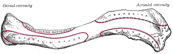
Anchored by muscles from above, the clavicle acts as a strut that reaches sideways to support the scapula (shoulder blade)2. This holds the shoulder joint up and out from the trunk, so the arm has the most freedom to move2. The bone also passes forces acting on the arm on to the sternum (breastbone) and the axial skeleton (the bones of the head and trunk)2. It protects the nerves and blood vessels that pass beneath it between the trunk and the arm2.
Its two ends
The inner, sternal end is triangular and meets the breastbone at the sternoclavicular joint2. This is the only bony joint between the shoulder girdle (the shoulder blades and collarbones) and the axial skeleton2. The joint allows considerable movement, so the clavicle and shoulder blade can move up and down and forwards and backwards2.
The outer, acromial end has a small, flat, oval surface that faces downwards at a slant6. It meets the acromion, the bony point at the top of the shoulder that is part of the shoulder blade6. The acromioclavicular joint page covers that joint.
Where the bone is weakest
A study of 15 adult clavicles found the central third of the bone denser, smaller and more circular in cross-section7. A model of the bones’ mechanical behaviour, built from their cross-sections, placed the weakest point in the central third7. That matches the fracture site most often reported clinically7.
An anatomical reason has also been offered for the middle being more open to trauma8. Strong ligaments (tough bands of tissue that hold bones together at a joint) and muscles firmly hold the inner and outer parts8. The middle part has no strong attachments8.
How it grows
The clavicle is the first bone in the body to begin ossifying (turning into bone), in the fifth week of development2,6. The clavicle does not finish ossifying until about age 252.
Breakdown
How often it breaks
Reports put clavicle fractures at 2.6% to 4% of all fractures, with about 80% of them in the middle third of the bone3. Three studies counted new fractures per 100,000 person-years, a count of people multiplied by the years each was followed9,10,11:
| Where the fractures were counted | Clavicle fractures per 100,000 person-years |
|---|---|
| US emergency department sample, 9,428 fractures in 5 years | 24.49 |
| US emergency department data, 2012–2015 | 22.410 |
| All adults in Sweden, 2001 | 35.611 |
| All adults in Sweden, 2012 | 59.311 |
Who breaks it, and where along the bone
In the US emergency sample of 9,428 fractures, incidence peaked between ages 10 and 199. Men were nearly three times as likely as women to be affected9.
The Swedish Fracture Register held 2,422 clavicle fractures in people aged 15 and over, 68% of them in males8. From age 65, females had more clavicle fractures than males8. Younger patients typically had fractures in the middle of the shaft (midshaft)8. Medial (inner) and lateral (outer) fractures were far more common in the older age groups8.
How it breaks
Sources give different accounts of the mechanism1,2,4:
- Anatomy references describe two: a fall onto the outstretched arms or hand, or a direct blow to the outer shoulder1,2.
- A consecutive series of 150 patients found the mechanism was known in 81%4. Of those, 94% had taken a direct blow on the shoulder and 6% had fallen on an outstretched hand4. The authors noted that this was at odds with commonly held views4.
In the Swedish register, the most common cause was a transport accident or a fall, generally one on the same level8. Among transport causes, bicycle accidents were by far the most frequent, at 20.5% of fractures8. The muscles attached to the clavicle often pull the main fragments out of place and shorten the bone, particularly in midshaft fractures2,8.
In sport
Clavicle fractures have been reported to make up as much as 10% of all sport-related fractures12. A systematic review, which sums up the research on a question, covered 12 studies of road cycling injuries13. It found the clavicle was the most common fracture13.
Stress fractures and lifting
A review of upper limb stress fractures (small cracks in a bone from repeated loading) describes those of the medial clavicle as rare14. The reported cases were in athletes aged 10 to 30 in sports that load the clavicle repeatedly, such as rowing, throwing, diving and gymnastics14.
Single case reports (reports on a single patient) describe clavicle stress fractures with two exercises15,16:
- One describes a healthy 34-year-old man who had done hundreds of push-ups each day for over 4 months15. The stress fracture was in the middle of the collarbone15.
- One describes a 13-year-old boy who felt a pop and sudden pain after about 20 bodyweight triceps dips16. The stress fracture was in the inner part of the clavicle16.
The review also defines distal clavicle osteolysis as chronic erosion and resorption (breakdown) of the outer end of the clavicle14. It follows repeated small injuries at the acromioclavicular joint14. These come as the shoulder repeatedly hyperextends (bends back past its usual range) in the bench press, overhead lifts and pushing14. The acromioclavicular joint page covers this in detail.
A scoping review (a review that maps what research exists on a topic) covered eight cohort studies of 483 patients17. The most common activity-based risk factor was bench pressing, at 49.1%17.
Recovery
How often the break fails to join
In a prospective cohort (one followed forward in time) of 868 patients managed without surgery, 6.2% of fractures had not united at 24 weeks5. That was 8.3% of medial end fractures, 4.5% of shaft fractures and 11.5% of lateral end fractures5. After a shaft fracture, the risk of nonunion (a broken bone that fails to join and heal) was significantly higher with four factors5. These were older age, female sex, displacement of the fracture and comminution5. Displacement means the bone ends have moved out of line, and comminution means the bone is in several pieces.
What trials of surgery against a sling found
A Cochrane review pooled 14 randomised and quasi-randomised studies of 1,469 people with acute middle-third fractures3. It compared surgery with conservative care: a sling, a figure-of-eight bandage or both3. It found low-quality evidence that surgery may not improve arm function at one year or later3. The pooled difference equalled 2.3 points on the 100-point Constant score (a score of shoulder pain, daily activity, movement and strength)3. The review did not count that as a clinically important difference3.
The review also found low-quality evidence that surgery may reduce the risk of a further, non-routine operation3. It reported these adverse outcomes3:
- Removal of the metal hardware for discomfort was common after surgery, at 10.2%3.
- Symptomatic malunion (a broken bone that heals out of line) was more common without surgery, at 11.3% against 1.2%3.
- Infection occurred only in the surgery group, at 3.2%3.
Almost all the studies had design features that carried a high risk of bias, and the review rated the evidence low or very low quality3.
Two multicentre randomised trials studied displaced midshaft fractures18,19:
- In one, of 301 adults, nonunion on X-ray at 3 months was 28% after surgery and 27% after nonoperative care18. At 9 months it was 0.8% after surgery and 11% after nonoperative care18. Function scores and satisfaction were significantly better after surgery at 6 weeks and 3 months18.
- The other studied plate fixation (surgery that holds a broken bone together with a metal plate) in 200 patients aged 16 to 6019. There was one nonunion after plate fixation and sixteen after nonoperative care19. Shoulder scores at one year were significantly better after plate fixation19. Once patients with nonunion were left out, there was no significant difference at any time point19.
A network meta-analysis, which compares many options at once, pooled 22 randomised trials in adults with displaced midshaft fractures20. It found union in 88.9% of patients managed without surgery and 96.7% of those who had surgery20. The gains in function scores with surgery were minor, and did not approach the 8 points the review set as the smallest meaningful change20.
In teenagers
A prospective cohort followed adolescents aged 10 to 18 with completely displaced midshaft fractures, 282 of them with two-year data21. Refracture, a repeat break, occurred in 2.6%, and delayed union, slower healing than expected, in 1.9%21. Nonunion and symptomatic malunion each occurred in 0.4%, and all four were rare with or without surgery21. Those who had surgery had more unexpected further surgery, at 10.4% against 1.4%21. Patient-reported scores did not differ between the 88 who had surgery and the 194 who did not21.
Getting back to sport
A systematic review of 33 clinical studies covered 1,087 athletes with clavicle fractures12. It found that 91% returned to play and 86% returned at the same level12. The mean time to return was 3.1 months after operative and 3.9 months after nonoperative management12.
The review of upper limb stress fractures covers the reported cases of medial clavicle stress fracture14. In all of them, management without surgery was followed by a complete return to sport, usually 8 weeks after the injury14. In the scoping review of distal clavicle osteolysis, most patients recovered without surgery, and 16% went on to surgery17.
Connections
Muscles and ligaments on the bone
These muscles and ligaments attach to the clavicle1,6:
| Structure | Where it attaches to the clavicle |
|---|---|
| Deltoid | The front of the outer third1,6 |
| Trapezius | The back of the outer third1,6 |
| Pectoralis major | A lower, oval area on the front of the inner part1,6 |
| Sternocleidomastoid | An upper area on the front of the inner part1,6 |
| Subclavius muscle | A groove along the underside1,6 |
| Costoclavicular ligament | The costal tuberosity, a broad rough area on the inner part of the underside; it runs to the first rib1,2,6 |
| Conoid ligament | The conoid tubercle, on the underside of the outer third1,6 |
| Trapezoid ligament | A ridge running forwards and outwards from the conoid tubercle1,6 |
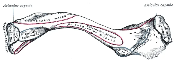
Joints, neighbours and exercises
The clavicle’s outer end meets the acromion of the scapula6. The acromioclavicular joint page covers that joint, and the AC separation page covers injuries to its ligaments. As a strut, the clavicle holds the shoulder joint up and out from the trunk2.
A review names the bench press in its account of distal clavicle osteolysis, and single case reports describe stress fractures with push-ups and dips14,15,16. Related exercise pages: bench press, push-up and dip.
Clavicle forms a joint with
Clavicle holds up and out from the trunk
Clavicle is attached to by
Clavicle forms the joint involved in
Clavicle is loaded by
Sources
- La Rose J, Charlick M, Varacallo MA (2026). Anatomy, Shoulder and Upper Limb, Clavicle.. StatPearls. PMID 30252246
- Betts JG, Young KA, Wise JA, Johnson E, Poe B, Kruse DH, Korol O, Johnson JE, Womble M, DeSaix P (2022). Anatomy and Physiology 2e. OpenStax. Link
- Lenza M, Buchbinder R, Johnston RV, Ferrari BA, Faloppa F (2019). Surgical versus conservative interventions for treating fractures of the middle third of the clavicle.. Cochrane Database of Systematic Reviews. doi:10.1002/14651858.CD009363.pub3 PMID 30666620
- Stanley D, Trowbridge EA, Norris SH (1988). The mechanism of clavicular fracture. A clinical and biomechanical analysis.. The Journal of Bone and Joint Surgery. British Volume. doi:10.1302/0301-620X.70B3.3372571 PMID 3372571
- Robinson CM, Court-Brown CM, McQueen MM, Wakefield AE (2004). Estimating the risk of nonunion following nonoperative treatment of a clavicular fracture.. The Journal of Bone and Joint Surgery. American Volume. doi:10.2106/00004623-200407000-00002 PMID 15252081
- Gray H; Lewis WH (editor) (1918). Anatomy of the Human Body. Lea & Febiger, Philadelphia (20th edition). Link
- Harrington MA, Keller TS, Seiler JG, Weikert DR, Moeljanto E, Schwartz HS (1993). Geometric properties and the predicted mechanical behavior of adult human clavicles.. Journal of Biomechanics. doi:10.1016/0021-9290(93)90005-y PMID 8478346
- Kihlström C, Möller M, Lönn K, Wolf O (2017). Clavicle fractures: epidemiology, classification and treatment of 2 422 fractures in the Swedish Fracture Register; an observational study.. BMC Musculoskeletal Disorders. doi:10.1186/s12891-017-1444-1 PMID 28202071
- Van Tassel D, Owens BD, Pointer L, Moriatis Wolf J (2014). Incidence of clavicle fractures in sports: analysis of the NEISS Database.. International Journal of Sports Medicine. doi:10.1055/s-0033-1345127 PMID 23771828
- DeFroda SF, Lemme N, Kleiner J, Gil J, Owens BD (2019). Incidence and mechanism of injury of clavicle fractures in the NEISS database: Athletic and non athletic injuries.. Journal of Clinical Orthopaedics and Trauma. doi:10.1016/j.jcot.2019.01.019 PMID 31528074
- Huttunen TT, Launonen AP, Berg HE, Lepola V, Felländer-Tsai L, Mattila VM (2016). Trends in the Incidence of Clavicle Fractures and Surgical Repair in Sweden: 2001-2012.. The Journal of Bone and Joint Surgery. American Volume. doi:10.2106/JBJS.15.01284 PMID 27807117
- Kilkenny CJ, Daly GR, Whelehan SP, Vukanic D, Alrawashdeh M, Boland F, Quinlan JF, Molony DC (2025). Return to play following clavicular fracture - A systematic review and meta analysis.. JSES Reviews, Reports, and Techniques. doi:10.1016/j.xrrt.2024.11.002 PMID 40321855
- Rooney D, Sarriegui I, Heron N (2020). 'As easy as riding a bike': a systematic review of injuries and illness in road cycling.. BMJ Open Sport & Exercise Medicine. doi:10.1136/bmjsem-2020-000840 PMID 34422283
- Koslosky EJ, Heath DM, Atkison CL, Dutta A, Brady CI (2024). Upper Extremity Stress Fractures.. Sports Medicine - Open. doi:10.1186/s40798-024-00769-7 PMID 39327396
- Ohnishi Y, Koulopoulos M, Scuderi M (2026). Middle-shaft clavicle stress fracture in a non-athlete adult after excessive daily push-ups: a case report.. Journal of rural medicine. doi:10.2185/jrm.2025-073 PMID 42487887
- Kang M, Kohli S, Naumowicz Z, Barlow I (2021). Stress fracture of the clavicle in a young adolescent male - A case report. Trauma Case Reports. doi:10.1016/j.tcr.2021.100558 PMID 34901371
- Wilkinson M, Groch N, Freestone CA, Clements A, Obst S (2026). Risk factors and management of atraumatic distal clavicular osteolysis: A scoping review.. Shoulder & elbow. doi:10.1177/17585732261479715 PMID 42656558
- Ahrens PM, Garlick NI, Barber J, Tims EM (2017). The Clavicle Trial. The Journal of Bone and Joint Surgery. American Volume. doi:10.2106/JBJS.16.01112 PMID 28816894
- Robinson CM, Goudie EB, Murray IR, Jenkins PJ, Ahktar MA, Read EO, Foster CJ, Clark K, Brooksbank AJ, Arthur A, Crowther MA, Packham I, Chesser TJ (2013). Open reduction and plate fixation versus nonoperative treatment for displaced midshaft clavicular fractures. The Journal of Bone and Joint Surgery. American Volume. doi:10.2106/JBJS.L.00307 PMID 24005198
- Axelrod DE, Ekhtiari S, Bozzo A, Bhandari M, Johal H (2020). What Is the Best Evidence for Management of Displaced Midshaft Clavicle Fractures? A Systematic Review and Network Meta-analysis of 22 Randomized Controlled Trials.. Clinical Orthopaedics and Related Research. doi:10.1097/CORR.0000000000000986 PMID 31574019
- Heyworth BE, Pennock AT, Li Y, Liotta ES, Dragonetti B, Williams D, Ellis HB, Nepple JJ, Spence D, Willimon SC, Perkins CA, Pandya NK, Kocher MS, Edmonds EW, Wilson PL, Busch MT, Sabatini CS, Farley F, Bae DS (2022). Two-Year Functional Outcomes of Operative vs Nonoperative Treatment of Completely Displaced Midshaft Clavicle Fractures in Adolescents: Results From the Prospective Multicenter FACTS Study Group.. The American Journal of Sports Medicine. doi:10.1177/03635465221114420 PMID 35984091
Changes
- Revised to report sources added after their published corrections were read