Acromioclavicular joint
Also called AC joint
The small joint where the collarbone meets the top of the shoulder blade. Which ligaments hold it, how it moves, how it is injured or worn, and what studies report about healing.
- Region
- Shoulder
- Muscles
- deltoid, trapezius
- Updated
- October 8, 2026
- Reviewed
- October 8, 2026 · how entries are checked
Function
Where the collarbone meets the shoulder blade
The joint lies between the outer clavicle and the acromion, part of the scapula (shoulder blade)1. It is a plane synovial joint, and it helps to stabilise the shoulder girdle (the shoulder blades and collarbones)1. A capsule (a sleeve of tough tissue) with a synovial lining, its thin inner lining, encloses the joint, and cartilage covers its surfaces2.
A 1918 anatomy text describes the joint’s articular disc, a pad between the joint surfaces, as frequently absent3. When present, the text says, it usually separates the joint surfaces only partly3. In 53 cadaver joints examined by eye, one had a complete disc, 25 a partial disc, 16 disc remnants and 11 no disc6. A cadaver is a body donated for study, and the ages of these cadavers were unknown6.
Two sets of ligaments
The capsule is quite thin, but four AC ligaments reinforce it: superior, inferior, anterior and posterior2. The superior ligament is a four-sided band across the top of the joint3. Its fibres interlace with the flat tendons of the trapezius and deltoid muscles3. The inferior ligament is somewhat thinner and covers the underside of the joint3.
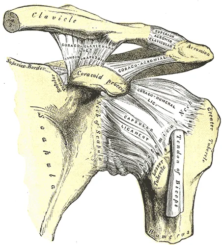
The coracoclavicular ligament joins the clavicle to the coracoid process, a hooked bony point at the front of the shoulder blade3. It is not strictly part of the joint, but it holds the clavicle against the acromion3. It has two parts3:
- The trapezoid ligament is the front and outer part, a broad, thin band3.
- The conoid ligament is the back and inner part, a dense, cone-shaped band3.
Which ligament resists which push
A review states the generally accepted principle2. The AC ligaments give front-to-back stability, and the coracoclavicular ligaments give vertical stability2. One ligament-cutting study pushed the clavicle with 50 newtons in 20 cadaver specimens7:
| Clavicle pushed | Primary stabiliser in that study | Share of the resistance |
|---|---|---|
| Backward | AC capsular ligament7 | 38.4%7 |
| Downward | AC capsular ligament7 | 33.8%7 |
| Upward | Conoid ligament7 | 45.6%7 |
| Forward | AC capsular ligament and conoid, about equally7 | 23% and 25.2%7 |
Two other cadaver studies reported on backward displacement of the clavicle. In a ligament-cutting study of 12 specimens, the AC ligament was a primary restraint to it8. In 10 shoulders under 70 newtons, the trapezoid was the primary restraint to it, with 55.8% of the resisting force9.
How it moves
The shoulder blade can move on the rib cage only when this joint and the sternoclavicular joint move together10. The sternoclavicular joint is where the collarbone meets the breastbone. One study fixed sensors to pins in the bones of 12 subjects without a shoulder abnormality10. As the arm rose overhead, the shoulder blade turned on the clavicle by these average amounts10:
- 19 degrees of backward tilt10
- 11 degrees of upward rotation, a turn that tilts the shoulder socket up10
- 8 degrees of internal rotation, a turn inward10
Breakdown
Sudden injuries
A review states that injury of the joint most commonly follows a direct impact2. In a case series of 158 adults with a dislocation of the joint, a direct force to the shoulder caused 150 cases (96%)11. A case series reports on a group of patients without a comparison group, and a dislocation is a bone forced fully out of place. The review adds that a fall on an outstretched hand can also injure the joint indirectly2.
As force on the top of the acromion grows, the soft tissues are assumed to fail in order11. The AC ligaments go first, then the coracoclavicular ligaments, then the fascia (a sheet of tough connective tissue) shared by the deltoid and trapezius11. The Rockwood classification is a six-part grading of AC joint separations, described in 198412. Its first three types differ in which ligaments are torn2:
- Type I: the AC ligaments are sprained and the joint is intact2.
- Type II: the AC ligaments are torn and the coracoclavicular ligaments are intact2. One account describes them as sprained instead12.
- Type III: both sets of ligaments are torn, and the deltoid and trapezius fascia stays intact2.
In types IV to VI the outer clavicle is displaced backward or downward, or the fascia is stripped off the bones2. The AC separation page covers the grading in full.
How often, in which groups
Each rate below comes from one study group, and the groups counted different things13,14. Incidence is how many new cases appear in a group over a set time. Person-years count people multiplied by the years each was followed. An athlete-exposure is one athlete taking part in one practice or game. The NCAA runs college sport in the United States.
| Group | What was counted | Incidence |
|---|---|---|
| One emergency department serving the city of Oslo | All AC joint injuries | 45 per 100,000 person-years13 |
| Three university hospitals in the capital region of Denmark, over one year | Acute dislocations | 19.3 per 100,000 person-years14 |
| 158 adults aged 18 to 75 from a general population | Dislocations | 2.0 per 10,000 person-years11 |
| United States Military Academy, 2005 to 2009 | All new AC joint injuries | 9.2 per 1,000 person-years15 |
| 25 NCAA sports, 2009-2010 to 2014-2015 | Sprains | 1.72 per 10,000 athlete-exposures16 |
| National Football League, 2000 to 2011 | Shoulder injuries involving the AC joint | 26.1 per 10,000 athlete-exposures17 |
In the Oslo group, 68% of the injuries were sprains and 32% were dislocations13. In the Military Academy group, 89% were low-grade sprains (types I and II) and 91% happened during athletics15.
Wear with age
In 168 joints dissected from 85 cadavers aged 18 to 92, the disc and joint cartilage showed age-related breakdown18. One review reports osteoarthritis in cadaver AC joints by age band4:
| Age of cadaver | Joints with osteoarthritis |
|---|---|
| 21 to 40 | 25%4 |
| 41 to 60 | 53%4 |
| Over 61 | 66%4 |
Two systematic reviews looked at imaging of shoulders without symptoms4,19. In a systematic review of people not seeking shoulder care, 70% of 210 symptom-free shoulder MRI scans showed changes consistent with osteoarthritis of this joint4. The other review covered 31 imaging studies of adults without shoulder symptoms19. Its reported rates varied widely, and it rated the certainty of the evidence very low19.
Eight studies in that review imaged shoulders both with and without symptoms19. Osteoarthritis of the joint was seen in 13 to 95% of shoulders without symptoms and 20 to 100% of those with symptoms19. The authors concluded that the prevalence is often comparable in the two19.
Wearing away of the outer collarbone
One series of athletes described osteolysis of the outer clavicle in 46 men with no history of acute injury to the area20. Every one was an athlete, and 45 lifted weights as part of their training20. A literature review gives the most widely accepted explanation: tiny breaks in the bone beneath the cartilage, then attempts at repair21. The review calls it most likely an overuse phenomenon21.
Three sources report which activities went with the condition5,22,23:
- A scoping review (a review that maps what research exists on a topic) included 8 cohort studies with 483 patients5. A cohort study follows a group of people over time. The most common activity-based risk factor was bench pressing (49.1%), then general or unspecified weight training (24.4%)5.
- A review of shoulder MRI reports compared 262 men aged 20 to 40 who had the condition with 227 who did not22. High-intensity bench pressing was a risk factor, and low-intensity bench pressing was not22.
- Among 1,432 consecutive shoulder MRI reports in patients aged 13 to 19, 6.5% showed the atraumatic condition23. Combining an overhead sport with supplemental weight training was a risk factor23.
The scoping review found that most of its 8 studies were retrospective (looking back at past records), with small or mixed samples5.
Recovery
How the torn ligament heals
The authors of a 2020 biopsy study state that, to their knowledge, how the human AC ligament heals on its own is not understood24. A biopsy is a small sample of tissue taken from the body for study. Their samples came from patients operated on for acute dislocations within 21 days of injury24. They describe three weeks:
- First week, 5 biopsies: cell activation and an early inflammatory response24.
- Second week: more fibroblast-like cells in the torn zone than in the intact zone, and a peak in type 3 collagen24. A fibroblast is a cell in connective tissue that makes collagen.
- Third week, 4 patients: signs of consolidation and early remodelling, the steady replacement of old tissue with new24.
The authors name the small sample and the lack of a healthy control group as major limitations24. A consensus among the European Shoulder Associates of ESSKA set the line between acute and chronic AC joint injuries at 3 weeks25. The AC separation page reports what trials and follow-up studies found after these injuries.
What followed osteolysis
In the series of 46 men, the 25 who did not have an operation improved20. They improved only after stopping or changing their sport and avoiding weight training20. In the scoping review of 483 patients, most recovered without an operation5. Sixteen percent went on to surgery after non-surgical care failed5.
One study followed teenagers aged 13 to 19 with the atraumatic condition and repeated their imaging after 2 years23. Osteoarthritis of the AC joint showed in 71% of them, against 35% of controls23.
Connections
Bones, joints and muscles
The joint links the clavicle to the acromion of the scapula, and it passes forces from the arm to the clavicle1,26. The glenohumeral joint (the ball and socket) is the main shoulder joint, and the AC joint is one of the girdle’s smaller joints27. The inferior AC ligament lies directly above the tendon of the supraspinatus, part of the rotator cuff3. The deltoid lies in front of the coracoclavicular ligaments and the trapezius behind them3.
The related condition page is AC separation, which sets out the Rockwood grades of injury to these ligaments2.
Exercises that load it
Related exercise pages: bench press28. Ten experienced strength athletes performed bench press variations, and a shoulder model estimated their joint loads28. Grips narrower than 1.5 times bi-acromial width (the width across the shoulders) decreased the modelled compression at the AC joint28. The authors suggest this may decrease the risk of distal clavicular osteolysis28. Another source notes that high-quality data on specific bench press injuries are lacking28.
Acromioclavicular joint is formed by
Acromioclavicular joint lies next to
Acromioclavicular joint is injured in
Acromioclavicular joint is loaded by
Sources
- La Rose J, Tiwari V (2026). Anatomy, Shoulder and Upper Limb, Acromioclavicular Joint.. StatPearls. PMID 29763033
- Saccomanno MF, DE Ieso C, Milano G (2014). Acromioclavicular joint instability: anatomy, biomechanics and evaluation.. Joints. doi:10.11138/jts/2014.2.2.087 PMID 25606549
- Gray H; Lewis WH (editor) (1918). Anatomy of the Human Body. Lea & Febiger, Philadelphia (20th edition). Link
- Rossano A, Manohar N, Veenendaal WJ, van den Bekerom MPJ, Ring D, Fatehi A (2022). Prevalence of acromioclavicular joint osteoarthritis in people not seeking care: A systematic review.. Journal of orthopaedics. doi:10.1016/j.jor.2022.05.009 PMID 35638093
- Wilkinson M, Groch N, Freestone CA, Clements A, Obst S (2026). Risk factors and management of atraumatic distal clavicular osteolysis: A scoping review.. Shoulder & elbow. doi:10.1177/17585732261479715 PMID 42656558
- Salter EG, Nasca RJ, Shelley BS (1987). Anatomical observations on the acromioclavicular joint and supporting ligaments.. The American journal of sports medicine. doi:10.1177/036354658701500301 PMID 3618870
- Lee J, El-Daou H, Alkoheji M, Carlos A, Di Mascio L, Amis A (2021). Ligamentous and capsular restraints to anterior-posterior and superior-inferior laxity of the acromioclavicular joint: a biomechanical study.. Journal of shoulder and elbow surgery. doi:10.1016/j.jse.2020.09.006 PMID 33010434
- Fukuda K, Craig EV, An KN, Cofield RH, Chao EY (1986). Biomechanical study of the ligamentous system of the acromioclavicular joint.. The Journal of bone and joint surgery. American volume. PMID 3949839
- Lee KW, Debski RE, Chen CH, Woo SL, Fu FH (1997). Functional evaluation of the ligaments at the acromioclavicular joint during anteroposterior and superoinferior translation.. The American journal of sports medicine. doi:10.1177/036354659702500622 PMID 9397278
- Ludewig PM, Phadke V, Braman JP, Hassett DR, Cieminski CJ, LaPrade RF (2009). Motion of the shoulder complex during multiplanar humeral elevation.. The Journal of bone and joint surgery. American volume. doi:10.2106/JBJS.G.01483 PMID 19181982
- Nordin JS, Olsson O, Lunsjö K (2020). Acromioclavicular joint dislocations: incidence, injury profile, and patient characteristics from a prospective case series.. JSES international. doi:10.1016/j.jseint.2020.01.009 PMID 32490410
- Gorbaty JD, Hsu JE, Gee AO (2017). Classifications in Brief: Rockwood Classification of Acromioclavicular Joint Separations.. Clinical orthopaedics and related research. doi:10.1007/s11999-016-5079-6 PMID 27637619
- Skjaker SA, Enger M, Engebretsen L, Brox JI, Bøe B (2021). Young men in sports are at highest risk of acromioclavicular joint injuries: a prospective cohort study.. Knee surgery, sports traumatology, arthroscopy. doi:10.1007/s00167-020-05958-x PMID 32270265
- Haugaard KB, Bak K, Seem K, Hölmich P, Barfod KW (2023). Rockwood type III is the most common type of acromioclavicular joint dislocation: A prospective cohort study investigating the incidence and epidemiology of acute acromioclavicular joint dislocations in an urban population.. Shoulder & elbow. doi:10.1177/17585732221123314 PMID 37811384
- Pallis M, Cameron KL, Svoboda SJ, Owens BD (2012). Epidemiology of acromioclavicular joint injury in young athletes.. The American journal of sports medicine. doi:10.1177/0363546512450162 PMID 22707749
- Hibberd EE, Kerr ZY, Roos KG, Djoko A, Dompier TP (2016). Epidemiology of Acromioclavicular Joint Sprains in 25 National Collegiate Athletic Association Sports. The American journal of sports medicine. doi:10.1177/0363546516643721 PMID 27159287
- Lynch TS, Saltzman MD, Ghodasra JH, Bilimoria KY, Bowen MK, Nuber GW (2013). Acromioclavicular joint injuries in the National Football League. The American journal of sports medicine. doi:10.1177/0363546513504284 PMID 24057030
- Petersson CJ (1983). Degeneration of the acromioclavicular joint. A morphological study.. Acta orthopaedica Scandinavica. doi:10.3109/17453678308996597 PMID 6858663
- Ibounig T, Rämö L, Haas R, Jones M, Järvinen TLN, Taimela S, Docking S, Sanders S, Buchbinder R (2025). Imaging abnormalities of the acromioclavicular joint and subacromial space are common in asymptomatic shoulders: a systematic review.. Journal of orthopaedic surgery and research. doi:10.1186/s13018-024-05378-4 PMID 39754140
- Cahill BR (1982). Osteolysis of the distal part of the clavicle in male athletes.. The Journal of bone and joint surgery. American volume. PMID 7118971
- Schwarzkopf R, Ishak C, Elman M, Gelber J, Strauss DN, Jazrawi LM (2008). Distal clavicular osteolysis: a review of the literature.. Bulletin of the NYU hospital for joint diseases. PMID 18537776
- Nevalainen MT, Ciccotti MG, Morrison WB, Zoga AC, Roedl JB (2016). Distal clavicular osteolysis in adults: association with bench pressing intensity.. Skeletal Radiology. doi:10.1007/s00256-016-2446-z PMID 27550324
- Roedl JB, Nevalainen M, Gonzalez FM, Dodson CC, Morrison WB, Zoga AC (2015). Frequency, imaging findings, risk factors, and long-term sequelae of distal clavicular osteolysis in young patients.. Skeletal radiology. doi:10.1007/s00256-014-2092-2 PMID 25560997
- Maier D, Tuecking LR, Bernstein A, Lang G, Wagner FC, Jaeger M, Ogon P, Südkamp NP, Izadpanah K (2020). The acromioclavicular ligament shows an early and dynamic healing response following acute traumatic rupture.. BMC musculoskeletal disorders. doi:10.1186/s12891-020-03614-6 PMID 32887599
- Rosso C, Martetschläger F, Saccomanno MF, Voss A, Lacheta L, Beitzel K, Milano G (2021). High degree of consensus achieved regarding diagnosis and treatment of acromioclavicular joint instability among ESA-ESSKA members.. Knee surgery, sports traumatology, arthroscopy. doi:10.1007/s00167-020-06286-w PMID 32980887
- Betts JG, Young KA, Wise JA, Johnson E, Poe B, Kruse DH, Korol O, Johnson JE, Womble M, DeSaix P (2022). Anatomy and Physiology 2e. OpenStax. Link
- Chang LR, Anand P, Varacallo MA (2025). Anatomy, Shoulder and Upper Limb, Glenohumeral Joint. StatPearls. PMID 30725703
- Noteboom L, Belli I, Hoozemans MJM, Seth A, Veeger HEJ, Van Der Helm FCT (2024). Effects of bench press technique variations on musculoskeletal shoulder loads and potential injury risk.. Frontiers in Physiology. doi:10.3389/fphys.2024.1393235 PMID 38974522
Changes
- Revised to report sources added after their published corrections were read