Supraspinatus
Also called supraspinatus muscle
The rotator cuff muscle along the top of the shoulder blade. How it helps raise the arm, how it wastes and fills with fat after a tendon tear, what studies found after repair, and what it works with.
- Region
- Shoulder
- Starts on
- the inner two-thirds of the supraspinous fossa, the hollow above the shoulder blade's ridge, and the fascia covering the muscle1,7
- Ends on
- the highest of three impressions on the greater tubercle, a bump on the outer side of the upper arm bone (Rotator cuff tendons)1,7
- Tendons
- Rotator cuff tendons
- Updated
- October 8, 2026
- Reviewed
- October 8, 2026 · how entries are checked
Function
Where the muscle sits
The supraspinatus fills the whole supraspinous fossa, a shallow hollow in the scapula (shoulder blade)1. The muscle starts on the inner two-thirds of the fossa and on the strong fascia, a sheet of tough connective tissue, that covers it1. This fascia closes over the fossa, so the muscle lies in a case of bone and fascia1.
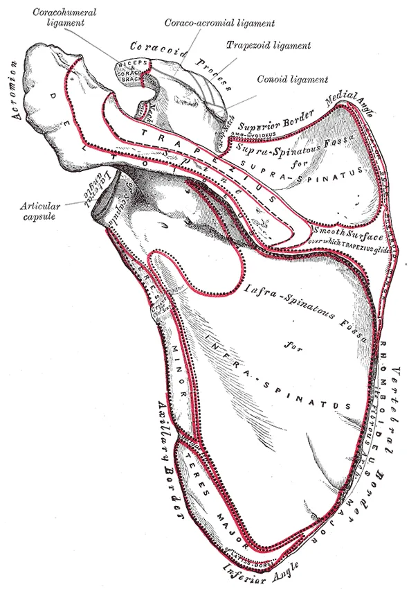
The fibres converge on a tendon, a tough cord of tissue that connects a muscle to a bone1. The tendon crosses the top of the shoulder joint1. It is closely bound to the joint capsule, the sleeve of tough tissue that encloses a joint1. It attaches to the highest of three impressions on the greater tubercle, a bump at the top of the humerus (upper arm bone)1. With three other tendons it forms the rotator cuff, a ring of tendons around the joint; that page covers tendon tears7.
Two regions in one muscle
A dissection study of ten embalmed male cadavers (bodies donated for study) found an anterior (front) and a posterior (back) region8. One paper describes the two regions this way9:
| Anterior region | Posterior region | |
|---|---|---|
| Size | 75–86% of the muscle by volume9 | Much smaller, and partly deep to the anterior region9 |
| Fibre bundles | Run at an angle into the anterior tendon9 | Parallel, attaching to the posterior tendon9 |
| Role | Produces most of the muscle’s force9 | Thought to have an important part in adjusting tension on the rotator cuff9 |
Its part in raising the arm
The supraspinatus helps the deltoid raise the arm from the side, and holds the head of the humerus in the socket1. Three studies measured its share of the lift10,11,12:
- Paralysing nerves in ten healthy volunteers. One study paralysed the suprascapular and axillary nerves in turn10. It concluded that the supraspinatus and deltoid are equally responsible for the torque (turning force) of two lifts10. These were raising the arm forward and raising it in the plane of the shoulder blade10.
- Nerve blocks and strength. Volunteers were given suprascapular nerve blocks, injections that numb a nerve for a short time11. Paralysing the supraspinatus and infraspinatus together removed about 75% of the strength of abduction, lifting the arm out to the side11. Paralysing the infraspinatus alone removed about 45%11.
- Five fresh cadaver shoulders. In these, adding simulated supraspinatus pull lowered the deltoid force needed to raise the arm by an average of 28%12.
Does it start the lift?
The authors of a 2013 study wrote that the supraspinatus is commonly said to initiate abduction3. They added that there was no direct evidence for the claim3. Four studies bear on the question2,3,11,13:
- No earlier than four other muscles. That study used electromyography (EMG), which measures a muscle’s activity from its electrical signals, in fourteen volunteers3. The supraspinatus switched on before the arm moved, but no earlier than the infraspinatus, deltoid, trapezius and serratus anterior3. The authors called the statement that it initiates abduction misleading3.
- Ahead of two others. An EMG study recorded 24 healthy shoulders raising a light dumbbell out to the side2. The supraspinatus became active 0.102 seconds before the arm moved2. That was ahead of the middle trapezius, at 0.019 seconds before, and the middle deltoid, at 0.014 seconds before2.
- A larger effect at the start, in a model. In a cadaver shoulder model, simulated supraspinatus paralysis doubled the middle deltoid force needed to start abduction13. It raised the force needed for full abduction by 12%13.
- The same loss at four angles. A suprascapular nerve block paralysed the supraspinatus and infraspinatus in volunteers11. The loss of abduction strength was the same at 0, 30, 60 and 90 degrees11.
How well it holds the humerus down
One study tested 10 fresh-frozen cadaver shoulders14. Each muscle pulled the humeral head (the ball at the top of the upper arm bone) down against an upward force14:
| Muscle | How far it pulled the head down |
|---|---|
| Latissimus dorsi | 5.6 mm14 |
| Teres major | 5.1 mm14 |
| Subscapularis | 4.7 mm14 |
| Infraspinatus | 4.6 mm14 |
| Supraspinatus | 2.0 mm14 |
The authors wrote that the supraspinatus may matter less for holding the head down than previously assumed14.
Its nerve
The fifth and sixth cervical (neck) nerves supply the supraspinatus through the suprascapular nerve1. This nerve reaches the top edge of the shoulder blade at the suprascapular notch, where it passes under a ligament15. It then runs along the supraspinous fossa and gives motor branches to the supraspinatus15.
Breakdown
Wasting and fat after a tendon tear
A review describes fatty degeneration of the rotator cuff muscles as a degenerative condition16. It is marked by wasting of muscle fibres, scarring, and fat building up in and around the muscles16. Studies measure wasting and fat in different ways17,18:
- Wasting is measured on a slice of an MRI (a scan that shows soft tissue in detail) as the occupation ratio17. This is the cross-section of the muscle belly divided by that of the supraspinous fossa17.
- Fat is graded in stages18. As applied in one MRI study, the modified Goutallier stages run from 0, normal muscle with no fatty streaks, to 4, more fat than muscle18.
In an MRI study of 55 shoulders, the mean occupation ratio was 0.7 in 15 controls17. It was 0.62 in 10 degenerative cuffs without tears and 0.44 in 30 operated tears17. Larger tears went with more wasting17.
In 1688 patients with rotator cuff tears, moderate fatty infiltration of the supraspinatus appeared on average 3 years after symptoms began4. Severe fatty infiltration appeared at 5 years4.
Why the changes are uneven
On MRI of 64 painful shoulders, forty had a full-thickness tear, a tear right through the supraspinatus tendon, and showed uneven changes in the muscle19. The superficial portion arising from the fascia had wasted by 43%, and the deep portion arising from the bone by 9%19. The deep portion mainly showed fatty infiltration19. The authors concluded that muscle changes after a tendon tear are not simply a result of disuse, but depend on changes in the muscle’s architecture19.
When the nerve is affected
A review of 59 studies describes suprascapular neuropathy as an uncommon cause of shoulder pain and weakness15. Neuropathy is damage to a nerve. The review says it is identified more often in overhead athletes and in people with massive rotator cuff tears15. The suprascapular notch is the most common place for the nerve to be compressed15. In this condition, MRI can show swelling, wasting and fatty degeneration of the supraspinatus and infraspinatus15.
Two studies bear on whether a torn, pulled-back muscle harms the nerve20,21. In twelve shoulders from six cadavers, pulling the supraspinatus tendon 2 to 3 cm toward the midline left the nerve’s first motor branch taut20. The authors suggested this tension may explain the wasting often seen after a massive tear20. In fifty patients with retracted tears of both the supraspinatus and infraspinatus, electrical testing of 49 shoulders found suprascapular neuropathy in one21. Results did not differ in shoulders with significant fatty infiltration21.
Recovery
What training studies measured
Thirteen participants were randomised to concentric or eccentric shoulder abduction training on a dynamometer, a machine that measures force23. They trained three times a week for eight weeks23. Concentric work is done while the muscle shortens, and eccentric work while it lengthens. Supraspinatus thickness, fibre angle and peak torque (the greatest turning force) each increased, with no significant difference between the two modes23. Fibre bundles became shorter with concentric training and were unchanged with eccentric training23.
Whether wasting and fat reverse after repair
Four studies followed the muscle after surgical repair of the tendon, and their results differ5,6,18,24:
| Who was studied | What was found |
|---|---|
| Patients in a 1994 CT study by Goutallier and colleagues | After an effective repair, moderate fatty degeneration of the supraspinatus regressed in six of 14 patients24 |
| 38 patients with MRI before and 1 year after repair | A successful repair did not improve or reverse wasting or fatty infiltration of the supraspinatus and infraspinatus5 |
| 94 patients whose repaired cuffs stayed intact | Supraspinatus wasting and fatty infiltration had improved significantly on MRI at 2 years, compared with MRI 2 weeks after surgery6 |
| 24 patients with massive rotator cuff tears | When the repair held, wasting improved in half of cases and fatty degeneration in a quarter; when the tendon retore, both got worse18 |
Fat in the muscle and later retears
A systematic review, a study that searches for the research on a question and sums it up, covered 11 studies and 925 shoulders25. Cuffs with Goutallier grades 2 to 4 before repair retore more often than those with grades 0 to 1, at 59% against 25%25.
After surgical release of the nerve
A 2018 systematic review of 21 studies, with 275 patients, reported that 92% of athletes returned to sport after surgical release of the suprascapular nerve26. A 2024 update covered 33 studies and 730 patients27. In it, 90 of 92 patients went back to sport or military duty, and 39 of 322 had continued symptoms27. A systematic review of six case series looked at keyhole release at the notch28. Muscle electrical activity recovered, but full strength did not return in most reported cases (60%), and fatty degeneration of the muscle bellies did not regress28.
Connections
The deltoid
When a suprascapular nerve block paralysed the supraspinatus and infraspinatus, activity rose in the front, middle and back of the deltoid29.
Exercises that load it
Related exercise pages: band external rotation and bench press30,31. EMG studies of shoulder exercises found these results:
- In 13 healthy men doing standing band external rotation, supraspinatus activity was higher with the arm raised 45 or 90 degrees than at 0 degrees30.
- In 15 volunteers, the authors read rotator cuff activity during the bench press as steadying the shoulder against front-to-back forces31.
- In 15 people with normal shoulders making maximal isometric efforts, supraspinatus activation did not differ between five positions32. Isometric means the muscle works but the joint does not move, and prone means face-down. The five were prone elevation, two external rotation positions, and the empty can and full can positions32.
In 28 people with subacromial impingement syndrome, the empty can drew higher pain ratings than the full can, by 1 point on an 11-point scale33. Subacromial impingement is pinching of tissue in the space beneath the acromion, the bony point at the top of the shoulder. The shoulder impingement page covers it.
Supraspinatus pulls through
Supraspinatus starts on
Supraspinatus ends on
Supraspinatus crosses the top of
Supraspinatus works with to raise the arm
Supraspinatus works with
Supraspinatus is studied in
Supraspinatus is loaded by
Sources
- Gray H; Lewis WH (editor) (1918). Anatomy of the Human Body. Lea & Febiger, Philadelphia (20th edition). Link
- Wickham J, Pizzari T, Stansfeld K, Burnside A, Watson L (2010). Quantifying 'normal' shoulder muscle activity during abduction.. Journal of Electromyography and Kinesiology. doi:10.1016/j.jelekin.2009.06.004 PMID 19625195
- Reed D, Cathers I, Halaki M, Ginn K (2013). Does supraspinatus initiate shoulder abduction?. Journal of Electromyography and Kinesiology. doi:10.1016/j.jelekin.2012.11.008 PMID 23265661
- Melis B, DeFranco MJ, Chuinard C, Walch G (2010). Natural history of fatty infiltration and atrophy of the supraspinatus muscle in rotator cuff tears.. Clinical Orthopaedics and Related Research. doi:10.1007/s11999-009-1207-x PMID 20094853
- Gladstone JN, Bishop JY, Lo IK, Flatow EL (2007). Fatty infiltration and atrophy of the rotator cuff do not improve after rotator cuff repair and correlate with poor functional outcome.. The American Journal of Sports Medicine. doi:10.1177/0363546506297539 PMID 17337727
- Hamano N, Yamamoto A, Shitara H, Ichinose T, Shimoyama D, Sasaki T, Kobayashi T, Kakuta Y, Osawa T, Takagishi K (2017). Does successful rotator cuff repair improve muscle atrophy and fatty infiltration of the rotator cuff? A retrospective magnetic resonance imaging study performed shortly after surgery as a reference.. Journal of Shoulder and Elbow Surgery. doi:10.1016/j.jse.2016.10.016 PMID 28214172
- Betts JG, Young KA, Wise JA, Johnson E, Poe B, Kruse DH, Korol O, Johnson JE, Womble M, DeSaix P (2022). Anatomy and Physiology 2e. OpenStax. Link
- Kim SY, Boynton EL, Ravichandiran K, Fung LY, Bleakney R, Agur AM (2007). Three-dimensional study of the musculotendinous architecture of supraspinatus and its functional correlations.. Clinical Anatomy. doi:10.1002/ca.20469 PMID 17352416
- Kim SY, Sachdeva R, Li Z, Lee D, Rosser BW (2015). Change in the Pathologic Supraspinatus: A Three-Dimensional Model of Fiber Bundle Architecture within Anterior and Posterior Regions.. BioMed Research International. doi:10.1155/2015/564825 PMID 26413533
- Howell SM, Imobersteg AM, Seger DH, Marone PJ (1986). Clarification of the role of the supraspinatus muscle in shoulder function.. The Journal of Bone and Joint Surgery. American Volume. PMID 3949834
- Gerber C, Blumenthal S, Curt A, Werner CM (2007). Effect of selective experimental suprascapular nerve block on abduction and external rotation strength of the shoulder.. Journal of Shoulder and Elbow Surgery. doi:10.1016/j.jse.2007.02.120 PMID 17931906
- Sharkey NA, Marder RA, Hanson PB (1994). The entire rotator cuff contributes to elevation of the arm.. Journal of Orthopaedic Research. doi:10.1002/jor.1100120513 PMID 7931787
- Thompson WO, Debski RE, Boardman ND 3rd, Taskiran E, Warner JJ, Fu FH, Woo SL (1996). A biomechanical analysis of rotator cuff deficiency in a cadaveric model.. The American Journal of Sports Medicine. doi:10.1177/036354659602400307 PMID 8734877
- Halder AM, Zhao KD, Odriscoll SW, Morrey BF, An KN (2001). Dynamic contributions to superior shoulder stability.. Journal of orthopaedic research : official publication of the Orthopaedic Research Society. doi:10.1016/S0736-0266(00)00028-0 PMID 11347692
- Bozzi F, Alabau-Rodriguez S, Barrera-Ochoa S, Ateschrang A, Schreiner AJ, Monllau JC, Perelli S (2020). Suprascapular Neuropathy around the Shoulder: A Current Concept Review.. Journal of Clinical Medicine. doi:10.3390/jcm9082331 PMID 32707860
- Osti L, Buda M, Del Buono A (2013). Fatty infiltration of the shoulder: diagnosis and reversibility.. Muscles, Ligaments and Tendons Journal. PMID 24596701
- Thomazeau H, Rolland Y, Lucas C, Duval JM, Langlais F (1996). Atrophy of the supraspinatus belly. Assessment by MRI in 55 patients with rotator cuff pathology.. Acta Orthopaedica Scandinavica. doi:10.3109/17453679608994685 PMID 8686465
- Yamaguchi H, Suenaga N, Oizumi N, Hosokawa Y, Kanaya F (2012). Will preoperative atrophy and Fatty degeneration of the shoulder muscles improve after rotator cuff repair in patients with massive rotator cuff tears?. Advances in Orthopedics. doi:10.1155/2012/195876 PMID 22288018
- Meyer DC, Pirkl C, Pfirrmann CW, Zanetti M, Gerber C (2005). Asymmetric atrophy of the supraspinatus muscle following tendon tear.. Journal of Orthopaedic Research. doi:10.1016/j.orthres.2004.06.010 PMID 15734234
- Albritton MJ, Graham RD, Richards RS 2nd, Basamania CJ (2003). An anatomic study of the effects on the suprascapular nerve due to retraction of the supraspinatus muscle after a rotator cuff tear.. Journal of Shoulder and Elbow Surgery. doi:10.1016/s1058-2746(03)00182-4 PMID 14564276
- Collin P, Treseder T, Lädermann A, Benkalfate T, Mourtada R, Courage O, Favard L (2014). Neuropathy of the suprascapular nerve and massive rotator cuff tears: a prospective electromyographic study.. Journal of Shoulder and Elbow Surgery. doi:10.1016/j.jse.2013.07.039 PMID 24090983
- Taneja AK, Kattapuram SV, Chang CY, Simeone FJ, Bredella MA, Torriani M (2014). MRI findings of rotator cuff myotendinous junction injury.. AJR. American Journal of Roentgenology. doi:10.2214/AJR.13.11474 PMID 25055277
- Kim SY, Ko JB, Farthing JP, Butcher SJ (2015). Investigation of supraspinatus muscle architecture following concentric and eccentric training.. Journal of Science and Medicine in Sport. doi:10.1016/j.jsams.2014.05.007 PMID 24969812
- Goutallier D, Postel JM, Bernageau J, Lavau L, Voisin MC (1994). Fatty muscle degeneration in cuff ruptures. Pre- and postoperative evaluation by CT scan.. Clinical Orthopaedics and Related Research. PMID 8020238
- Khair MM, Lehman J, Tsouris N, Gulotta LV (2016). A Systematic Review of Preoperative Fatty Infiltration and Rotator Cuff Outcomes.. HSS Journal. doi:10.1007/s11420-015-9465-5 PMID 27385947
- Momaya AM, Kwapisz A, Choate WS, Kissenberth MJ, Tolan SJ, Lonergan KT, Hawkins RJ, Tokish JM (2018). Clinical outcomes of suprascapular nerve decompression: a systematic review.. Journal of Shoulder and Elbow Surgery. doi:10.1016/j.jse.2017.09.025 PMID 29221575
- Sandler AB, Wells ME, Tran C, Arakawa R, Klahs KJ, Scanaliato JP, Green CK, Hettrich CM, Dunn JC, Adler A, Parnes N (2024). High rates of return to sport after suprascapular nerve decompression: an updated systematic review.. JSES Reviews, Reports, and Techniques. doi:10.1016/j.xrrt.2024.05.007 PMID 39474168
- von Knoch M, Frosch S, Baums MH, Lehmann W (2021). Motor Recovery of the Suprascapular Nerve after Arthroscopic Decompression in the Scapular Notch - a Systematic Review.. Zeitschrift für Orthopädie und Unfallchirurgie. doi:10.1055/a-1128-0557 PMID 32392599
- McCully SP, Suprak DN, Kosek P, Karduna AR (2007). Suprascapular nerve block results in a compensatory increase in deltoid muscle activity.. Journal of Biomechanics. doi:10.1016/j.jbiomech.2006.07.010 PMID 17034796
- Sung JH, Kim GT, Shin H, Lee D, Wang J (2026). The effects of shoulder abduction angle and elastic band resistance on shoulder muscle activity during external rotation exercises.. Scientific Reports. doi:10.1038/s41598-025-34767-w PMID 41559205
- Wattanaprakornkul D, Halaki M, Cathers I, Ginn KA (2011). Direction-specific recruitment of rotator cuff muscles during bench press and row.. Journal of Electromyography and Kinesiology. doi:10.1016/j.jelekin.2011.09.002 PMID 21978788
- Boettcher CE, Ginn KA, Cathers I (2009). Which is the optimal exercise to strengthen supraspinatus?. Medicine and Science in Sports and Exercise. doi:10.1249/MSS.0b013e3181a740a7 PMID 19812522
- Timmons MK, Ericksen JJ, Yesilyaprak SS, Michener LA (2016). Empty can exercise provokes more pain and has undesirable biomechanics compared with the full can exercise.. Journal of Shoulder and Elbow Surgery. doi:10.1016/j.jse.2015.08.046 PMID 26577128
Changes
- Revised to report sources added after their published corrections were read