Subscapularis
Also called subscapularis muscle
The large rotator cuff muscle on the front of the shoulder blade that turns the arm inward. Where it attaches, how its two parts work, how it strains and wastes, and what studies of its repair report.
- Region
- Shoulder
- Starts on
- the inner two-thirds of the subscapular fossa, the hollow on the front of the shoulder blade1,2,9
- Ends on
- the lesser tubercle, a bump on the front of the upper arm bone, and the front of the shoulder capsule (Rotator cuff tendons)1,2,9
- Tendons
- Rotator cuff tendons
- Updated
- October 8, 2026
- Reviewed
- October 8, 2026 · how entries are checked
Function
Where it sits and attaches
The subscapularis is a large, triangular muscle1,2. It fills the subscapular fossa, the hollow on the front of the scapula (shoulder blade)1,2. Its fibres arise from the inner two-thirds of that hollow1,2. They converge into a tendon, a tough cord of tissue that connects a muscle to a bone1,2.
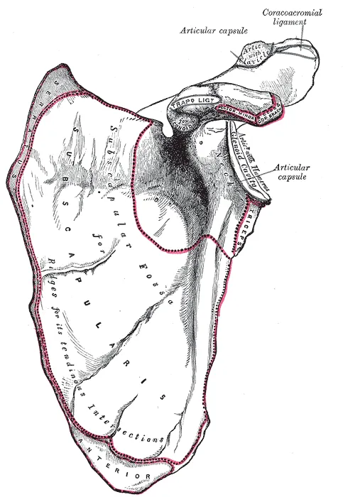
The tendon attaches to the lesser tubercle, a small, rounded bony bump on the humerus (upper arm bone)1,2. It also attaches to the front of the joint capsule, the sleeve of tough tissue that encloses the shoulder joint1,2. A large bursa (a fluid-filled sac that cushions tissues where they rub) separates the tendon from the neck of the shoulder blade1.
In 40 shoulders from cadavers (bodies donated for study), the upper part of the attachment was tendon and the lower part was muscle7. Its mean top-to-bottom length was 39.5 mm, of which the tendon part was 26.3 mm7.
What it does
The main action of the subscapularis is to rotate the humerus inward1,2. It can also help draw the arm toward the body1,2. A classic anatomy text calls it a powerful guard to the front of the shoulder joint, holding the head of the humerus in place1.
A cadaver study cut the subscapularis and the glenohumeral ligaments, the ligaments of the main shoulder joint, in different orders8. It concluded that the muscle does much of the work of steadying the front of the joint with the arm at the side8. At 45 degrees of abduction (lifting the arm out to the side), the muscle shares that work with ligaments8. As the arm nears 90 degrees, the inferior glenohumeral ligament takes over8.
How it compares with the other cuff muscles
The subscapularis is one of the four muscles of the rotator cuff, a group that crosses the shoulder joint and helps hold it steady9. One study scanned the right shoulders of 20 young adults3. The subscapularis was the largest cuff muscle in each participant3:
| Cuff muscle | Share of total cuff muscle volume |
|---|---|
| Subscapularis | 43%3 |
| Infraspinatus | 34%3 |
| Supraspinatus | 15%3 |
| Teres minor | 8%3 |
One study of 10 cadaver specimens used physiological cross-sectional area, a measure of a muscle’s size taken straight across its fibres10. It pointed to the subscapularis having the greatest force-producing capacity of the cuff muscles10. In five cadaver shoulders, the subscapularis was the most powerful cuff muscle, contributing 53% of the cuff moment11. A moment is the turning effect of a force around a joint.
How its upper and lower parts differ
In ten cadavers, a distinct sheet of fascia (tough connective tissue that wraps or joins muscles) divided the subscapularis into upper and lower compartments4. The territories of the upper and lower subscapular nerves mostly matched those two compartments4. In a cadaver study, type 1 fibres (slow-contracting muscle fibres) made up about 22% of the upper compartment and about 81% of the lower4.
Studies using electromyography (EMG), which measures a muscle’s activity from its electrical signals, have compared the two parts12,13:
- In 20 adults without shoulder problems, the upper part was more active in maximal inward rotation and the lower part in maximal outward rotation12. The authors suggest the upper part mainly rotates the arm inward, while the lower part has more of a stabilising role12.
- In 24 subjects with normal shoulders, the lower part was significantly more active than the upper during abduction and flexion (raising the arm forward)13. This held in both the lifting and the lowering phases13.
Two studies disagree on timing13,14. In eight subjects, the upper part switched on significantly earlier than the lower during forward flexion and abduction14. In the study of 24 subjects, the upper part switched on significantly later than the lower13.
Its nerve supply
The upper and lower subscapular nerves supply the muscle, carrying fibres of the fifth and sixth cervical nerves1,15. One dissection covered 62 adult cadaver upper limbs16. In 97%, the upper nerve came off the posterior cord of the brachial plexus, the network of nerves serving the arm16. The lower nerve did so in 79%, and arose from the axillary nerve in the other 21%16.
Breakdown
Strains in baseball players
Two reports from professional baseball describe strains (overstretched or torn muscle) of the subscapularis17,18. Among 191 players on one Japanese team over ten years, 8 (4.2%) had a subscapularis muscle strain17. The strains were on the dominant side17. Most were at the myotendinous junction, where muscle fibres join their tendon, in the lower half of the muscle17.
A second report reviewed 133 shoulder MRI scans from one professional baseball organisation18. Eleven showed changes suggesting subscapularis injury, each in the lower half of the muscle at that junction18. The risk of a throwing-related subscapularis injury rose in those players as outward rotation of the throwing arm fell18.
Wasting and fatty change in the muscle
One study looked at 153 patients having repair of upper and back cuff tears19. No subscapularis tear was seen at arthroscopy, keyhole surgery inside a joint using a small camera19. Even so, 73.9% had wasting and fatty change of the subscapularis at Goutallier grade 1 or 219. Female sex, advanced wasting and fatty change of the supraspinatus, and joint-lining inflammation were independent factors for it19.
Another study compared 11 MRI scans of patients with subscapularis wasting after an isolated tendon tear against 49 control scans20. In the patients, the upper half of the muscle made up a significantly smaller share of its cross-section20.
When its tendon tears
The rotator cuff tendons page covers cuff tendon tears; a few findings are specific to the subscapularis5. A systematic review, a study that searches for the research on a question and sums it up, counted its tears5. In series of people with rotator cuff disease or scheduled for surgery, a mean of 34% had one5. In series of people presenting with shoulder pain, the mean was 15%5. In 348 keyhole cuff repairs, the authors report that subscapularis tears usually appeared together with supraspinatus or infraspinatus tears21.
Two reviews describe the cause differently22,23. A 2012 review states that subscapularis tears are generally not traumatic22. It says they arise from degeneration within the tendon, or from pinching at the front and top of the joint or under the coracoid process22. The coracoid process is a hooked bony point at the front of the shoulder blade. A systematic review of nine studies of repaired isolated tears concluded that these often occur as a traumatic injury in a younger population23.
Recovery
After a muscle strain
The 8 professional baseball players with a subscapularis strain returned to play after a mean of 55.3 days (range 7 to 120)17. At a mean of 22.7 months, no player had injured the muscle again17.
After a tendon repair
Two systematic reviews have summed up studies of repaired isolated subscapularis tears6,23. The first, from 2012, covered nine studies and reported healing in 90% to 95% of shoulders23. It noted that biceps procedures done at the same operation can affect the results23.
The second, from 2026, pooled 17 non-randomised studies of keyhole repair, with 537 patients6. It found fewer positive lift-off, belly-press and bear-hug tests after surgery than before6. These are physical examination tests used for subscapularis tears5. The review rated the certainty of evidence as low to very low for each outcome6. It concluded that how far muscle strength is restored remains uncertain6.
How fat in the muscle relates to results
Three studies report on fatty infiltration, fat building up inside a muscle, and results after repair19,24,25:
- A case series (a report on a group of patients, without a comparison group) followed 30 patients after open repair of an isolated traumatic tear24. Six (20%) still had a positive lift-off or belly-press test, which was more likely with more fatty infiltration before surgery24.
- A cohort of 52 patients had isolated full-thickness tears and advanced fatty infiltration, at grade 3 or 425. Of the 28 repaired tendons, 22 (78.6%) had torn again25.
- In the 153 patients with no subscapularis tear, its wasting and fatty change did not affect results after repair of their other cuff tears19. The results were range of motion, pain, function and belly-press strength19.
Connections
The rest of the rotator cuff
The tendons of the subscapularis, supraspinatus, infraspinatus and teres minor form the rotator cuff9. They connect the scapula to the humerus9. In the scans of 20 young adults, the physiological cross-sectional area of the subscapularis equalled that of the infraspinatus and teres minor together3. The authors note that these muscles, when active together, can produce torques (turning forces) that balance each other and steady the humeral head3.
When the deltoid contracts, it tends to slide the humeral head upward26. The subscapularis, infraspinatus and teres minor counter this with their downward pull26.
The long head of the biceps tendon
In 33 embalmed shoulder girdles, the top attachment of the subscapularis tendon touched the underside of the long head biceps tendon27. The contact was where that tendon turns into its groove27. The authors of a study of 10 autopsy samples describe the tendon’s superficial layer as forming a fibrous ring around the biceps tendon28. They describe the ring as steadying the biceps tendon in its groove28.
In 435 keyhole cuff repairs, no shoulder with an unstable biceps tendon had an intact subscapularis tendon27.
The serratus anterior
The serratus anterior, a muscle on the side of the chest, attaches along the front of the inner border of the shoulder blade29. That attachment lies close to the attachments of the subscapularis and often interconnects with them29.
Exercises and positions studied
Related exercise pages: push-up, bench press and band external rotation30,31,32. EMG studies have recorded the muscle in these and other movements31:
| Movement | What the study found |
|---|---|
| Push-up plus, a push-up that ends by pushing the upper back higher | In 15 healthy subjects doing seven exercises, it and a diagonal exercise consistently produced the most activity in both parts of the muscle30 |
| Bench press | In 15 volunteers, the infraspinatus was significantly more active than the subscapularis31 |
| A row | In 15 volunteers, subscapularis activity was higher than infraspinatus activity, and the pattern did not change with load31 |
| Outward rotation lying face-down, arm at 90 degrees of abduction | In a 30-person study, it drew the most subscapularis activity of the five exercises studied32 |
One EMG study recorded 16 healthy volunteers in the belly-press and lift-off positions33. Both positions activated the upper and lower subscapularis more than the five other muscles recorded33. The belly press activated the upper part significantly more, and the lift-off challenged the lower part significantly more33.
Subscapularis pulls through
Subscapularis forms part of the sling around
Subscapularis attaches to
Subscapularis steadies the front of
Subscapularis works with
Subscapularis lies next to
Subscapularis is sometimes supplied by a branch of
Subscapularis is loaded by
Sources
- Gray H; Lewis WH (editor) (1918). Anatomy of the Human Body. Lea & Febiger, Philadelphia (20th edition). Link
- Aguirre K, Mudreac A, Kiel J (2023). Anatomy, Shoulder and Upper Limb, Subscapularis Muscle.. StatPearls. PMID 30020716
- Zhang Y, Herbert RD, Bilston LE, Bolsterlee B (2024). Three-dimensional architecture and moment arms of human rotator cuff muscles in vivo: Interindividual, intermuscular, and intramuscular variations.. Journal of Anatomy. doi:10.1111/joa.14050 PMID 38690607
- Cho TH, Hong JE, Yang HM (2023). Neuromuscular compartmentation of the subscapularis muscle and its clinical implication for botulinum neurotoxin injection.. Scientific Reports. doi:10.1038/s41598-023-38406-0 PMID 37430018
- Lädermann A, Collin P, Zbinden O, Meynard T, Saffarini M, Chiu JC (2021). Diagnostic Accuracy of Clinical Tests for Subscapularis Tears: A Systematic Review and Meta-analysis.. Orthopaedic Journal of Sports Medicine. doi:10.1177/23259671211042011 PMID 35146034
- Fernández-Cortiñas AB, Seoane-Pillado T, Lopiz Y, Hermida M, Caeiro-Rey JR, Marco F (2026). Postoperative outcomes after arthroscopic repair of isolated subscapularis tears: a systematic review and meta-analysis.. Journal of Orthopaedic Surgery and Research. doi:10.1186/s13018-026-07088-5 PMID 42469843
- Ide J, Tokiyoshi A, Hirose J, Mizuta H (2008). An anatomic study of the subscapularis insertion to the humerus: the subscapularis footprint.. Arthroscopy. doi:10.1016/j.arthro.2008.02.009 PMID 18589262
- Turkel SJ, Panio MW, Marshall JL, Girgis FG (1981). Stabilizing mechanisms preventing anterior dislocation of the glenohumeral joint.. The Journal of Bone and Joint Surgery. American Volume. PMID 7287791
- Betts JG, Young KA, Wise JA, Johnson E, Poe B, Kruse DH, Korol O, Johnson JE, Womble M, DeSaix P (2022). Anatomy and Physiology 2e. OpenStax. Link
- Ward SR, Hentzen ER, Smallwood LH, Eastlack RK, Burns KA, Fithian DC, Friden J, Lieber RL (2006). Rotator cuff muscle architecture: implications for glenohumeral stability.. Clinical Orthopaedics and Related Research. PMID 16826111
- Keating JF, Waterworth P, Shaw-Dunn J, Crossan J (1993). The relative strengths of the rotator cuff muscles. A cadaver study.. The Journal of Bone and Joint Surgery. British Volume. doi:10.1302/0301-620X.75B1.8421011 PMID 8421011
- Rathi S, Taylor NF, Green RA (2017). The upper and lower segments of subscapularis muscle have different roles in glenohumeral joint functioning.. Journal of Biomechanics. doi:10.1016/j.jbiomech.2017.08.007 PMID 28867549
- Wickham J, Pizzari T, Balster S, Ganderton C, Watson L (2014). The variable roles of the upper and lower subscapularis during shoulder motion.. Clinical Biomechanics. doi:10.1016/j.clinbiomech.2014.07.003 PMID 25172119
- O'Connell NE, Cowan J, Christopher T (2006). An investigation into EMG activity in the upper and lower portions of the subscapularis muscle during normal shoulder motion.. Physiotherapy Research International. doi:10.1002/pri.336 PMID 17019944
- Kasper JC, Itamura JM, Tibone JE, Levin SL, Stevanovic MV (2008). Human cadaveric study of subscapularis muscle innervation and guidelines to prevent denervation.. Journal of Shoulder and Elbow Surgery. doi:10.1016/j.jse.2007.11.013 PMID 18472282
- Tubbs RS, Loukas M, Shahid K, Judge T, Pinyard J, Shoja MM, Slappey JB, McEvoy WC, Oakes WJ (2007). Anatomy and quantitation of the subscapular nerves.. Clinical Anatomy. doi:10.1002/ca.20478 PMID 17352414
- Komatsu S, Nagashima M, Kaneko H (2024). Characteristics of subscapularis muscle strain in professional baseball players: a case series.. Journal of Shoulder and Elbow Surgery. doi:10.1016/j.jse.2023.05.036 PMID 37422132
- Polster JM, Lynch TS, Bullen JA, Soloff L, Ilaslan H, Subhas N, Schickendantz MS (2016). Throwing-related injuries of the subscapularis in professional baseball players.. Skeletal Radiology. doi:10.1007/s00256-015-2239-9 PMID 26305059
- Kim SC, Yoo SJ, Jo JH, Lee JH, Baek E, Lee SM, Yoo JC (2024). The impact of supraspinatus tear on subscapularis muscle atrophy and fatty infiltration.. Clinics in Shoulder and Elbow. doi:10.5397/cise.2024.00521 PMID 39558566
- Seppel G, Voss A, Henderson DJH, Waldt S, Haller B, Forkel P, Reuter S, Holzapfel BM, Plath JE, Imhoff AB (2021). Atrophy patterns in isolated subscapularis lesions.. BMC Musculoskeletal Disorders. doi:10.1186/s12891-021-04241-5 PMID 33888115
- Garavaglia G, Ufenast H, Taverna E (2011). The frequency of subscapularis tears in arthroscopic rotator cuff repairs: A retrospective study comparing magnetic resonance imaging and arthroscopic findings.. International Journal of Shoulder Surgery. doi:10.4103/0973-6042.91000 PMID 22223958
- Longo UG, Berton A, Marinozzi A, Maffulli N, Denaro V (2012). Subscapularis tears.. Medicine and Sport Science. doi:10.1159/000328886 PMID 21986050
- Mall NA, Chahal J, Heard WM, Bach BR, Bush-Joseph CA, Romeo AA, Verma NN (2012). Outcomes of arthroscopic and open surgical repair of isolated subscapularis tendon tears.. Arthroscopy. doi:10.1016/j.arthro.2012.02.018 PMID 22607828
- Bartl C, Scheibel M, Magosch P, Lichtenberg S, Habermeyer P (2011). Open repair of isolated traumatic subscapularis tendon tears.. The American Journal of Sports Medicine. doi:10.1177/0363546510388166 PMID 21189357
- Yoon TH, Kim SJ, Choi YR, Keum HS, Chun YM (2021). Clinical Outcomes for Isolated Subscapularis Tears With Advanced Fatty Infiltration: Nonoperative Treatment Versus Arthroscopic Single-Row Repair.. Orthopaedic Journal of Sports Medicine. doi:10.1177/2325967120975754 PMID 33614807
- Elder A, Powers CM (2025). Scapular Stabilization for Shoulder Pain: Putting the Cart Before the Horse?. International Journal of Sports Physical Therapy. doi:10.26603/001c.128049 PMID 39906061
- Arai R, Sugaya H, Mochizuki T, Nimura A, Moriishi J, Akita K (2008). Subscapularis tendon tear: an anatomic and clinical investigation.. Arthroscopy. doi:10.1016/j.arthro.2008.04.076 PMID 18760206
- Tebaa E, Tantot J, Isaac-Pinet S, Nové-Josserand L (2019). Histologic characteristics of the subscapularis tendon from muscle to bone: reference to subscapularis lesions.. Journal of Shoulder and Elbow Surgery. doi:10.1016/j.jse.2018.11.045 PMID 30711396
- Neumann DA, Camargo PR (2019). Kinesiologic considerations for targeting activation of scapulothoracic muscles - part 1: serratus anterior.. Brazilian Journal of Physical Therapy. doi:10.1016/j.bjpt.2019.01.008 PMID 30737019
- Decker MJ, Tokish JM, Ellis HB, Torry MR, Hawkins RJ (2003). Subscapularis muscle activity during selected rehabilitation exercises.. The American Journal of Sports Medicine. doi:10.1177/03635465030310010601 PMID 12531769
- Wattanaprakornkul D, Halaki M, Cathers I, Ginn KA (2011). Direction-specific recruitment of rotator cuff muscles during bench press and row.. Journal of Electromyography and Kinesiology. doi:10.1016/j.jelekin.2011.09.002 PMID 21978788
- Alizadehkhaiyat O, Hawkes DH, Kemp GJ, Frostick SP (2015). Electromyographic Analysis of the Shoulder Girdle Musculature During External Rotation Exercises.. Orthopaedic Journal of Sports Medicine. doi:10.1177/2325967115613988 PMID 26740950
- Tokish JM, Decker MJ, Ellis HB, Torry MR, Hawkins RJ (2003). The belly-press test for the physical examination of the subscapularis muscle: electromyographic validation and comparison to the lift-off test.. Journal of Shoulder and Elbow Surgery. doi:10.1016/s1058-2746(03)00047-8 PMID 14564261
Changes
- Revised to report sources added after their published corrections were read