Brachial plexus
The network of nerves that runs from the side of the neck, behind the collarbone, into the armpit, and serves the whole arm. How it is built, where it can be stretched or squeezed, and what studies report about recovery.
- Region
- Neck
- Updated
- October 28, 2026
- Reviewed
- October 28, 2026 · how entries are checked
Function
How the network is built
In anatomy, a plexus is a network of nerve fibres with no nerve cell bodies in it1. Gray’s Anatomy (1918) describes this one running from the lower side of the neck to the armpit3. It forms from the front branches of the spinal nerves labelled C5 to T12,3.
The fibres regroup in stages on the way to the arm3,4:
- Roots to trunks. C5 and C6 join as the upper trunk, C7 runs alone as the middle trunk, and C8 and T1 join as the lower trunk3,4.
- Trunks to divisions. Under the collarbone each trunk splits into a front and a back division, making six3,4.
- Divisions to cords. Three cords form, named for where they lie beside the axillary artery, the main artery of the armpit3.
- Cords to end branches. The plexus ends as five nerves4.
| Cord | Formed from | End branches |
|---|---|---|
| Lateral | Front divisions of the upper and middle trunks3 | Musculocutaneous nerve; one root of the median nerve4 |
| Medial | Front division of the lower trunk3 | Ulnar nerve; the other root of the median nerve4 |
| Posterior | All three back divisions3 | Axillary and radial nerves4 |
Because of this regrouping, each nerve leaving the plexus carries motor fibres from several spinal levels4.
What it supplies
The plexus drives every muscle of the arm, from the chest and shoulder-blade muscles down to the hand4. It also carries feeling from the skin of the limb4. Several nerves leave before the five end branches4:
| Where it leaves | Nerve | Goes to |
|---|---|---|
| Roots (C5–C7) | Long thoracic | Serratus anterior4 |
| Root (C5) | Dorsal scapular | Levator scapulae and rhomboids4 |
| Upper trunk | Suprascapular | Supraspinatus and infraspinatus4 |
| Cords | Lateral and medial pectoral | The pectoral muscles4 |
| Posterior cord | Upper and lower subscapular | Subscapularis and teres major4 |
| Posterior cord | Thoracodorsal | Latissimus dorsi4 |
| Medial cord | Two skin nerves (C8, T1) | Feeling only (no muscles), inner arm and forearm4 |
Where it runs
A 2022 European neurosurgical consensus describes the plexus and the subclavian artery, the large artery beneath the collarbone, passing through the scalene triangle7. That gap lies between two neck muscles, the anterior and middle scalene, and the first rib8,9. Gray describes the plexus then passing behind the collarbone and into the armpit3. There it surrounds the axillary artery, with one cord on each side and one behind3.
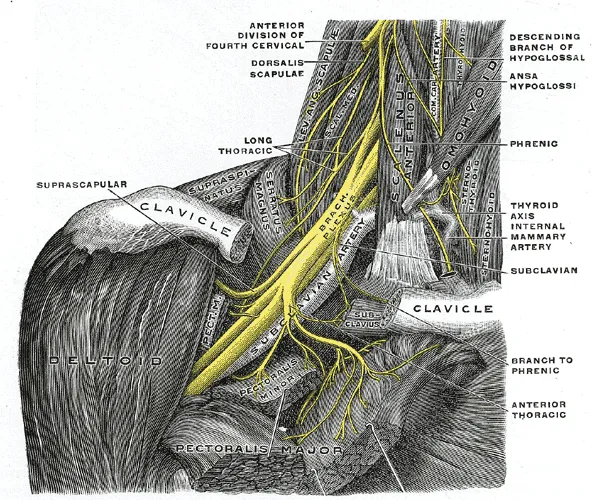
A 2025 review describes Erb’s point, where C5 and C6 join, as about 2–3 cm above the collarbone11. It lies at the back edge of the sternocleidomastoid, a muscle at the front and side of the neck11.
How much its layout varies
- In a meta-analysis of 40 studies (3,055 arms), the roots formed the trunks in the usual way in 84%5. The cords formed in the usual way in 96%5.
- The same analysis found the usual link to the axillary artery in 96% and to the scalenes in 86%5.
- In 51 cadavers (bodies donated for study), the textbook path between the anterior and middle scalene was found in only 60%12.
How arm and neck positions load it
In 25 men without symptoms, bending the neck away and turning the shoulder outward each reduced the range in an arm nerve test13. Bending the wrist back did too, and straightening the elbow loaded the nerves a great deal13. Positions added together had a cumulative effect, while easing a neighbouring joint offset it13.
In 6 cadaver arms, a pull of 9 to 15 newtons (a unit of force), depending on the cord, stretched the cords by 10%14. That is the threshold for stretch injury seen in animal models14. Raising the arm out to the side beyond 70° pushed the medial cord past that threshold14. In 9 cadavers, arm nerve tests moved the C5–C8 spinal nerves about 2 to 4.4 mm15. They stretched them by about 7% to 13%15.
Breakdown
How injuries are sorted
Injuries are called pre-ganglionic when they lie nearer the spinal cord than the dorsal root ganglion11. That ganglion is where the cell bodies of sensory nerves sit; injuries beyond it are post-ganglionic11. Injuries are also graded by how much of the nerve is damaged, as the nerve tissue page describes16.
In sport
A 2020 review describes severe plexus injuries as rare in sport4. It describes neurapraxia, the mildest grade of nerve injury, as apparently the most common sport injury of the plexus4. It adds that almost all studies count only injuries that needed surgery, so milder injuries from sport may be undercounted4.
A 2024 study of National Football League injuries describes the stinger as a short-lived nerve injury of the plexus or neck nerve roots17. A 2024 review describes three main ways the plexus is hurt in contact sport18:
- a direct blow above the collarbone18;
- a pull when the shoulder is pushed down and the neck bends the other way18;
- pressure on nerve roots when the neck is forced forward or back18.
A 2025 review describes Erb’s point as fixed toward the spine but free to be pulled further out, a common site of that pull11. The same review says the arm is usually down at the side in contact sports, so upper trunk injuries are more frequent11. It adds that lower trunk injuries are far less common, partly because that trunk lies deeper11.
In NCAA men’s wrestling records from 2009 to 2014, plexus injury was the most common type of spine injury recorded, at 20%19.
Severe injuries
A meta-analysis of 10 studies covered 3,032 adults operated on for severe plexus injury6. In all, 93% were closed injuries, most often from motorcycle accidents (67%) and car crashes (14%)6. Of the patients, 93% were male6. Nine in ten injuries were above the collarbone, or both above and below it6.
| Part of the plexus injured, in the same 3,032 adults | Share of patients (%) |
|---|---|
| Whole plexus | 536 |
| Upper plexus | 396 |
| Lower plexus | 66 |
From 84 patients who had plexus surgery, a 2014 paper concluded that serious plexus injury usually comes with other severe injuries in high-energy accidents20. Below the collarbone, a series of 101 patients found four patterns, most often after a forward shoulder dislocation (55 patients)21.
Squeezing at the thoracic outlet
A 2022 consensus names three places in the thoracic outlet where narrowing can squeeze the plexus or the vessels beside it7:
- the scalene triangle7;
- the space between the collarbone and first rib7;
- the space beneath the pectoralis minor, a smaller chest muscle7.
Reporting standards from 2016 describe thoracic outlet syndrome as a group of disorders sharing squeezing there, with the plexus one structure at risk22. A 2017 review describes squeezing under the pectoralis minor as giving symptoms like squeezing above the collarbone23.
An MRI study scanned 35 healthy volunteers and 54 people with outlet symptoms, with arms down and raised24. No artery or nerve squeeze was seen in the volunteers24. With the arm raised, vein squeeze was common in both groups. Among the patients, nerve squeeze was seen in 7% and artery squeeze in 72%24.
In 25 cadavers, tissue changes consistent with long-term squeezing were found at two sites25. One was where the upper trunk meets the anterior scalene; the other, where the lower trunk roots meet the first rib25.
Recovery
What the grade predicts
A 2020 review states that in neurapraxia the nerve fibres survive, so the muscle does not waste4. Function comes back fully, usually within 2 weeks4. A 2025 review sets out the expected course for each grade11:
| Grade | Expected course |
|---|---|
| First degree (neurapraxia) | Full recovery within about 2 weeks11 |
| Second degree (axonotmesis: fibres damaged, wrappings whole) | Full recovery, at about 1 mm a day11 |
| Third degree | Recovery may be incomplete11 |
| Fourth and fifth degree | Poor outlook11 |
The same review links more severe grades to a worse outlook for recovery on its own11. It explains that more of the tissue and blood supply guiding regrowth is disrupted11. It describes pre-ganglionic injuries as having a poor outlook, often with the nerve roots torn from the spinal cord and little chance of regrowth11. The 2020 review states that neurotmesis, where every part of the nerve is damaged, cannot recover on its own, and is common in severe injuries4.
How muscles get their nerve supply back
The 2020 review describes two routes: the damaged axons (nerve fibres) can regrow to the muscle, or nearby healthy axons can sprout and take over4. Regrowth depends on the distance to the muscle and is usually slow4. Another review gives about 1 mm a day, so a plexus injury up to a metre away needs more than 2–3 years26. It adds that there may then be little or no return of function26.
Timing after an injury in sport
A 2025 review notes that weakness can fade or newly appear over the first 1 to 3 days11. EMG (electromyography) measures a muscle’s activity from its electrical signals. The review states that needle EMG shows signs of axon breakdown around 10–14 days in muscles near the trunk11. In muscles further down the limb, they take about 3–6 weeks11.
A 2024 scoping review found no trials, cohort studies or registry studies on managing plexus injury in contact sport18. A 1977 report of 13 cases of axonotmesis, all in the upper trunk, found all but one recovered within 3 to 42 weeks27. The scoping review found only 8 case reports, covering 10 athletes, as published data on managing these injuries18. A 2025 review concludes that most such injuries are mild and settle, but severe, possibly permanent damage can occur11.
Connections
Nerves and muscles
Its five end branches are the axillary, median, musculocutaneous, radial and ulnar nerves4. Its roots give off the long thoracic nerve to the serratus anterior, a muscle on the side of the chest4.
The scalenes receive nerves from this plexus and the cervical plexus, a network of nerves in the neck28. The commonest extra scalene muscle, the scalenus minimus, passes between the subclavian artery and the plexus and may irritate its lower trunk10. Erb’s point lies at the back edge of the sternocleidomastoid11. In 66% of 25 cadavers, the lower trunk formed on or beside the first of the ribs25. The cords form in the space behind the pectoralis minor29.
Stretches that load it
In 25 men, bending the neck away reduced the range in a plexus nerve test13. In cadavers with the arm in a nerve-test position, the same neck movement strained the median nerve30. Related stretch page: upper trapezius stretch30.
Brachial plexus ends in five branches, one of which is
Brachial plexus gives off, before its trunks form,
Brachial plexus passes between two of
Brachial plexus forms its upper trunk near the back edge of
Brachial plexus forms its lower trunk, in 66% of 25 cadavers, on or beside the first of
Brachial plexus has cords that form in the space behind
Brachial plexus is briefly injured in
Brachial plexus is compressed in
Brachial plexus is compressed, below the collarbone, in
Brachial plexus is loaded, with the arm in a nerve-test position, by the neck side-bend of
Sources
- Betts JG, Young KA, Wise JA, Johnson E, Poe B, Kruse DH, Korol O, Johnson JE, Womble M, DeSaix P (2022). Anatomy and Physiology 2e. OpenStax. Link
- Polcaro L, Charlick M, Daly DT (2023). Anatomy, Head and Neck: Brachial Plexus.. StatPearls. PMID 30285368
- Gray H; Lewis WH (editor) (1918). Anatomy of the Human Body. Lea & Febiger, Philadelphia (20th edition). Link
- Belviso I, Palermi S, Sacco AM, Romano V, Corrado B, Zappia M, Sirico F (2020). Brachial Plexus Injuries in Sport Medicine: Clinical Evaluation, Diagnostic Approaches, Treatment Options, and Rehabilitative Interventions.. Journal of functional morphology and kinesiology. doi:10.3390/jfmk5020022 PMID 33467238
- Benes M, Kachlik D, Belbl M, Kunc V, Havlikova S, Whitley A, Kunc V (2021). A meta-analysis on the anatomical variability of the brachial plexus: Part I - Roots, trunks, divisions and cords. Annals of anatomy = Anatomischer Anzeiger : official organ of the Anatomische Gesellschaft. doi:10.1016/j.aanat.2021.151751 PMID 33940116
- Kaiser R, Waldauf P, Ullas G, Krajcová A (2018). Epidemiology, etiology, and types of severe adult brachial plexus injuries requiring surgical repair: systematic review and meta-analysis.. Neurosurgical review. doi:10.1007/s10143-018-1009-2 PMID 30014280
- Dengler NF, Ferraresi S, Rochkind S, Denisova N, Garozzo D, Heinen C, Alimehmeti R, Capone C, Barone DG, Zdunczyk A, Pedro MT, Antoniadis G, Kaiser R, Dubuisson A, Kretschmer T, Rasulic L (2022). Thoracic Outlet Syndrome Part I: Systematic Review of the Literature and Consensus on Anatomy, Diagnosis, and Classification of Thoracic Outlet Syndrome by the European Association of Neurosurgical Societies' Section of Peripheral Nerve Surgery.. Neurosurgery. doi:10.1227/neu.0000000000001908 PMID 35319532
- Nito T, Satake H, Tsuchiya M, Hanaka N, Takagi M (2025). Anatomical and histological analysis of the scalene triangle in a Japanese population: implications for thoracic outlet syndrome diagnosis and management. BMC musculoskeletal disorders. doi:10.1186/s12891-025-09048-2 PMID 40847307
- Boezaart AP, Haller A, Laduzenski S, Koyyalamudi VB, Ihnatsenka B, Wright T (2010). Neurogenic thoracic outlet syndrome: A case report and review of the literature. International journal of shoulder surgery. doi:10.4103/0973-6042.70817 PMID 21072145
- Connolly MR, Auchincloss HG (2021). Anatomy and Embryology of the Thoracic Outlet. Thoracic surgery clinics. doi:10.1016/j.thorsurg.2020.09.007 PMID 33220766
- Windmueller RA, Gbayisomore OO, Mead RL, Desai MJ, Bowman EN (2025). Brachial plexus injuries in the contact athlete: a narrative review.. Annals of joint. doi:10.21037/aoj-24-67 PMID 40385690
- Harry WG, Bennett JD, Guha SC (1997). Scalene muscles and the brachial plexus: anatomical variations and their clinical significance. Clinical anatomy (New York, N.Y.). doi:10.1002/(SICI)1098-2353(1997)10:4<250::AID-CA6>3.0.CO;2-W PMID 9213042
- Coppieters MW, Van de Velde M, Stappaerts KH (2002). Positioning in anesthesiology: toward a better understanding of stretch-induced perioperative neuropathies.. Anesthesiology. doi:10.1097/00000542-200207000-00011 PMID 12131106
- Kam AW, Lam PH, Haen PSWA, Tan M, Shamsudin A, Murrell GAC (2018). Preventing brachial plexus injury during shoulder surgery: a real-time cadaveric study.. Journal of shoulder and elbow surgery. doi:10.1016/j.jse.2017.11.030 PMID 29370965
- Lohman Bonfiglio CM, Gilbert KK, Brismée JM, Sobczak S, Hixson KM, James CR, Sizer PJ (2021). Upper limb neurodynamic testing with radial and ulnar nerve biases: An analysis of cervical spinal nerve mechanics.. Musculoskeletal science & practice. doi:10.1016/j.msksp.2021.102320 PMID 33513560
- Menorca RM, Fussell TS, Elfar JC (2013). Nerve physiology. Hand Clinics. doi:10.1016/j.hcl.2013.04.002 PMID 23895713
- Lamplot JD, Petit C, Lee R, Mack CD, Herzog MM, Solomon GS, et al. (2024). Epidemiology of Stingers in the National Football League, 2015-2019.. Sports health. doi:10.1177/19417381231223413 PMID 38229225
- Armstrong R, McKeever T, Leavitt M, McLelland C, Hamilton DF (2024). Rehabilitation of brachial plexus injury in contact sport: Where are the data that underpin clinical management? A scoping review.. PloS one. doi:10.1371/journal.pone.0298317 PMID 38913647
- Brinkman JC, Tummala SV, McQuivey KS, Hassebrock JD, Pagdilao C, Makovicka JL, et al. (2021). Epidemiology of Spine Injuries in National Collegiate Athletic Association Men's Wrestling Athletes.. Orthopaedic journal of sports medicine. doi:10.1177/23259671211032007 PMID 34497862
- Kaiser R, Mencl L, Haninec P (2014). Injuries associated with serious brachial plexus involvement in polytrauma among patients requiring surgical repair.. Injury. doi:10.1016/j.injury.2012.05.013 PMID 22658417
- Hems TE, Mahmood F (2012). Injuries of the terminal branches of the infraclavicular brachial plexus. The Journal of bone and joint surgery. British volume. doi:10.1302/0301-620x.94b6.28286 PMID 22628595
- Illig KA, Donahue D, Duncan A, Freischlag J, Gelabert H, Johansen K, Jordan S, Sanders R, Thompson R (2016). Reporting standards of the Society for Vascular Surgery for thoracic outlet syndrome.. Journal of Vascular Surgery. doi:10.1016/j.jvs.2016.04.039 PMID 27565607
- Sanders RJ, Annest SJ (2017). Pectoralis Minor Syndrome: Subclavicular Brachial Plexus Compression.. Diagnostics (Basel). doi:10.3390/diagnostics7030046 PMID 28788065
- Demondion X, Bacqueville E, Paul C, Duquesnoy B, Hachulla E, Cotten A (2003). Thoracic outlet: assessment with MR imaging in asymptomatic and symptomatic populations.. Radiology. doi:10.1148/radiol.2272012111 PMID 12637678
- Tiongco RFP, Dellon AL (2023). Histologic evidence of brachial plexus compression sites at the thoracic inlet and variations in formation of the lower trunk in cadavers.. Microsurgery. doi:10.1002/micr.31037 PMID 37042225
- Grinsell D, Keating CP (2014). Peripheral nerve reconstruction after injury: a review of clinical and experimental therapies.. BioMed Research International. doi:10.1155/2014/698256 PMID 25276813
- Clancy WG Jr, Brand RL, Bergfield JA (1977). Upper trunk brachial plexus injuries in contact sports.. The American journal of sports medicine. doi:10.1177/036354657700500508 PMID 907035
- Bordoni B, Jozsa F, Varacallo MA (2025). Anatomy, Head and Neck, Scalenus Muscle. StatPearls. PMID 30085600
- Ahmed AS, Graf AR, Karzon AL, Graulich BL, Egger AC, Taub SM, Gottschalk MB, Bowers RL, Wagner ER (2022). Pectoralis minor syndrome - review of pathoanatomy, diagnosis, and management of the primary cause of neurogenic thoracic outlet syndrome.. JSES reviews, reports, and techniques. doi:10.1016/j.xrrt.2022.05.008 PMID 37588453
- Bueno-Gracia E, Pérez-Bellmunt A, Estébanez-de-Miguel E, López-de-Celis C, Caudevilla-Polo S, Shacklock M, González-Rueda V (2020). Effect of cervical contralateral lateral flexion on displacement and strain in the median nerve and flexor digitorum superficialis at the wrist during the ULNT1 - Cadaveric study.. Musculoskeletal science & practice. doi:10.1016/j.msksp.2020.102244 PMID 32906084
Changes
- First published.