Rhomboids
Also called rhomboid major, rhomboid minor, rhomboid muscles
Two muscles that run from the spine to the inner border of the shoulder blade. How they move and hold the shoulder blade, how their nerve and the muscles themselves are injured, what studies of recovery found, and what they work with.
- Region
- Shoulder
- Starts on (rhomboid minor)
- the lower part of the neck ligament and the spinous processes of the seventh neck and first thoracic vertebrae (Cervical vertebrae)3,4
- Starts on (rhomboid major)
- the spinous processes of the second to fifth thoracic vertebrae3,4
- Ends on (rhomboid minor)
- the inner border of the shoulder blade, at the base of the ridge across its back3,4,28
- Ends on (rhomboid major)
- the inner border of the shoulder blade, from the ridge across its back down to its lower tip3,4,28
- Updated
- October 8, 2026
- Reviewed
- October 8, 2026 · how entries are checked
Function
Where the two muscles sit
The rhomboids are a group of two muscles, the rhomboid major and the rhomboid minor1. They lie beneath the middle part of the trapezius, the large muscle across the upper back2. Both start on the spine and end on the inner border of the scapula (shoulder blade)3,4.
A spinous process is the bony point that sticks out from the back of a vertebra, one of the spine’s bones. Thoracic vertebrae are the spine bones that carry the ribs. The spine of the scapula is the ridge across the back of the shoulder blade, where the rhomboid minor ends3,4.
| Muscle | Starts on | Ends on |
|---|---|---|
| Rhomboid minor | The lower part of the neck’s ligament, plus the spinous processes of the last neck vertebra (C7) and the first thoracic one3,4 | The inner border of the shoulder blade, at the base of the spine of the scapula3,4 |
| Rhomboid major | The spinous processes of the second to fifth thoracic vertebrae3,4 | The inner border of the shoulder blade, from where the spine of the scapula begins down to the lower corner3,4 |
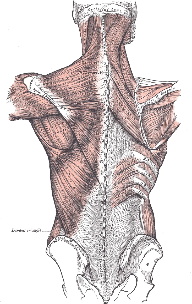
A slight gap usually separates the two muscles, but their neighbouring edges are occasionally joined4. The levator scapulae is a muscle running from the neck to the top of the shoulder blade. One dissection study of body donors found it and the rhomboid minor linked and wrapped in connective tissue8. The two formed a unit separate from the rhomboid major8.
How they move and hold the shoulder blade
The two rhomboids work together to draw the shoulder blade back toward the spine, raise it and rotate its outer border downward3. They draw the shoulder blade back together with the middle and lower fibres of the trapezius4. Authors of a 2016 study note that the trapezius and rhomboids both do this, but the rhomboids also rotate the shoulder blade downward9.
One review of scapular winging names three muscles that help keep the shoulder blade’s inner border against the back of the chest wall3. They are the rhomboids, the trapezius and the serratus anterior, a muscle on the side of the chest3.
Their nerve and blood supply
The dorsal scapular nerve supplies both rhomboids1,4. It arises from the C5 nerve root, the part of a nerve where it leaves the spinal cord6. It pierces the middle scalene muscle in the neck, then runs deep to the levator scapulae and the rhomboids6. The muscles take their blood from the dorsal scapular artery1.
Studies of cadavers (bodies donated for study) differ on how reliably the nerve reaches the rhomboids10,11:
- In 70 adult Japanese cadavers, the nerve supplied both rhomboids and the levator scapulae in every case10.
- Of 23 nerves from 20 embalmed adult cadavers, about 52% supplied both the levator scapulae and the rhomboids11. About 48% supplied the levator scapulae alone11.
- A systematic review states that the nerve reliably supplies both rhomboids and may variably supply the levator scapulae5.
Breakdown
How the nerve is squeezed or injured
Entrapment means a nerve being squeezed by the tissue around it. A systematic review names the middle scalene, which the dorsal scapular nerve often passes through, as the most common site of its entrapment5. It also reports that the nerve can be pressed against the shoulder blade’s upper inner border in people doing repetitive or strenuous overhead activity5. It names painters, electricians, and volleyball and basketball players5.
In six fresh cadaver dissections, the upper part of the shoulder blade’s inner border pressed on the nerve when the arm was raised13. The winging review also reports rhomboid paralysis after direct nerve injury, including a shoulder dislocation in a judo player3.
The systematic review found this literature to be mainly reports on one patient or a group, cadaver studies, technical notes and narrative reviews5. It identified no randomised controlled trials, which assign people to groups by chance5. It reports that people with this entrapment typically have pain along the inner border of the shoulder blade, rhomboid weakness and subtle winging5.
How the shoulder blade sits when the rhomboids are paralysed
Palsy is loss of power in a muscle from a nerve problem. The winging review states that paralysis of any of the three holding muscles lets the shoulder blade’s inner border lift off3. Sources describe the position in rhomboid palsy, and label it, in different ways3,5,13:
- The winging review describes it as very subtle, with the shoulder blade shifted outward and its lower corner rotated outward3. It labels the winging of trapezius or rhomboid paralysis “lateral”, in contrast to the “medial” winging of serratus anterior paralysis3.
- A systematic review states that rhomboid weakness can produce “medial” scapular winging5. It calls this subtler than the winging that weakness of the serratus anterior or trapezius causes5.
- A third source says rhomboid paralysis makes the inner border of the shoulder blade stand out, with the lower corner rotated outward13.
Two accounts of the shoulder blade’s height differ. A review of long thoracic nerve palsy attributes “lateral” winging to trapezius dysfunction or rhomboid paralysis14. In its account the shoulder droops and the shoulder blade sits lower14. The systematic review states that in rhomboid weakness, unlike trapezius palsy, the shoulder blade does not drop and may sit higher5. The long thoracic nerve page covers the winging of serratus anterior palsy.
A review of scapular winging calls rhomboid paralysis hard to recognise, because the muscles lie deep to the trapezius and the winging is subtle3. In one cohort it cites, 25 of 36 patients were first given another explanation for their symptoms3. It reports that the winging may show more as the arm comes down from a fully raised position3. The review found no published report of how often rhomboid paralysis causes winging, only anecdotal statements calling it rare3.
Tears and strain
Authors of a 2023 case report describe rhomboid tears as rare7. They write that tears can present with pain around the shoulder blade, but that how they present and are recognised remains largely unknown7. Their report describes a 28-year-old man whose pain at the inner border of the right shoulder blade began after carrying a heavy box7. MRI, a scan that shows soft tissue in detail, showed the rhomboid major torn where it attaches to the shoulder blade7.
Another report covers 72 patients who had the lower trapezius and rhomboids surgically reattached to the shoulder blade15. A 2019 review states that sudden injury tears of these muscles or the serratus anterior are important, under-recognised causes of winging16.
The winging review reports that muscle injury in overhead sport has been implicated in rhomboid problems3. In its account, slowing the shoulder blade during a pitcher’s or volleyball player’s follow-through can strain the rhomboids3.
Trigger points
One study compared 17 people who had pain in the upper thoracic spine with 17 controls free of pain17. Active trigger points (tender knots) in the rhomboid were the most prevalent, found in 75% of the patients17.
Recovery
After nerve palsy
The winging review reports that an injured dorsal scapular nerve or rhomboid muscle is usually managed without surgery3. It states that the middle part of the trapezius can make up for rhomboid weakness or paralysis3.
Two reports give outcomes13,18:
- In a series of four patients with isolated rhomboid deficiency, winging was not completely corrected in any patient, and three had residual pain13. One of the four had follow-up electrical testing, which no longer showed signs that the rhomboids had lost their nerve supply13.
- Another report looked back at 21 consecutive patients who had surgery to take pressure off the nerve18. Their mean pain score around the shoulder blade fell from 54.0 before surgery to 26.8 at six months18. Fifteen of the 21 had a good outcome18. Longer-lasting symptoms predicted a poor outcome18.
The systematic review concluded that the evidence for this surgery remains limited and varied5.
After a tear
In the case report of the 28-year-old man, symptoms did not improve over two months without surgery7. The muscle was then stitched back to the shoulder blade, and he had a satisfactory functional outcome with no re-tear7. In the 72 patients with reattached muscles, the time from injury to care averaged 52 months15. Their average shoulder score rose from 39 at first assessment to 63 at discharge, on average 7.4 months after surgery15.
How the muscles change with use
Authors of a 2023 study of professional pitchers wrote that no earlier study had evaluated how the shoulder blade’s rear muscles adapt to throwing19. Six crewmembers flew 4- to 6-month space missions with a resistance exercise device20. After flight, MRI showed an average 23.1% increase in the rhomboid minor’s cross-sectional area, the size of a slice cut straight across20. The change differed between people: three had increases on both sides and two had decreases on both sides20.
Connections
The muscles around the shoulder blade
The trapezius rotates the shoulder blade against the pull of the levator scapulae and rhomboids21. In 12 healthy participants, each of four positions of drawing the shoulder blade back produced activity in both the middle trapezius and the rhomboid major9. In maximal efforts, the rhomboid major was more active relative to the middle trapezius in a rowing position, elbow bent, than with the elbow straight and the shoulder turned outward9.
Exercises that load them
Related exercise pages: shrug, band external rotation, push-up and overhead press22,23,24,25.
| Exercise | What studies found |
|---|---|
| Shrug | In 26 people, rhomboid major activity was significantly lower in an arms-overhead shrug than in a weighted shrug, arms at the sides22 |
| Band external rotation | In a 30-person study of five exercises, turning the arm out while side-lying, a towel under the arm, drew the most rhomboid activity23 |
| Push-up | With the hands fixed, the serratus anterior lifts the trunk, which requires the rhomboids and three other muscles to steady the shoulder blade24 |
| Overhead press | In 12 men pressing half their one-lift maximum, an unstable bar hung with kettlebells increased rhomboid activation over a stable barbell25 |
| Lawnmower and robbery | In 23 healthy men, these two exercises activated the rhomboid major to 59% and 66% of its hardest voluntary effort26 |
| Seated row | Twelve trained men wore a skin sensor over the middle trapezius and rhomboid region27. It recorded more activity in a held seated row, shoulder blades not drawn back, than in a reverse-grip pulldown27 |
Rhomboids draws the shoulder blade back with
Rhomboids holds the shoulder blade to the chest wall with
Rhomboids ends on
Rhomboids is compared in studies of winging with the muscle supplied by
Rhomboids is loaded by
Sources
- Farrell C, Kiel J (2023). Anatomy, Back, Rhomboid Muscles.. StatPearls. PMID 30521277
- Yi KH, Lee HJ, Choi YJ, Lee JH, Hu KS, Kim HJ (2020). Intramuscular Neural Distribution of Rhomboid Muscles: Evaluation for Botulinum Toxin Injection Using Modified Sihler's Method.. Toxins. doi:10.3390/toxins12050289 PMID 32375284
- Martin RM, Fish DE (2008). Scapular winging: anatomical review, diagnosis, and treatments.. Current Reviews in Musculoskeletal Medicine. doi:10.1007/s12178-007-9000-5 PMID 19468892
- Gray H; Lewis WH (editor) (1918). Anatomy of the Human Body. Lea & Febiger, Philadelphia (20th edition). Link
- Afshar A, Tabrizi A, Shariyate MJ (2026). Dorsal scapular nerve entrapment: a systematic review.. JSES reviews, reports, and techniques. doi:10.1016/j.xrrt.2026.100807 PMID 42502829
- Bishop KN, Varacallo MA (2023). Anatomy, Shoulder and Upper Limb, Dorsal Scapular Nerve.. StatPearls. PMID 29083775
- Furuhata R, Yamaguchi S, Tanji A (2023). Surgical Repair for Rhomboid Major Tear: A Case Report.. Clinical medicine insights. Arthritis and musculoskeletal disorders. doi:10.1177/11795441231219009 PMID 38115856
- Kulow C, Theopold JD, Ehrlich A, Hobusch C, Steinke H (2022). Levator scapulae and rhomboid minor are united.. Annals of anatomy = Anatomischer Anzeiger : official organ of the Anatomische Gesellschaft. doi:10.1016/j.aanat.2022.151938 PMID 35367623
- Fennell J, Phadke CP, Mochizuki G, Ismail F, Boulias C (2016). Shoulder Retractor Strengthening Exercise to Minimize Rhomboid Muscle Activity and Subacromial Impingement.. Physiotherapy Canada. Physiotherapie Canada. doi:10.3138/ptc.2014-83 PMID 27504044
- Tetsu S, Terayama H, Qu N, Yamazaki H, Sakamoto R, Tanaka O, Suyama K, Takenaka M, Suzuki T, Sakabe K (2018). Anatomical variants of dorsal scapular nerve in relation to the middle scalene muscle in Japanese population.. Medicine. doi:10.1097/MD.0000000000013349 PMID 30461656
- Nguyen VH, Liu HH, Rosales A, Reeves R (2016). A Cadaveric Investigation of the Dorsal Scapular Nerve.. Anatomy research international. doi:10.1155/2016/4106981 PMID 27597900
- Smith J, Padgett DJ, Kaufman KR, Harrington SP, An KN, Irby SE (2004). Rhomboid muscle electromyography activity during 3 different manual muscle tests.. Archives of physical medicine and rehabilitation. doi:10.1016/s0003-9993(03)00618-x PMID 15179655
- Morante de Los Reyes A, Bacle G, Chaves C, Tranier M, Jacquot A, Corcia P, Laulan J, Roulet S (2022). Scapular winging due to rhomboid muscle paralysis: clinical assessment of 4 cases and anatomic study of the dorsal scapular nerve.. Journal of shoulder and elbow surgery. doi:10.1016/j.jse.2022.05.011 PMID 35718255
- Wu F, Ng CY (2023). Long Thoracic Nerve Palsy: When Is Decompression Indicated.. Journal of hand surgery global online. doi:10.1016/j.jhsg.2022.11.006 PMID 37521538
- Kibler WB, Sciascia A, Uhl T (2014). Medial scapular muscle detachment: clinical presentation and surgical treatment.. Journal of shoulder and elbow surgery. doi:10.1016/j.jse.2013.05.008 PMID 23867169
- Didesch JT, Tang P (2019). Anatomy, Etiology, and Management of Scapular Winging.. The Journal of hand surgery. doi:10.1016/j.jhsa.2018.08.008 PMID 30292717
- Ortega-Santiago R, Maestre-Lerga M, Fernández-de-Las-Peñas C, Cleland JA, Plaza-Manzano G (2019). Widespread Pressure Pain Sensitivity and Referred Pain from Trigger Points in Patients with Upper Thoracic Spine Pain.. Pain medicine (Malden, Mass.). doi:10.1093/pm/pnz020 PMID 30821833
- Ottestad E, Wilson TJ (2023). Assessment of outcomes in consecutive patients undergoing dorsal scapular nerve decompression.. Journal of neurosurgery. doi:10.3171/2022.7.JNS221223 PMID 36087331
- Valle O, Sheridan SS, Rauch JJ, Sarver JJ, Paul RW, Thomas SJ (2023). Chronic Effects of Pitching on Muscle Thickness and Strength of the Scapular Stabilizers in Professional Baseball Players.. Sports health. doi:10.1177/19417381221085004 PMID 35466817
- McNamara KP, Greene KA, Tooze JA, Dang J, Khattab K, Lenchik L, Weaver AA (2019). Neck Muscle Changes Following Long-Duration Spaceflight.. Frontiers in physiology. doi:10.3389/fphys.2019.01115 PMID 31572205
- Ourieff J, Scheckel B, Agarwal A (2023). Anatomy, Back, Trapezius.. StatPearls. PMID 30085536
- Castelein B, Cools A, Parlevliet T, Cagnie B (2016). Modifying the shoulder joint position during shrugging and retraction exercises alters the activation of the medial scapular muscles.. Manual Therapy. doi:10.1016/j.math.2015.09.005 PMID 26409441
- Alizadehkhaiyat O, Hawkes DH, Kemp GJ, Frostick SP (2015). Electromyographic Analysis of the Shoulder Girdle Musculature During External Rotation Exercises.. Orthopaedic Journal of Sports Medicine. doi:10.1177/2325967115613988 PMID 26740950
- Neumann DA, Camargo PR (2019). Kinesiologic considerations for targeting activation of scapulothoracic muscles - part 1: serratus anterior.. Brazilian Journal of Physical Therapy. doi:10.1016/j.bjpt.2019.01.008 PMID 30737019
- Williams MR Jr, Hendricks DS, Dannen MJ, Arnold AM, Lawrence MA (2020). Activity of Shoulder Stabilizers and Prime Movers During an Unstable Overhead Press.. Journal of Strength and Conditioning Research. doi:10.1519/JSC.0000000000002660 PMID 29889776
- Berckmans K, Castelein B, Borms D, Palmans T, Parlevliet T, Cools A (2020). Analysis of Scapular Kinematics and Muscle Activity by Use of Fine-Wire Electrodes During Shoulder Exercises.. The American journal of sports medicine. doi:10.1177/0363546520908604 PMID 32176519
- Lehman GJ, Buchan DD, Lundy A, Myers N, Nalborczyk A (2004). Variations in muscle activation levels during traditional latissimus dorsi weight training exercises: An experimental study.. Dynamic medicine : DM. doi:10.1186/1476-5918-3-4 PMID 15228624
- Betts JG, Young KA, Wise JA, Johnson E, Poe B, Kruse DH, Korol O, Johnson JE, Womble M, DeSaix P (2022). Anatomy and Physiology 2e. OpenStax. Link
Changes
- Revised to report sources added after their published corrections were read