Scalenes
Also called scalene muscles, scalene, anterior scalene, middle scalene, posterior scalene, scalenus anterior, scalenus medius, scalenus posterior
Three muscles deep in the side of the neck, running from the neck bones to the top two ribs. How they move the neck and help breathing, and how the nerves to the arm pass between them.
- Region
- Neck
- Starts on (anterior scalene)
- the front bumps (anterior tubercles) on the side spurs (transverse processes) of the third to sixth neck vertebrae5,12
- Starts on (middle scalene)
- the back bumps (posterior tubercles) on the side spurs (transverse processes) of the second to seventh neck vertebrae5,12
- Starts on (posterior scalene)
- the fourth or fifth to the sixth neck vertebrae, in one dissection of 10 cadavers3,13
- Ends on (anterior scalene)
- a point on the upper surface of the first rib (the scalene tubercle)4,5,12
- Ends on (middle scalene)
- the upper surface of the first rib, behind the groove for the subclavian artery4,5,12
- Ends on (posterior scalene)
- the second rib3
- Muscles
- anterior scalene, middle scalene, posterior scalene
- Updated
- October 28, 2026
- Reviewed
- October 28, 2026 · how entries are checked
Function
Three muscles from neck to ribs
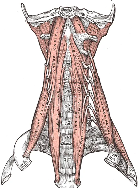
There are three scalene muscles on each side of the neck1. The anterior scalene sits in front, the middle scalene between, and the posterior scalene behind1. The middle one is the longest and the posterior one the smallest1. The scalenes begin as one shared muscle in the eighth week of development, which the growing brachial plexus splits into anterior and middle parts3.
The anterior and middle scalenes start on the transverse processes (small bony spurs on each side of a vertebra) of the neck vertebrae5,12:
| Muscle | Where it starts | Where it ends |
|---|---|---|
| Anterior | The front knobs of the spurs on the third to sixth neck vertebrae5,12 | The scalene tubercle, a point on the upper surface of the first rib5,12 |
| Middle | The back knobs of the spurs on the second to seventh; another account gives the first to seventh5,12 | The first rib, behind the groove made by the subclavian artery, the large artery beneath the collarbone5,7,12 |
| Posterior | In 10 cadavers (bodies donated for study), the fourth to sixth in half and the fifth to sixth in half13 | The second rib3 |
In the same 10 cadavers, the levels varied: the anterior scalene reached the seventh vertebra in 30%, and the middle scalene in 60%13.
Where they meet the first rib
On top of the first rib, the scalene tubercle lies between two grooves5. The subclavian artery makes the outer groove, and the subclavian vein, the large vein beneath the collarbone, makes the inner one5,7. In a study of 42 Japanese cadavers, no prominent raised bump was seen where the anterior scalene met the rib in any of them4. In that study, the anterior scalene attached to the rib’s upper, back and lower surfaces, and the middle scalene to its upper surface only4. Both attached by fibrous tissue, with no fibrocartilage (a tough, fibrous kind of cartilage) at either site4.
What they move
Working together, the scalenes bend the head and neck forward, bend them to the side and turn them1,2. They also help keep the head and neck steady2. One review describes the anterior and middle scalenes lifting the first rib and resisting forces that pull it down14.
How they help breathing
The scalenes are described as accessory (helper) breathing muscles, which raise the upper rib cage3. In humans, they are among the muscles that widen the rib cage on a breath in by lifting the ribs15. Studies in healthy adults found16,17,18:
- In needle recordings from five people, the scalenes were always active in quiet breathing, usually switching on as a breath in began16.
- In the same five, breathing in with the diaphragm (the main breathing muscle) alone cut scalene activity, and the upper ribs moved inward16.
- With skin sensors in 10 people, averaging many breaths showed a low burst of activity with each breath in, in eight17.
- In 13 people breathing against a rising load, activity rose with the load, with no electrical signs of fatigue18.
Their nerves and fibres
Nerves reach the scalenes from two networks: the brachial plexus and the cervical plexus (a network of nerves in the neck)2. In 26 cadavers, the anterior scalene took inner front branches from the front roots of the neck nerves, and the middle scalene outer back branches19. In four elderly male cadavers, 71.9% of anterior scalene fibres were slow (type 1) fibres, the most of the neck muscles studied20.
Breakdown
The gap the nerves pass through
The scalene triangle is the space bounded by the anterior scalene, the middle scalene and the first rib4,5. The brachial plexus runs through it, behind the subclavian artery and in front of the middle scalene3. The anterior scalene separates the subclavian vein, in front of it, from the artery behind it3. The phrenic nerve, which supplies the diaphragm, runs down the front of the anterior scalene2,3. In 42 Japanese cadavers, the triangle’s base averaged 8.2 mm wide, against 10.7 mm reported in Western groups4.
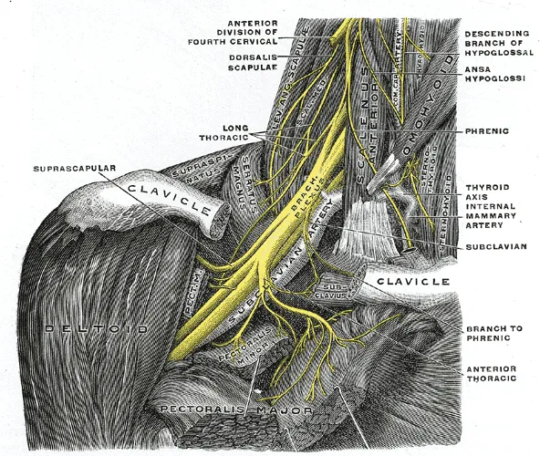
This triangle, above the collarbone, is one of the thoracic outlet’s three areas3. A 2022 consensus calls the nerve form of thoracic outlet syndrome the most common7. One study says it typically comes from nerves and vessels pinched between the anterior and middle scalene8.
Causes one review lists
A case report with a literature review lists causes of one subtype of the nerve form, involving the upper brachial plexus5:
- an enlarged scalene5;
- the brachial plexus passing through the anterior scalene5;
- pressure on the middle scalene from a long spur on the seventh neck vertebra5;
- a cervical rib (an extra rib in the neck)5.
Here, an enlarged scalene as a cause of this subtype rests on that list5. Separately, a systematic review (a summary of the research on a question) covers the nerve to the rhomboids21. It reports an enlarged middle scalene, particularly in bodybuilders, may press on that nerve21,22.
How often the textbook layout holds
In 51 adult cadavers, the textbook path of the brachial plexus between the anterior and middle scalene was seen in only 60% of cases11. Other cadaver studies also found variations8,23:
| Study | What it found |
|---|---|
| 51 adult cadavers | The fifth and sixth neck nerve roots pierced the anterior scalene: fused first in 15%, the fifth alone in 13%, separately in 6%11 |
| 65 cadavers | 31 variations (47.7%); in 27, part or all of the plexus’s upper trunk (its top nerve bundle) pierced the anterior scalene8 |
| 93 cadavers (186 sides) | The plexus’s upper trunk took an unusual course on 12.9% of sides, most often through the muscle23 |
| Meta-analysis of 40 studies (3,055 arms) | The usual relationship in 86% of cases, with a 95% confidence interval (a range showing how precise the result is) of 66% to 98%6 |
The authors of the 65-cadaver study suggest that people whose plexus pierces the muscle would not show positive signs on the traditional arm-position tests8. Their reason is that those tests rely on the artery being squeezed8.
An extra scalene
The scalenus minimus is the commonest extra scalene muscle3. It passes between the subclavian artery and the brachial plexus, and may irritate the plexus’s lower trunk (its lowest main nerve bundle)3. It was present in 46% of 51 adult cadavers, but in three of 73 Greek cadavers (4.11%)10,11. The Greek study’s authors found published rates from 7.8% to 71.7%10. In 200 operations for compression at the thoracic outlet, surgeons found 20 extra scalene muscles24.
Injury, whiplash and neck pain
One review describes neck hyperextension injuries (bending the neck back past its normal limit) as tearing some anterior and middle scalene fibres3. In 45 scalenes removed from people with thoracic outlet syndrome after an injury, connective tissue averaged 36%, against under 15% in control muscles25. Slow fibres made up 78% of fibres, against 53% in controls25.
One systematic review gathered what is known about the scalenes after whiplash26:
- It says strain on the scalene triangle during whiplash would in theory put the thoracic outlet at risk, but needs more study26.
- The same review found five studies reporting thoracic outlet syndrome in 31% to over 74% of people with chronic whiplash, from very low quality evidence26.
- It found trigger points (tender muscle knots) in the scalenes and nearby ones in 48% to 65%, on low or very low quality evidence26.
A study following 110 people after injury to the upper body classed 39 as having the definite nerve form of thoracic outlet syndrome27. Its authors suggest that cases after injury relate to direct damage to the scalenes27.
In 10 people with chronic neck pain and 10 matched controls, the neck was held bent forward at 25% and 50% of maximum effort28. The anterior scalene showed more electrical signs of fatigue in the neck pain group28. In another study, people with neck pain used accessory neck muscles, including the anterior scalene, more than people without symptoms during a repeated arm task29.
Recovery
Healing after a tear
One review describes torn scalene fibres healing with scar tissue3. That scarred muscle is less pliable and can press on the brachial plexus, the review says3. Separately, in anterior scalene samples from people with thoracic outlet syndrome, 85.1% of fibres were slow fibres, and those fibres were enlarged30. After the anterior scalene’s tendon was cut in people with thoracic outlet syndrome, the share of slow fibres fell, they shrank, and fast fibres increased30.
How training changes them
In a randomised trial of 58 women with chronic neck pain, six weeks of endurance-strength training of the neck flexors raised neck flexion strength31. It also reduced electrical signs of fatigue in the anterior scalene and the sternocleidomastoid31. In a trial of 46 people with chronic neck pain, six weeks of practising a small nod lowered anterior scalene activity in a nod test32. In a trial of 25 healthy men, five weeks of breathing-muscle training or a sham (a dummy version) left scalene activity in exercise unchanged33.
A trial injected botulinum toxin (a drug that relaxes the muscle it is injected into) into the scalenes of 38 people with thoracic outlet syndrome34. It brought no clinically or statistically significant gain in pain, tingling or function over a placebo (a dummy injection)34.
Connections
Nerves and vessels beside them
The brachial plexus and the subclavian artery pass between the anterior and middle scalene and the first rib, according to a 2022 consensus7. In 18 cadaver sides, the long thoracic nerve lay between the middle and posterior scalene in 56%35. The dorsal scapular nerve, which supplies the rhomboids, pierces the middle scalene21. A systematic review names the middle scalene as that nerve’s most common site of entrapment22.
Bones they pull on
The scalenes start on the neck vertebrae and end on the ribs3. One review describes a hypothesis that opposing pulls from the scalenes (upward) and the serratus anterior (downward) cause most first-rib stress fractures in baseball pitchers14.
Muscles they work with
In 12 control subjects (people studied for comparison) moving one arm fast, the anterior scalene switched on less than 50 ms after the deltoid36. So did the sternocleidomastoid and the deep neck flexors36. In 10 healthy volunteers, a conventional neck flexion hold used the anterior scalene and sternocleidomastoid significantly more than a small nod, at each effort tested37. Related exercise pages: neck isometric hold37.
Scalenes is loaded by
Scalenes lies next to
Scalenes attaches to
Scalenes starts on
Scalenes has been measured in studies with
Scalenes works with
Scalenes is pierced by the nerve of
Scalenes has been measured in studies of
Scalenes has been studied in trials reported on
Sources
- Betts JG, Young KA, Wise JA, Johnson E, Poe B, Kruse DH, Korol O, Johnson JE, Womble M, DeSaix P (2022). Anatomy and Physiology 2e. OpenStax. Link
- Bordoni B, Jozsa F, Varacallo MA (2025). Anatomy, Head and Neck, Scalenus Muscle. StatPearls. PMID 30085600
- Connolly MR, Auchincloss HG (2021). Anatomy and Embryology of the Thoracic Outlet. Thoracic surgery clinics. doi:10.1016/j.thorsurg.2020.09.007 PMID 33220766
- Nito T, Satake H, Tsuchiya M, Hanaka N, Takagi M (2025). Anatomical and histological analysis of the scalene triangle in a Japanese population: implications for thoracic outlet syndrome diagnosis and management. BMC musculoskeletal disorders. doi:10.1186/s12891-025-09048-2 PMID 40847307
- Boezaart AP, Haller A, Laduzenski S, Koyyalamudi VB, Ihnatsenka B, Wright T (2010). Neurogenic thoracic outlet syndrome: A case report and review of the literature. International journal of shoulder surgery. doi:10.4103/0973-6042.70817 PMID 21072145
- Benes M, Kachlik D, Belbl M, Kunc V, Havlikova S, Whitley A, Kunc V (2021). A meta-analysis on the anatomical variability of the brachial plexus: Part I - Roots, trunks, divisions and cords. Annals of anatomy = Anatomischer Anzeiger : official organ of the Anatomische Gesellschaft. doi:10.1016/j.aanat.2021.151751 PMID 33940116
- Dengler NF, Ferraresi S, Rochkind S, Denisova N, Garozzo D, Heinen C, Alimehmeti R, Capone C, Barone DG, Zdunczyk A, Pedro MT, Antoniadis G, Kaiser R, Dubuisson A, Kretschmer T, Rasulic L (2022). Thoracic Outlet Syndrome Part I: Systematic Review of the Literature and Consensus on Anatomy, Diagnosis, and Classification of Thoracic Outlet Syndrome by the European Association of Neurosurgical Societies' Section of Peripheral Nerve Surgery.. Neurosurgery. doi:10.1227/neu.0000000000001908 PMID 35319532
- Leonhard V, Smith R, Caldwell G, Smith HF (2016). Anatomical variations in the brachial plexus roots: implications for diagnosis of neurogenic thoracic outlet syndrome. Annals of anatomy = Anatomischer Anzeiger : official organ of the Anatomische Gesellschaft. doi:10.1016/j.aanat.2016.03.011 PMID 27133185
- Illig KA, Donahue D, Duncan A, Freischlag J, Gelabert H, Johansen K, Jordan S, Sanders R, Thompson R (2016). Reporting standards of the Society for Vascular Surgery for thoracic outlet syndrome.. Journal of Vascular Surgery. doi:10.1016/j.jvs.2016.04.039 PMID 27565607
- Natsis K, Totlis T, Didagelos M, Tsakotos G, Vlassis K, Skandalakis P (2013). Scalenus minimus muscle: overestimated or not? An anatomical study. The American surgeon. PMID 23574846
- Harry WG, Bennett JD, Guha SC (1997). Scalene muscles and the brachial plexus: anatomical variations and their clinical significance. Clinical anatomy (New York, N.Y.). doi:10.1002/(SICI)1098-2353(1997)10:4<250::AID-CA6>3.0.CO;2-W PMID 9213042
- Elliott JM, Cornwall J, Kennedy E, Abbott R, Crawford RJ (2018). Towards defining muscular regions of interest from axial magnetic resonance imaging with anatomical cross-reference: part II - cervical spine musculature. BMC musculoskeletal disorders. doi:10.1186/s12891-018-2074-y PMID 29807530
- Rusnak-Smith S, Moffat M, Rosen E (2001). Anatomical variations of the scalene triangle: dissection of 10 cadavers. The Journal of orthopaedic and sports physical therapy. doi:10.2519/jospt.2001.31.2.70 PMID 11232741
- Schowalter S, Le B, Creps J, McInnis KC (2022). Rib Fractures in Professional Baseball Pitchers: Mechanics, Epidemiology, and Management. Open access journal of sports medicine. doi:10.2147/OAJSM.S288882 PMID 36248020
- De Troyer A, Boriek AM (2011). Mechanics of the respiratory muscles. Comprehensive physiology. doi:10.1002/cphy.c100009 PMID 23733642
- De Troyer A, Estenne M (1984). Coordination between rib cage muscles and diaphragm during quiet breathing in humans. Journal of applied physiology: respiratory, environmental and exercise physiology. doi:10.1152/jappl.1984.57.3.899 PMID 6238017
- Hug F, Raux M, Prella M, Morelot-Panzini C, Straus C, Similowski T (2006). Optimized analysis of surface electromyograms of the scalenes during quiet breathing in humans. Respiratory physiology & neurobiology. doi:10.1016/j.resp.2005.04.008 PMID 16448935
- Basoudan N, Rodrigues A, Gallina A, Garland J, Guenette JA, Shadgan B, Road J, Reid WD (2020). Scalene and sternocleidomastoid activation during normoxic and hypoxic incremental inspiratory loading. Physiological reports. doi:10.14814/phy2.14522 PMID 32726513
- Sakamoto Y (2012). Spatial relationships between the morphologies and innervations of the scalene and anterior vertebral muscles. Annals of anatomy = Anatomischer Anzeiger : official organ of the Anatomische Gesellschaft. doi:10.1016/j.aanat.2011.11.004 PMID 22209543
- Cornwall J, Kennedy E (2015). Fiber types of the anterior and lateral cervical muscles in elderly males. European spine journal : official publication of the European Spine Society, the European Spinal Deformity Society, and the European Section of the Cervical Spine Research Society. doi:10.1007/s00586-015-3795-3 PMID 25681116
- Bishop KN, Varacallo MA (2023). Anatomy, Shoulder and Upper Limb, Dorsal Scapular Nerve.. StatPearls. PMID 29083775
- Afshar A, Tabrizi A, Shariyate MJ (2026). Dorsal scapular nerve entrapment: a systematic review.. JSES reviews, reports, and techniques. doi:10.1016/j.xrrt.2026.100807 PMID 42502829
- Natsis K, Totlis T, Tsikaras P, Anastasopoulos N, Skandalakis P, Koebke J (2006). Variations of the course of the upper trunk of the brachial plexus and their clinical significance for the thoracic outlet syndrome: a study on 93 cadavers. The American surgeon. PMID 16536255
- Makhoul RG, Machleder HI (1992). Developmental anomalies at the thoracic outlet: an analysis of 200 consecutive cases. Journal of vascular surgery. PMID 1404675
- Sanders RJ, Jackson CG, Banchero N, Pearce WH (1990). Scalene muscle abnormalities in traumatic thoracic outlet syndrome. American journal of surgery. doi:10.1016/s0002-9610(05)80269-7 PMID 2301718
- Heneghan NR, Smith R, Tyros I, Falla D, Rushton A (2018). Thoracic dysfunction in whiplash associated disorders: A systematic review. PloS one. doi:10.1371/journal.pone.0194235 PMID 29570722
- Kai Y, Oyama M, Kurose S, Inadome T, Oketani Y, Masuda Y (2001). Neurogenic thoracic outlet syndrome in whiplash injury. Journal of spinal disorders. doi:10.1097/00002517-200112000-00004 PMID 11723397
- Falla D, Rainoldi A, Merletti R, Jull G (2003). Myoelectric manifestations of sternocleidomastoid and anterior scalene muscle fatigue in chronic neck pain patients. Clinical neurophysiology : official journal of the International Federation of Clinical Neurophysiology. doi:10.1016/s1388-2457(02)00418-2 PMID 12705429
- Falla D, Bilenkij G, Jull G (2004). Patients with chronic neck pain demonstrate altered patterns of muscle activation during performance of a functional upper limb task. Spine. doi:10.1097/01.brs.0000128759.02487.bf PMID 15223935
- Machleder HI, Moll F, Verity MA (1986). The anterior scalene muscle in thoracic outlet compression syndrome. Histochemical and morphometric studies. Archives of surgery (Chicago, Ill. : 1960). doi:10.1001/archsurg.1986.01400100047009 PMID 3767646
- Falla D, Jull G, Hodges P, Vicenzino B (2006). An endurance-strength training regime is effective in reducing myoelectric manifestations of cervical flexor muscle fatigue in females with chronic neck pain. Clinical neurophysiology : official journal of the International Federation of Clinical Neurophysiology. doi:10.1016/j.clinph.2005.12.025 PMID 16490395
- Jull GA, Falla D, Vicenzino B, Hodges PW (2009). The effect of therapeutic exercise on activation of the deep cervical flexor muscles in people with chronic neck pain.. Manual therapy. doi:10.1016/j.math.2009.05.004 PMID 19632880
- Ramsook AH, Molgat-Seon Y, Schaeffer MR, Wilkie SS, Camp PG, Reid WD, Romer LM, Guenette JA (2017). Effects of inspiratory muscle training on respiratory muscle electromyography and dyspnea during exercise in healthy men. Journal of applied physiology (Bethesda, Md. : 1985). doi:10.1152/japplphysiol.00046.2017 PMID 28255085
- Finlayson HC, O'Connor RJ, Brasher PMA, Travlos A (2011). Botulinum toxin injection for management of thoracic outlet syndrome: a double-blind, randomized, controlled trial.. Pain. doi:10.1016/j.pain.2011.04.027 PMID 21628084
- Tubbs RS, Salter EG, Custis JW, Wellons JC, Blount JP, Oakes WJ (2006). Surgical anatomy of the cervical and infraclavicular parts of the long thoracic nerve.. Journal of neurosurgery. doi:10.3171/jns.2006.104.5.792 PMID 16703885
- Falla D, Jull G, Hodges PW (2004). Feedforward activity of the cervical flexor muscles during voluntary arm movements is delayed in chronic neck pain.. Experimental brain research. doi:10.1007/s00221-003-1814-9 PMID 14762639
- O'Leary S, Falla D, Jull G, Vicenzino B (2007). Muscle specificity in tests of cervical flexor muscle performance.. Journal of Electromyography and Kinesiology. doi:10.1016/j.jelekin.2005.10.006 PMID 16423538
Changes
- First published.