Deep neck flexors
Also called deep cervical flexors, prevertebral muscles, longus colli, longus capitis, rectus capitis anterior, rectus capitis lateralis
Four muscles that lie in front of the neck bones. Where they attach, what they move, how they differ in people with neck pain, and how they change with training.
- Region
- Neck
- Starts on (longus colli, upper oblique part)
- the front bumps (anterior tubercles) on the side spurs (transverse processes) of the third, fourth and fifth neck vertebrae2
- Starts on (longus colli, vertical part)
- the front of the bodies of the upper three thoracic and lower three neck vertebrae2
- Starts on (longus colli, lower oblique part)
- the front of the bodies of the first two or three thoracic vertebrae2
- Starts on (longus capitis)
- the front bumps (anterior tubercles) on the side spurs (transverse processes) of the third to sixth neck vertebrae2
- Starts on (rectus capitis anterior)
- the atlas, the first neck vertebra, at the front of its lateral mass and the root of its side spur (Cervical vertebrae)2
- Starts on (rectus capitis lateralis)
- the upper surface of the side spur (transverse process) of the atlas, the first neck vertebra (Cervical vertebrae)2
- Ends on (longus colli, upper oblique part)
- the tubercle on the front arch of the atlas, the first neck vertebra, by a narrow tendon (Cervical vertebrae)2
- Ends on (longus colli, vertical part)
- the front of the bodies of the second, third and fourth neck vertebrae2
- Ends on (longus colli, lower oblique part)
- the front bumps (anterior tubercles) on the side spurs (transverse processes) of the fifth and sixth neck vertebrae2
- Ends on (longus capitis)
- the underside of the basilar part of the occipital bone, at the base of the skull2
- Ends on (rectus capitis anterior)
- the underside of the basilar part of the occipital bone, just in front of the foramen magnum2
- Ends on (rectus capitis lateralis)
- the underside of the jugular process of the occipital bone2
- Muscles
- longus colli, longus capitis, rectus capitis anterior, rectus capitis lateralis
- Updated
- October 9, 2026
- Reviewed
- October 9, 2026 · how entries are checked
Function
Where the four muscles sit
An anatomy reference names four prevertebral muscles of the head and neck1. They are the longus colli, longus capitis, rectus capitis anterior and rectus capitis lateralis1. They lie in front of the cervical (neck) part of the vertebral column, the spine1. One paper on a neck test names two of them, the longus capitis and longus colli, as the deep cervical flexors8.
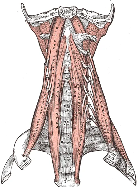
The longus colli lies on the front of the spine, between the atlas (the first neck vertebra) and the third thoracic vertebra2. A vertebra is one of the bones stacked to form the spine, and the thoracic vertebrae are those of the chest. The muscle is broad in the middle, pointed at each end, and has three parts2,3. The longus capitis is broad and thick at the top and narrow lower down2.
The rectus capitis anterior is a short, flat muscle directly behind the top of the longus capitis2. Several of the muscles attach to transverse processes, the small spurs of bone that stick out to each side of a vertebra2. Some attach to tubercles, which are small, rounded bony bumps2.
| Muscle or part | Where it starts | Where it ends |
|---|---|---|
| Longus colli, upper slanted (oblique) part | Front tubercles on the transverse processes of neck vertebrae three, four and five2 | The tubercle on the front arch of the atlas, by a narrow tendon2 |
| Longus colli, vertical part | The front surfaces of the bodies of the top three thoracic and bottom three neck vertebrae2 | The front surfaces of the bodies of neck vertebrae two, three and four2 |
| Longus colli, lower slanted (oblique) part | The front surfaces of the bodies of the top two or three thoracic vertebrae2 | Front tubercles on the transverse processes of neck vertebrae five and six2 |
| Longus capitis | Four tendon slips from the front tubercles on the transverse processes of neck vertebrae three to six2 | The underside of part of the occipital bone, at the base of the skull2 |
| Rectus capitis anterior | The atlas: the front of its lateral mass and the root of its transverse process2 | The underside of the occipital bone2 |
| Rectus capitis lateralis | The top of the atlas’s transverse process2 | The underside of the occipital bone2 |
What they move
One anatomy reference gives the group’s main function as spanning the vertebrae, acting with and against the deep muscles of the upper back1. Each muscle has its own actions:
- The longus colli flexes (bends forward) the neck part of the spine and rotates it slightly2,3. One review adds that it bends the neck to its own side3.
- The longus capitis and rectus capitis anterior flex the head2. Because they run at a slant, they also turn the face to one side or the other2.
- The rectus capitis lateralis bends the head sideways when one side works alone2.
The longus capitis and rectus capitis anterior directly oppose the muscles behind the neck2. After the head has been drawn backward, they return it to its natural position2.
One study scanned 19 healthy people with functional MRI after three neck exercises9. Functional MRI in muscle studies is a scan taken after exercise to show which muscles worked. One exercise was craniocervical flexion, a small nod of the head on the top of the neck9. The scans also covered the sternocleidomastoid, a muscle at the front and side of the neck9.
| Exercise | What the scans showed |
|---|---|
| The nod | A significantly larger rise in the activity measure in the longus capitis than in the longus colli or sternocleidomastoid9 |
| Conventional neck flexion | A larger rise in the longus colli and sternocleidomastoid than after the nod, and no significant difference for the longus capitis9 |
| The two combined | The largest rise in each of the three muscles9 |
How much they steady the neck
Sources differ on this4,5. Some describe a supporting role:
- Authors of one study describe the deep neck muscles as fundamentally important for supporting the neck’s lordosis, its inward curve, and its joints4.
- In 36 healthy people, the cross-sectional area of the longus colli was correlated with a measure of that curve10. Cross-sectional area is the size of a slice cut straight across a muscle, and correlation is how closely two measures rise and fall together.
- The authors of that study concluded that the longus colli counters the extra curve produced by the head’s weight and by the back-of-neck muscles contracting10.
Authors of another study note that the longus capitis and longus colli are proposed to help stabilise the neck5. They add that it is not clear the muscles are suited to that role5. MRI is a scan that shows soft tissue such as muscle in detail. Their study combined dissection of 7 cadaver specimens (donated bodies), MRI scans of 6 young healthy volunteers and a model5.
Estimated peak compression, the squeezing force along the spine, was under 80 newtons5. The authors concluded that the muscles have a limited capacity to add to neck stability through traditional mechanisms5.
Fibres, sensors and timing
Two studies examined neck muscle taken at autopsy from 16 people, comparing the longus colli with the multifidus, a deep muscle running along the spine11,12. The longus colli had a high density of muscle spindles, and the multifidus had a low density11. Muscle spindles are sensors inside a muscle that detect stretch. The longus colli also had a significantly greater share of type II fibres, a fast-contracting kind of muscle fibre12.
One study measured timing in 12 control subjects, people studied for comparison, as they moved one arm rapidly while standing4. The deep flexors became active less than 50 milliseconds after the deltoid, the rounded muscle that caps the shoulder4.
Nerve and blood supply
Two anatomy sources name the cervical (neck) nerves that supply each muscle1,2. They differ for the longus colli.
| Muscle | Gray’s Anatomy | StatPearls anatomy chapter |
|---|---|---|
| Longus capitis | Branches from the first, second and third2 | Direct branches from the first to the third1 |
| Longus colli | Branches from the second to the seventh2 | Branches from the second to the sixth1 |
| Both rectus muscles | The loop between the first and second2 | Branches of the first1 |
The StatPearls chapter says three arteries bring blood to the longus capitis and longus colli, the vertebral artery among them1.
Breakdown
How studies test these muscles
The craniocervical flexion test is a low-load test done lying face-up8. The person nods through five stages, guided by a pressure sensor behind the neck8. The sensor picks up the slight flattening of the neck’s curve that happens when the longus colli contracts13.
What studies found in people with neck pain
A review of mechanical neck pain reports changes in the neck muscles’ structure and in their behaviour15. The neck pain page covers the condition itself.
| Who was studied | What was found |
|---|---|
| 10 people with chronic neck pain and 10 controls | Deep flexor activity during the nod was lower in the neck pain group, significantly so at the higher stages6 |
| 10 patients with chronic neck pain and 12 controls | The deep flexors switched on significantly later in the neck pain group when they rapidly raised an arm4 |
| 32 women with neck pain for more than 6 months | Higher average pain intensity was correlated with lower deep flexor activity and with later onset16. Neither was significantly correlated with how long the pain had lasted16 |
Size and fat
An ultrasound study compared 20 people with chronic mechanical neck pain on both sides and 20 controls17. Ultrasound is a scan made with sound waves. The neck pain group had a smaller longus colli cross-sectional area on both sides17.
A systematic review of 14 papers looked at neck muscle form in chronic neck pain18. It found that fatty infiltration, fat building up inside a muscle, seems to occur only in people with whiplash-associated disorders18. These are the symptoms and effects that can follow a whiplash injury. The review concluded that some evidence exists for changes in muscle form, and that more high-quality research is needed to confirm them18.
Two MRI studies of chronic whiplash-associated disorders reached different results19,20. In 78 women with the disorders and 31 healthy women, the whiplash group had significantly more fat in both longus muscles19. In 31 patients and 31 matched controls scanned at one level of the neck, those muscles did not differ in size or fat20. A systematic review with meta-analysis of MRI studies found inconsistent evidence that neck muscle size or fat content changes after whiplash21.
Calcific tendinitis of the longus colli
Acute calcific tendinitis of the longus colli is a painful and relatively rare condition3. Tendinitis is pain and swelling of a tendon. The condition is marked by neck pain, neck stiffness and painful swallowing3. It is believed to be caused by crystals deposited in the muscle’s upper slanted fibres3.
An observational study at one centre covered an urban adult population from 2008 to 201122. It put the mean yearly rate at 0.50 cases per 100,000 person-years22. Person-years count people multiplied by the years each was followed. The authors concluded that the condition is probably under-recognised22.
Recovery
How the muscles respond to training
A systematic review of 14 articles looked at neck exercise in people with chronic non-specific neck pain23. It found moderate-certainty evidence that craniocervical flexion training can bring about neural adaptations in the neck muscles23.
The same review found only one study that measured the deep flexors directly during the test after exercise23. In that trial, 46 people with chronic neck pain were randomly assigned to six weeks of training7. Craniocervical flexion training increased deep flexor activity during the test and decreased sternocleidomastoid activity7. Strength training did not change deep flexor activity7.
A randomised trial of 60 people with chronic neck pain measured muscle size with ultrasound24. Ten weeks of craniocervical flexion exercise increased longus colli size compared with neck flexion exercise24. Neck flexion exercise increased the thickness of the sternocleidomastoid24.
How calcific tendinitis runs its course
A review describes calcific tendinitis of the longus colli as often self-limiting3. It reports that the condition resolves in one to three weeks in patients aged 30 to 603.
Connections
The longus capitis and rectus capitis anterior work against the muscles behind the neck2. In 10 volunteers, deep flexor activity rose in a straight line across the five stages of the craniocervical flexion test25. Related exercise pages: neck isometric hold.
Deep neck flexors is loaded by
Deep neck flexors has been measured in studies of
Sources
- McDavid LJ, Khan YS (2023). Anatomy, Head and Neck, Prevertebral Muscles.. StatPearls. PMID 32809404
- Gray H; Lewis WH (editor) (1918). Anatomy of the Human Body. Lea & Febiger, Philadelphia (20th edition). Link
- Combalia A, Zelada-Castro K, Alemany X, Vargas-Reverón C, Muñoz-Mahamud E (2026). Longus colli calcific tendinitis, an uncommon cause of neck pain. A short series of cases and review of literature.. International orthopaedics. doi:10.1007/s00264-025-06713-y PMID 41410695
- Falla D, Jull G, Hodges PW (2004). Feedforward activity of the cervical flexor muscles during voluntary arm movements is delayed in chronic neck pain.. Experimental brain research. doi:10.1007/s00221-003-1814-9 PMID 14762639
- Kennedy E, Albert M, Nicholson H (2017). Do longus capitis and colli really stabilise the cervical spine? A study of their fascicular anatomy and peak force capabilities.. Musculoskeletal science & practice. doi:10.1016/j.msksp.2017.10.005 PMID 29107220
- Falla DL, Jull GA, Hodges PW (2004). Patients with neck pain demonstrate reduced electromyographic activity of the deep cervical flexor muscles during performance of the craniocervical flexion test.. Spine. doi:10.1097/01.brs.0000141170.89317.0e PMID 15454700
- Jull GA, Falla D, Vicenzino B, Hodges PW (2009). The effect of therapeutic exercise on activation of the deep cervical flexor muscles in people with chronic neck pain.. Manual therapy. doi:10.1016/j.math.2009.05.004 PMID 19632880
- Jull GA, O'Leary SP, Falla DL (2008). Clinical assessment of the deep cervical flexor muscles: the craniocervical flexion test.. Journal of manipulative and physiological therapeutics. doi:10.1016/j.jmpt.2008.08.003 PMID 18804003
- Cagnie B, Dickx N, Peeters I, Tuytens J, Achten E, Cambier D, Danneels L (2008). The use of functional MRI to evaluate cervical flexor activity during different cervical flexion exercises.. Journal of applied physiology. doi:10.1152/japplphysiol.00918.2007 PMID 17991788
- Mayoux-Benhamou MA, Revel M, Vallée C, Roudier R, Barbet JP, Bargy F (1994). Longus colli has a postural function on cervical curvature.. Surgical and radiologic anatomy. doi:10.1007/BF01627655 PMID 7725191
- Boyd-Clark LC, Briggs CA, Galea MP (2002). Muscle spindle distribution, morphology, and density in longus colli and multifidus muscles of the cervical spine.. Spine. doi:10.1097/00007632-200204010-00005 PMID 11923661
- Boyd-Clark LC, Briggs CA, Galea MP (2001). Comparative histochemical composition of muscle fibres in a pre- and a postvertebral muscle of the cervical spine.. Journal of anatomy. doi:10.1046/j.1469-7580.2001.19960709.x PMID 11787824
- Jull G, Kristjansson E, Dall'Alba P (2004). Impairment in the cervical flexors: a comparison of whiplash and insidious onset neck pain patients.. Manual therapy. doi:10.1016/S1356-689X(03)00086-9 PMID 15040968
- Romeo A, Baccini M, Carreras G, Sagripanti M, Ruggeri M, Pillastrini P, Di Bari M (2022). Reliability, Validity, and Responsiveness of the Craniocervical Flexion Test in People Who Are Asymptomatic and Patients With Nonspecific Neck Pain: A Systematic Review and Meta-Analysis.. Physical therapy. doi:10.1093/ptj/pzac054 PMID 35554598
- O'Leary S, Falla D, Elliott JM, Jull G (2009). Muscle dysfunction in cervical spine pain: implications for assessment and management.. The Journal of orthopaedic and sports physical therapy. doi:10.2519/jospt.2009.2872 PMID 19411767
- Falla D, O'Leary S, Farina D, Jull G (2011). Association between intensity of pain and impairment in onset and activation of the deep cervical flexors in patients with persistent neck pain.. The Clinical journal of pain. doi:10.1097/AJP.0b013e31820212cf PMID 21178596
- Javanshir K, Rezasoltani A, Mohseni-Bandpei MA, Amiri M, Ortega-Santiago R, Fernández-de-Las-Peñas C (2011). Ultrasound assessment of bilateral longus colli muscles in subjects with chronic bilateral neck pain.. American journal of physical medicine & rehabilitation. doi:10.1097/PHM.0b013e31820173e5 PMID 21173685
- De Pauw R, Coppieters I, Kregel J, De Meulemeester K, Danneels L, Cagnie B (2016). Does muscle morphology change in chronic neck pain patients? - A systematic review.. Manual therapy. doi:10.1016/j.math.2015.11.006 PMID 26724855
- Elliott JM, O'Leary S, Sterling M, Hendrikz J, Pedler A, Jull G (2010). Magnetic resonance imaging findings of fatty infiltrate in the cervical flexors in chronic whiplash.. Spine. doi:10.1097/BRS.0b013e3181bb0e55 PMID 20118837
- Peolsson A, Karlsson A, Peterson G, Borén H, Zsigmond P, Elliott JM, Leinhard OD (2022). Morphology and composition of the ventral neck muscles in individuals with chronic whiplash related disorders compared to matched healthy controls: a cross-sectional case-control study.. BMC musculoskeletal disorders. doi:10.1186/s12891-022-05811-x PMID 36114530
- Owers DS, Perriman DM, Smith PN, Neeman T, Webb AL (2018). Evidence for cervical muscle morphometric changes on magnetic resonance images after whiplash: A systematic review and meta-analysis.. Injury. doi:10.1016/j.injury.2017.12.001 PMID 29269107
- Horowitz G, Ben-Ari O, Brenner A, Fliss DM, Wasserzug O (2013). Incidence of retropharyngeal calcific tendinitis (longus colli tendinitis) in the general population.. Otolaryngology--head and neck surgery. doi:10.1177/0194599813482289 PMID 23525848
- Dirito AM, Abichandani D, Jadhakhan F, Falla D (2024). The effects of exercise on neuromuscular function in people with chronic neck pain: A systematic review and meta-analysis.. PloS one. doi:10.1371/journal.pone.0315817 PMID 39700104
- Javanshir K, Amiri M, Mohseni Bandpei MA, De las Penas CF, Rezasoltani A (2015). The effect of different exercise programs on cervical flexor muscles dimensions in patients with chronic neck pain.. Journal of back and musculoskeletal rehabilitation. doi:10.3233/BMR-150593 PMID 25812548
- Falla D, Jull G, Dall'Alba P, Rainoldi A, Merletti R (2003). An electromyographic analysis of the deep cervical flexor muscles in performance of craniocervical flexion.. Physical therapy. PMID 14519061
Changes
- First published.