Scapula
Also called shoulder blade
The flat, triangular bone on the back of the shoulder. How it anchors the arm's muscles and moves as the arm rises, how often it breaks, and what it connects to.
- Region
- Shoulder
- Muscles
- deltoid, trapezius, serratus anterior, triceps brachii, Biceps brachii, Latissimus dorsi, Supraspinatus, Infraspinatus, Teres minor, Subscapularis, Rhomboids, Levator scapulae, Pectoralis minor
- Updated
- September 30, 2026
- Reviewed
- September 30, 2026 · how entries are checked
Function
Its shape and landmarks
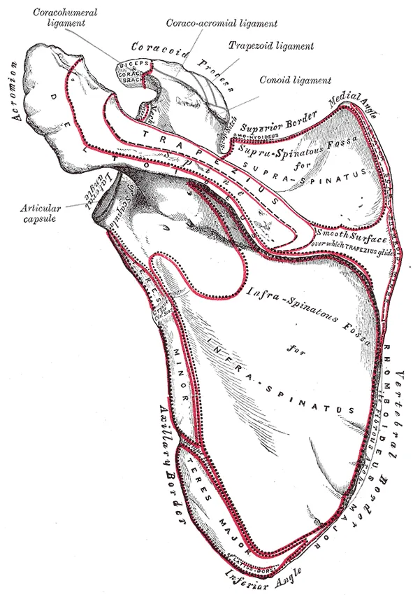
The shoulder blade lies on the back of the shoulder1. It is a flat, triangular bone with a raised ridge across its back surface1. It sits on the back of the rib cage, in the upper thoracic (chest) region2.
The bone has three borders: the superior (top), medial (inner) and lateral (outer) borders1. The medial border ends in a superior angle at the top and an inferior angle at the bottom1. The inferior angle is the bone’s lowest point1. Several powerful muscles that move the shoulder and arm attach there1.
The ridge is called the spine of the scapula, and it runs across the upper part of the back surface1. At its outer end it flattens into the acromion, the bony tip of the shoulder1. The coracoid process is a hook-like point that juts forward from the top of the bone1. Muscles from the front of the chest and arm attach to it1.
Three hollows for muscle
The shoulder blade has three hollows, called fossae1:
| Hollow | Where it sits | Width |
|---|---|---|
| Supraspinous fossa | Above the spine, on the back | Narrow1 |
| Infraspinous fossa | Below the spine, on the back | Broad1 |
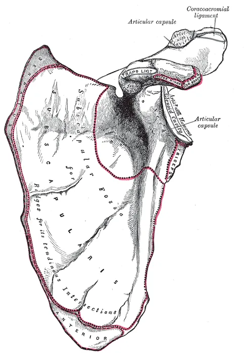
| Subscapular fossa | On the front, facing the rib cage | Broad1 |
All three give large surfaces for muscles that cross the shoulder and act on the humerus1.
The shoulder socket
The glenoid cavity is a shallow dip at the upper outer corner of the shoulder blade1. It meets the humerus to form the glenohumeral joint, the main shoulder joint1. The head of the humerus, a large, smooth, rounded surface, fits against it as a ball and socket1,7. Small bumps just above and below the socket, the supraglenoid and infraglenoid tubercles, anchor muscles of the arm1.
A bone held in place by muscle
The shoulder blade has muscle on both its front and back surfaces, so it does not form a joint with the ribs1. Together with the clavicle it makes up the pectoral girdle1. This girdle attaches the arm to the sternum (breastbone)1.
The acromioclavicular joint, where the collarbone meets the top of the shoulder blade, passes forces from the arm to the clavicle1. The ligaments around this joint are fairly weak1. Its main support is the strong coracoclavicular ligament, which ties the coracoid process to the underside of the clavicle1.
Six muscles position the girdle1:
- On the front of the chest: the subclavius, pectoralis minor and serratus anterior1.
- On the back: the trapezius, rhomboid major and rhomboid minor1.
By holding the girdle steady, these muscles give other muscles a firm base from which to move the arm1. When the rhomboids contract, the shoulder blade moves toward the spine1. This can pull the shoulder and arm backward1.
How it moves as the arm rises
One narrative review states that the shoulder blade turns upward and tilts backward on the rib cage as the arm is raised overhead3. It names upward rotation as the main movement3. Turning inward and outward varies more between people and between studies, the same review states3. It adds that shrugging and gliding forward and back actually come from the clavicle moving at the sternoclavicular joint, where the collarbone meets the breastbone3.
Two studies measured the bone’s motion directly, with pins or sensors fixed into the bone8,9:
- In 8 healthy volunteers raising the arm in the shoulder blade’s plane, the bone turned upward about 50 degrees and tilted back about 308.
- In the same 8 people, the shoulder joint moved on average 1.7 times as far as the shoulder blade8.
- In people without symptoms reaching freely, the shoulder joint rose 2.3 times as far as the blade turned upward9. When lowering the arm, it was 2.7 times9.
- In the same study, the ratio was greatest early in the lift, and upward rotation and backward tilt grew steadily to full height9.
Breakdown
How often it breaks
Fractures of the shoulder blade are fairly uncommon4. They make up under 1% of all fractures and about 3 to 5% of shoulder girdle fractures4. One anatomy chapter states that 17 muscles attach to the bone2.
Two studies counted them10,11:
- A 1995 study counted 338 fractures over 10 years in two Swedish counties10. That is an incidence (new cases per year) of 10 per 100,000 people10.
- In that Swedish study, 30% of the fractures involved the glenoid cavity10.
- Among all patients in the US National Trauma Data Bank from 2002 to 2012, 1.74% had a shoulder-blade fracture11.
Who breaks it, and how
In the Swedish Fracture Register from 2011 to 2020, 3,930 adults had 3,973 shoulder-blade fractures4. Their average age was 58, and 64% were men4. The most common breaks were of the glenoid rim, the edge of the socket (32%), and the body of the bone (28%)4.
Sources describe these fractures differently4,12. A 2006 review states that they typically follow high-energy trauma, and that about 90% of patients have other injuries too12. In the Swedish register, 22% came from high-energy trauma and 21% came with at least one other fracture4. There, high-energy injury was most common in fractures of the neck of the socket and of the body4. Falls on level ground caused most fractures of the glenoid rim and inside the joint4.
When the shoulder blade moves differently
Scapular dyskinesis means abnormal control of the shoulder blade as it moves6. It is commonly linked with shoulder problems but also appears in people without symptoms6. Studies vary on whether it is a cause or a symptom6.
One review covered 34 studies of 2,365 people in both groups, another 12 studies of 1,401 athletes5,6:
| Group studied | Share with dyskinesis |
|---|---|
| People with shoulder symptoms | 60%5 |
| People without shoulder symptoms | 48%5 |
| Overhead athletes | 61%6 |
| Non-overhead athletes | 33%6 |
The review of 34 studies concluded that many people with shoulder symptoms do not have dyskinesis5. It may be a normal finding in nearly half of people without symptoms5.
Two meta-analyses, which pool the results of earlier studies, looked at what happened next in athletes13,14. Both pooled prospective studies, which follow people forward in time13,14:
- In 419 athletes without symptoms, followed for 9 to 24 months, 35% of those with dyskinesis developed shoulder pain13. So did 25% of those without it, a 43% higher risk with dyskinesis13.
- In 923 athletes, 46% had dyskinesis, and it had no statistically significant link with later shoulder injury14.
A 2013 international consensus meeting on the scapula concluded that dyskinesis is present in a high share of most shoulder injuries15. It added that its exact role in causing or worsening shoulder problems is not clearly defined15.
Recovery
How bone answers load
A review of bone mechanics states that bone adapts its structure to the size, rate, frequency and spread of the strains it usually bears16. Too little loading may lead to bone loss, and too much to a stress reaction, an early bone stress injury before a crack forms16.
A meta-analysis of 10 articles on 761 males aged 10 to 26 looked at tennis17. Tennis raised bone mineral content in the dominant arm but had no significant effect on the other arm17.
How a broken bone heals
A review of fracture healing states that repair usually restores bone to its structure and function before the injury18. About 10% of fractures do not heal normally18.
In any broken bone, an anatomy textbook states, clotting blood first forms a fracture hematoma, then calluses of cartilage and bone steady the broken ends1. Later, dead bone is cleared and the cartilage is replaced by trabecular (spongy) bone, then compact bone, the dense outer layer1. Remodelling often leaves no outward sign of the break1.
What studies found after shoulder-blade fractures
A systematic review of 35 papers covered 822 shoulder-blade fracture cases19. The great majority healed, and most people eventually returned to work19. Lasting pain was common, and shoulder motion and strength were lower than on the other side19. The most common complications were malunion (a broken bone that heals out of line), further surgery, and arthritis after the injury19. Nonunion, where a broken bone fails to join and heal, was exceptionally rare, with or without surgery19.
Other studies looked at how these fractures were managed4,20:
- In the Swedish register, glenoid rim fractures and fractures inside the joint were operated on in over 30% of cases4. One year on, 4% of the whole group had died4.
- A meta-analysis of 26 studies covered 601 patients with fractures outside the joint20. It found no significant overall benefit of surgery over non-surgical care, though differences between studies limited the evidence20.
- A systematic review found ten reported sport-related fractures of the bone’s body21. Of these, 90% were managed without surgery, with no reported complications21. The average time back to sport was 2.5 months21.
Exercise and shoulder-blade movement
A systematic review of 20 clinical trials looked at exercise in people with scapular dyskinesis22. It found conflicting evidence, and concluded there is a lack of evidence that exercise changes shoulder-blade position and motion22. In people with subacromial impingement, pinching of tissue beneath the acromion, exercise reduced pain and disability22.
Connections
The upper arm bone and the rotator cuff
The shoulder blade joins the humerus at the glenohumeral joint and the clavicle at the acromioclavicular joint2. The tendons of four muscles connect it to the humerus and form the rotator cuff around the shoulder joint1. They are the subscapularis, supraspinatus, infraspinatus and teres minor1. The infraspinatus fills much of the infraspinous fossa23.
Muscles that start on it
Among the muscles that cross the shoulder and move the humerus, two are axial muscles, of the trunk: the pectoralis major and latissimus dorsi1. The rest start on the shoulder blade1. Examples include:
- The middle part of the deltoid, which starts from the acromion, and its back part, from the spine24.
- The long head of the triceps, which starts from the infraglenoid tubercle25.
The muscles that turn it
The trapezius steadies and moves the shoulder blade26. It and the serratus anterior are considered the only muscles that turn the shoulder blade upward27. The serratus anterior fans out from the upper eight or nine ribs28. It attaches along the shoulder blade’s medial border and at both of its angles28.
In 30 people, tiring the serratus anterior reduced backward tilt of the shoulder blade in the middle of the arm’s rise29. It also increased inward rotation as the arm went up29. Its upward rotation did not change29.
Exercises studied
One study took 17 people with scapular downward rotation syndrome, a shoulder blade that sits turned downward at rest30. It compared three shrugs with the arm 30 degrees out to the side30. A “stabilization shrug” turned the shoulder blade upward significantly more than the other two30. Related exercise page: shrug30.
Scapula forms the shoulder joint with
Scapula is attached to by
Scapula is moved by
Sources
- Betts JG, Young KA, Wise JA, Johnson E, Poe B, Kruse DH, Korol O, Johnson JE, Womble M, DeSaix P (2022). Anatomy and Physiology 2e. OpenStax. Link
- Miniato MA, Mudreac A, Borger J (2023). Anatomy, Thorax, Scapula.. StatPearls. PMID 30855903
- Ludewig PM, Reynolds JF (2009). The association of scapular kinematics and glenohumeral joint pathologies.. Journal of Orthopaedic and Sports Physical Therapy. doi:10.2519/jospt.2009.2808 PMID 19194022
- Schmidt V, Mukka S, Bergdahl C, Ekholm C, Brüggemann A, Wolf O (2025). Epidemiology, treatment, and mortality of 3973 scapula fractures from the Swedish fracture register.. Journal of Shoulder and Elbow Surgery. doi:10.1016/j.jse.2024.03.024 PMID 38688421
- Salamh PA, Hanney WJ, Boles T, Holmes D, McMillan A, Wagner A, Kolber MJ (2023). Is it Time to Normalize Scapular Dyskinesis? The Incidence of Scapular Dyskinesis in Those With and Without Symptoms: a Systematic Review of the Literature.. International Journal of Sports Physical Therapy. doi:10.26603/001c.74388 PMID 37425102
- Burn MB, McCulloch PC, Lintner DM, Liberman SR, Harris JD (2016). Prevalence of Scapular Dyskinesis in Overhead and Nonoverhead Athletes. Orthopaedic Journal of Sports Medicine. doi:10.1177/2325967115627608 PMID 26962539
- Mostafa E, Imonugo O, Varacallo MA (2023). Anatomy, Shoulder and Upper Limb, Humerus.. StatPearls. PMID 30521242
- McClure PW, Michener LA, Sennett BJ, Karduna AR (2001). Direct 3-dimensional measurement of scapular kinematics during dynamic movements in vivo.. Journal of Shoulder and Elbow Surgery. doi:10.1067/mse.2001.112954 PMID 11408911
- Braman JP, Engel SC, Laprade RF, Ludewig PM (2009). In vivo assessment of scapulohumeral rhythm during unconstrained overhead reaching in asymptomatic subjects.. Journal of Shoulder and Elbow Surgery. doi:10.1016/j.jse.2009.02.001 PMID 19395283
- Ideberg R, Grevsten S, Larsson S (1995). Epidemiology of scapular fractures. Incidence and classification of 338 fractures.. Acta Orthopaedica Scandinavica. doi:10.3109/17453679508995571 PMID 7484114
- Tatro JM, Schroder LK, Molitor BA, Parker ED, Cole PA (2019). Injury mechanism, epidemiology, and Hospital trends of scapula fractures: A 10-year retrospective study of the National Trauma Data Bank.. Injury. doi:10.1016/j.injury.2019.01.017 PMID 30661668
- Zlowodzki M, Bhandari M, Zelle BA, Kregor PJ, Cole PA (2006). Treatment of scapula fractures: systematic review of 520 fractures in 22 case series.. Journal of Orthopaedic Trauma. doi:10.1097/00005131-200603000-00013 PMID 16648708
- Hickey D, Solvig V, Cavalheri V, Harrold M, Mckenna L (2018). Scapular dyskinesis increases the risk of future shoulder pain by 43% in asymptomatic athletes: a systematic review and meta-analysis.. British Journal of Sports Medicine. doi:10.1136/bjsports-2017-097559 PMID 28735288
- Hogan C, Corbett JA, Ashton S, Perraton L, Frame R, Dakic J (2021). Scapular Dyskinesis Is Not an Isolated Risk Factor for Shoulder Injury in Athletes: A Systematic Review and Meta-analysis.. The American Journal of Sports Medicine. doi:10.1177/0363546520968508 PMID 33211975
- Kibler WB, Ludewig PM, McClure PW, Michener LA, Bak K, Sciascia AD (2013). Clinical implications of scapular dyskinesis in shoulder injury: the 2013 consensus statement from the 'Scapular Summit'.. British Journal of Sports Medicine. doi:10.1136/bjsports-2013-092425 PMID 23580420
- Hart NH, Nimphius S, Rantalainen T, Ireland A, Siafarikas A, Newton RU (2017). Mechanical basis of bone strength: influence of bone material, bone structure and muscle action.. Journal of musculoskeletal & neuronal interactions. PMID 28860414
- Huiwen H, Jun H, Dong F, Yan F, Tao W (2025). The effect of tennis on male bone mineral density: A meta-analysis.. PLOS ONE. doi:10.1371/journal.pone.0328636 PMID 40853978
- Einhorn TA, Gerstenfeld LC (2015). Fracture healing: mechanisms and interventions.. Nature reviews. Rheumatology. doi:10.1038/nrrheum.2014.164 PMID 25266456
- Sernandez HC, Riehl JT, Fogel J (2024). Sling and forget it? A systematic review of operative versus nonoperative outcomes for scapula fractures.. Journal of Shoulder and Elbow Surgery. doi:10.1016/j.jse.2024.05.042 PMID 39048030
- Stehling L, Ricker E, Klute L, Henßler L, Knüttel H, Zeman F, Alt V, Kerschbaum M (2026). Surgical vs. nonsurgical treatment of extra-articular scapula fractures: a systematic review and meta-analysis.. JSES Reviews, Reports, and Techniques. doi:10.1016/j.xrrt.2026.100666 PMID 41809069
- Neral M, Knapik DM, Wetzel RJ, Salata MJ, Voos JE (2018). Scapular Body Fracture in the Athlete: A Systematic Review.. HSS Journal: The Musculoskeletal Journal of Hospital for Special Surgery. doi:10.1007/s11420-018-9624-6 PMID 30258341
- Nodehi Moghadam A, Rahnama L, Noorizadeh Dehkordi S, Abdollahi S (2020). Exercise therapy may affect scapular position and motion in individuals with scapular dyskinesis: a systematic review of clinical trials.. Journal of Shoulder and Elbow Surgery. doi:10.1016/j.jse.2019.05.037 PMID 31420226
- Williams JM, Sinkler MA, Obremskey W (2023). Anatomy, Shoulder and Upper Limb, Infraspinatus Muscle.. StatPearls. PMID 30020627
- Stokey PJ, Kaur S, Lee A, Behrens K, Ebraheim N (2024). Anatomy and Deficiency of the Deltoid Muscle: A Review of Literature.. Orthopedic Reviews. doi:10.52965/001c.115352 PMID 38562147
- Tiwana MS, Sinkler MA, Bordoni B (2023). Anatomy, Shoulder and Upper Limb, Triceps Muscle.. StatPearls. PMID 30725681
- Ourieff J, Scheckel B, Agarwal A (2023). Anatomy, Back, Trapezius.. StatPearls. PMID 30085536
- Ekstrom RA, Donatelli RA, Soderberg GL (2003). Surface electromyographic analysis of exercises for the trapezius and serratus anterior muscles.. Journal of Orthopaedic and Sports Physical Therapy. doi:10.2519/jospt.2003.33.5.247 PMID 12774999
- Lung K, St Lucia K, Lui F (2024). Anatomy, Thorax, Serratus Anterior Muscles.. StatPearls. PMID 30285352
- Harrison N, Garrett WZ, Timmons MK (2021). Serratus Anterior Fatigue Reduces Scapular Posterior Tilt and External Rotation During Arm Elevation.. Journal of Sport Rehabilitation. doi:10.1123/jsr.2021-0010 PMID 34294607
- Lee JH, Cynn HS, Choi WJ, Jeong HJ, Yoon TL (2016). Various shrug exercises can change scapular kinematics and scapular rotator muscle activities in subjects with scapular downward rotation syndrome.. Human Movement Science. doi:10.1016/j.humov.2015.11.016 PMID 26625348
Changes
- First published.