Teres minor
Also called teres minor muscle
The narrow rotator cuff muscle on the outer edge of the shoulder blade that helps turn the arm outward. Where it attaches, how much it adds, how it wastes, and what it does when its larger neighbour fails.
- Region
- Shoulder
- Starts on
- the back of the shoulder blade's outer border, along its upper two-thirds, by one account5; the middle third of the shoulder blade's outer border, by another account6
- Ends on (upper fibres)
- the lowest of three impressions on the greater tubercle, a bump on the outer side of the upper arm bone (Rotator cuff tendons)5,6,28
- Ends on (lowest fibres)
- directly onto the upper arm bone, just below the lowest impression on the greater tubercle5
- Tendons
- Rotator cuff tendons
- Updated
- October 8, 2026
- Reviewed
- October 8, 2026 · how entries are checked
Function
Where it sits
The teres minor is a narrow muscle that runs from the outer border of the scapula (shoulder blade) to the humerus (upper arm bone)1. It is part of the rotator cuff, a group of four muscles that cross the shoulder joint and help hold it steady1. Sources differ on where it starts5,6:
- Gray’s Anatomy has it arising from the back surface of the scapula’s outer border, along its upper two-thirds5.
- A 2026 clinical commentary places the origin on the middle third of that border6.
The muscle ends on the greater tubercle, a bump at the top of the humerus1. Its upper fibres end in a tendon, a tough cord of tissue that connects a muscle to a bone5. That tendon attaches to the lowest of three impressions on the greater tubercle5. The lowest fibres attach straight to the humerus just below it5. The 2026 commentary notes that the attachment to the humerus is mostly muscle rather than tendon6.
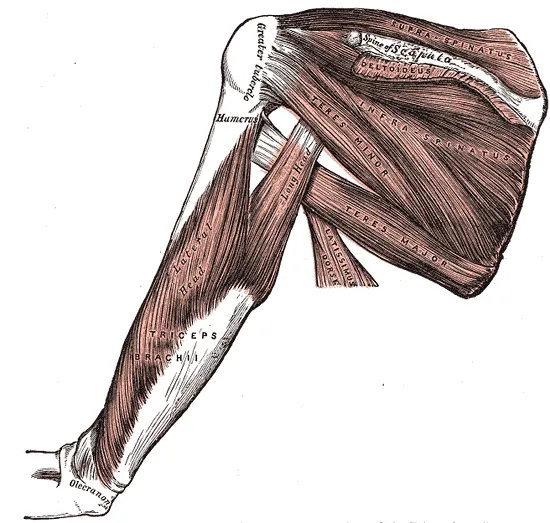
The 2026 commentary calls the teres minor the smallest of the rotator cuff muscles6. In 20 young adults whose right shoulders were scanned, it made up 8% of total cuff muscle volume7. In 40 fresh cadavers (bodies donated for study), it was a thick, spindle-shaped muscle with parallel fibres8.
What it does
The teres minor works with the infraspinatus, a rotator cuff muscle on the back of the shoulder blade5. Together they rotate the head of the humerus outward and help carry the arm backward5. Gray’s Anatomy describes the two muscles as supporting the shoulder joint from behind5.
Two cadaver studies tested how the cuff steadies the joint9,10:
- In five fresh cadaver shoulders, the arm was raised without the infraspinatus, teres minor and subscapularis9. The subscapularis is a rotator cuff muscle on the front of the shoulder blade, next to the ribs9. The humeral head (the ball at the top of the upper arm bone) shifted significantly upward on the socket9.
- Ten cadaver shoulders had all soft tissue except the rotator cuff removed10. The arm was placed in the end-range position where the joint tends to slip forward10. There the infraspinatus, teres minor and subscapularis did significantly more to steady the joint than the supraspinatus10. The supraspinatus is a rotator cuff muscle along the top of the shoulder blade10.
Its share of outward turning
Studies that used different methods give different pictures of how much the teres minor adds to external rotation (turning the arm outward)2,11,12,13,14:
| Method | Who or what was studied | What was found |
|---|---|---|
| Physiological cross-sectional area, a measure of a muscle’s size taken straight across all of its fibres | 10 cadaver specimens | The teres minor had the least capacity to produce force of the four cuff muscles11 |
| Moment arms, a measure of a muscle’s leverage | Eight cadaver upper limbs | Of 18 muscle sub-regions, the lower infraspinatus and the teres minor had the largest moment arms for external rotation12 |
| Moment arms | 10 shoulder muscles, four arm positions | With the arm at the side or raised, the infraspinatus was potentially the strongest external rotator. The teres minor and the back of the deltoid (the rounded muscle that caps the shoulder) came next13 |
| Nerve block, an injection that numbs a nerve for a short time | Volunteers, complete in 9 people | Paralysing the infraspinatus and supraspinatus removed about 80% of external rotation strength2 |
| Positron emission tomography (PET, a type of body scan) | Seven healthy volunteers | After elastic-band external rotation, the infraspinatus was the most active muscle with the arm at the side. At 90° of abduction (the arm lifted out to the side), the teres minor was14 |
A nerve-block study, complete in nine volunteers, concluded that the teres minor gives no more than 20% of external rotation strength in the short term2.
Its nerve
The teres minor is supplied by the fifth cervical (neck) nerve through the axillary nerve5. The infraspinatus is supplied through a different nerve, the suprascapular nerve5. In 40 fresh cadaver dissections, the teres minor received one branch from the axillary nerve8.
The axillary nerve divides in the quadrilateral space, a gap between muscles at the back of the shoulder15. It divides into an anterior (front) and a posterior (back) branch15. The motor branch to the teres minor is one of the posterior branch’s three end branches15. The teres minor forms the upper border of the space15.
Breakdown
How often scans show wasting
The authors of a combined cadaver and MRI study name three settings for teres minor atrophy (wasting)16. It occurs on its own, alongside disorders of the other rotator cuff muscles, or in quadrilateral space syndrome16. In that syndrome, a compressed axillary nerve is the likely cause16. Three MRI studies counted how often scans show it3,17,18:
| Scans studied | Share with the finding |
|---|---|
| 1,264 shoulder MRI scans | 6.2% showed isolated teres minor atrophy3 |
| All 2,563 shoulder MRI examinations done at one institution over two years | 3% showed isolated teres minor denervation, or loss of nerve supply17 |
| 528 shoulders of people aged 10 to 39, scanned for shoulder disorders | 2.1% showed teres minor atrophy18 |
| 884 shoulders of people aged 60 to 69, scanned for shoulder disorders | 6.7% showed teres minor atrophy18 |
In the first study, the atrophy was complete in 41% of the 78 affected shoulders and partial in 59%3. Most partial cases mainly involved the medial-dorsal part of the muscle, the part toward the midline and the back3.
What goes with it
The studies do not point to one cause3,16,18. In the 1,264 scans, isolated atrophy showed no significant association with previous trauma, shoulder instability, osteoarthritis or a previous operation3. The authors of the age study suggest rotator cuff tears may be the common cause18. The authors of a combined cadaver and MRI study describe the exact cause of isolated atrophy as unknown16.
Those authors dissected nine shoulder specimens16. From that, they suggest wide variation in the origin and length of the nerve to the muscle raises the risk of it being pinched16. In 31 dissected cadaver shoulders, the main nerve to the teres minor bent around a stout sling of fascia (tough connective tissue) before entering it19. The authors of that study suggest the sling may be where the nerve is most likely to be compressed or tethered19.
Quadrilateral space syndrome
The cadaver and MRI authors give a compressed axillary nerve as the likely cause of the atrophy in quadrilateral space syndrome16. A 2026 clinical commentary explains why the teres minor can be affected alone6. The axillary nerve is the muscle’s only motor supply, and it reaches the muscle by way of the space6. Compression or traction there can leave the teres minor fatty and wasted while the rest of the cuff is unaffected6. In long-standing compression of the axillary nerve, wasting of the deltoid and teres minor may be seen15.
The authors of a combined cadaver and MRI study say isolated teres minor atrophy appears to be clinically distinct from quadrilateral space syndrome16. In the 2,563 examinations, no patient with isolated denervation had a history suggesting the classic syndrome17. Nor did any have a structural lesion inside the space17. One review says that, when the syndrome’s symptoms are absent, teres minor atrophy on MRI is to be read cautiously as a nonspecific finding15.
Recovery
When other cuff tendons are torn
One series covered 1,332 CT scans (detailed X-ray pictures) and 240 MRI scans of rotator cuff tears20. The teres minor looked normal in 90.8%20. It was enlarged in 5.8% and wasted in 3.2%, and 0.2% had no teres minor20.
An MRI study of 279 people with and without cuff tears looked at when it enlarges21. The teres minor appeared enlarged in tears involving the infraspinatus tendon21. Its share of the external-rotator muscle area rose as the infraspinatus share fell21.
One study measured strength in 35 patients with a tear at the back and top of the cuff and a wasted infraspinatus4. It compared external rotation strength with the arm at the side against the healthy shoulder4:
| State of the teres minor | External rotation strength, as a share of the healthy side |
|---|---|
| Enlarged (17 patients) | 60%4 |
| Normal (10 patients) | 33%4 |
| Deficient (8 patients) | 7%4 |
One review calls the teres minor often overlooked in a normal shoulder22. It says the muscle changes from an insignificant external rotator to the most significant one when there is major cuff damage22. Another review states that, with a complete infraspinatus tear, a healthy, intact teres minor matters because the shoulder joint has no other external rotator23.
Sport and stretching
Three studies cited here measured the teres minor in sport or after stretching25,26,27. Among 35 symptom-free elite adolescent tennis players, the playing arm had larger infraspinatus and teres minor muscles than the other arm25.
Two studies measured stiffness, meaning how much force it takes to stretch something by a given amount26,27. One trial randomly assigned 24 college baseball players with limited inward shoulder rotation to one of two stretches for four weeks26. Teres minor stiffness fell in the modified cross-body stretch group26. In the modified sleeper stretch group, infraspinatus stiffness fell26.
The other was a crossover study, in which each person tries every option in turn, of twenty healthy men27. Stiffness was measured before and immediately after stretching27. A cross-body stretch done without the shoulder blade held still changed none of the measured muscles, the teres minor included27.
Connections
The infraspinatus and the rest of the cuff
The teres minor and the infraspinatus both lie behind the shoulder joint5. Gray’s Anatomy notes that the teres minor is sometimes inseparable from the infraspinatus5. In 40 fresh cadaver dissections, a sheet of fascia separated the two in about two-thirds of specimens8. Their tendons, with those of the supraspinatus and subscapularis, form the rotator cuff around the joint28.
The deltoid and the quadrilateral space
The teres minor shares its nerve with the deltoid, the rounded muscle that caps the shoulder15. The posterior branch of the axillary nerve supplies both the back of the deltoid and the teres minor15.
Other structures border the quadrilateral space with the teres minor15. The surgical neck of the humerus, the narrow part just below its rounded top, lies to the outside15. The long head of the triceps, the three-headed muscle on the back of the upper arm, lies to the inside15. The teres major, a thick shoulder muscle, and the latissimus dorsi lie below15.
Exercises and stretches
Two studies recorded the teres minor’s electrical activity during resisted shoulder movements29,30. In 10 healthy people, electrodes inside the muscles recorded seven shoulder exercises29. Side-lying external rotation with the arm at the side drew the most teres minor activity, 67% of maximum29.
In 18 collegiate baseball players lying face down, teres minor activity was lower than infraspinatus activity when external rotation was resisted by hand30. It was higher when horizontal abduction (drawing the raised arm backward) was resisted30. Related exercise and stretch pages: band external rotation, sleeper stretch.
Teres minor works with to turn the arm outward
Teres minor is supplied by
Teres minor pulls through
Teres minor starts on
Teres minor attaches to
Teres minor shares a nerve with
Teres minor works with to hold the arm bone down
Teres minor is loaded by
Teres minor has been measured in studies of
Sources
- Juneja P, Hubbard JB (2023). Anatomy, Shoulder and Upper Limb, Teres Minor Muscle.. StatPearls. PMID 30020696
- Gerber C, Blumenthal S, Curt A, Werner CM (2007). Effect of selective experimental suprascapular nerve block on abduction and external rotation strength of the shoulder.. Journal of Shoulder and Elbow Surgery. doi:10.1016/j.jse.2007.02.120 PMID 17931906
- Kang Y, Ahn JM, Chee CG, Lee E, Lee JW, Kang HS (2019). The pattern of idiopathic isolated teres minor atrophy with regard to its two-bundle anatomy.. Skeletal Radiology. doi:10.1007/s00256-018-3038-x PMID 30091009
- Kikukawa K, Ide J, Terakawa Y, Takada K, Morita M, Hashimoto K, Mizuta H (2016). Hypertrophic teres minor restores shoulder strength and range of external rotation in posterosuperior rotator cuff tears.. Journal of Shoulder and Elbow Surgery. doi:10.1016/j.jse.2016.04.016 PMID 27374237
- Gray H; Lewis WH (editor) (1918). Anatomy of the Human Body. Lea & Febiger, Philadelphia (20th edition). Link
- Manske RC, Wolfe C, Page P, Voight M (2026). Diagnostic Musculoskeletal Ultrasound in the Evaluation of the Teres Minor.. International Journal of Sports Physical Therapy. doi:10.26603/001c.163510 PMID 42404359
- Zhang Y, Herbert RD, Bilston LE, Bolsterlee B (2024). Three-dimensional architecture and moment arms of human rotator cuff muscles in vivo: Interindividual, intermuscular, and intramuscular variations.. Journal of Anatomy. doi:10.1111/joa.14050 PMID 38690607
- Bacle G, Gregoire JM, Patat F, Clavert P, de Pinieux G, Laulan J, Lakhal W, Favard L (2017). Anatomy and relations of the infraspinatus and the teres minor muscles: a fresh cadaver dissection study.. Surgical and Radiologic Anatomy. doi:10.1007/s00276-016-1707-9 PMID 27286948
- Sharkey NA, Marder RA (1995). The rotator cuff opposes superior translation of the humeral head.. The American Journal of Sports Medicine. doi:10.1177/036354659502300303 PMID 7661251
- Lee SB, Kim KJ, O'Driscoll SW, Morrey BF, An KN (2000). Dynamic glenohumeral stability provided by the rotator cuff muscles in the mid-range and end-range of motion. A study in cadavera.. The Journal of Bone and Joint Surgery. American Volume. doi:10.2106/00004623-200006000-00012 PMID 10859105
- Ward SR, Hentzen ER, Smallwood LH, Eastlack RK, Burns KA, Fithian DC, Friden J, Lieber RL (2006). Rotator cuff muscle architecture: implications for glenohumeral stability.. Clinical Orthopaedics and Related Research. PMID 16826111
- Ackland DC, Pandy MG (2011). Moment arms of the shoulder muscles during axial rotation.. Journal of Orthopaedic Research. doi:10.1002/jor.21269 PMID 21064160
- Kuechle DK, Newman SR, Itoi E, Niebur GL, Morrey BF, An KN (2000). The relevance of the moment arm of shoulder muscles with respect to axial rotation of the glenohumeral joint in four positions.. Clinical Biomechanics. doi:10.1016/s0268-0033(99)00081-9 PMID 10758292
- Kurokawa D, Sano H, Nagamoto H, Omi R, Shinozaki N, Watanuki S, Kishimoto KN, Yamamoto N, Hiraoka K, Tashiro M, Itoi E (2014). Muscle activity pattern of the shoulder external rotators differs in adduction and abduction: an analysis using positron emission tomography.. Journal of Shoulder and Elbow Surgery. doi:10.1016/j.jse.2013.12.021 PMID 24613183
- Ayik G, Kolac UC, Kaymakoglu M, McFarland E, Huri G (2025). Dark side of the shoulder: suprascapular and axillary nerve compressions.. International Orthopaedics. doi:10.1007/s00264-025-06465-9 PMID 40082300
- Friend J, Francis S, McCulloch J, Ecker J, Breidahl W, McMenamin P (2010). Teres minor innervation in the context of isolated muscle atrophy.. Surgical and Radiologic Anatomy. doi:10.1007/s00276-009-0605-9 PMID 20020125
- Sofka CM, Lin J, Feinberg J, Potter HG (2004). Teres minor denervation on routine magnetic resonance imaging of the shoulder.. Skeletal Radiology. doi:10.1007/s00256-004-0809-3 PMID 15221220
- Takeuchi Y, Sugaya H, Takahashi N, Matsuki K, Tokai M, Morioka T, Ueda Y, Hoshika S, Kawashima I (2022). Incidence of teres minor muscle atrophy in young and middle-aged populations.. JSES International. doi:10.1016/j.jseint.2022.01.003 PMID 35572432
- Chafik D, Galatz LM, Keener JD, Kim HM, Yamaguchi K (2013). Teres minor muscle and related anatomy.. Journal of Shoulder and Elbow Surgery. doi:10.1016/j.jse.2011.12.005 PMID 22521388
- Melis B, DeFranco MJ, Lädermann A, Barthelemy R, Walch G (2011). The teres minor muscle in rotator cuff tendon tears.. Skeletal Radiology. doi:10.1007/s00256-011-1178-3 PMID 21604212
- Kikukawa K, Ide J, Kikuchi K, Morita M, Mizuta H, Ogata H (2014). Hypertrophic changes of the teres minor muscle in rotator cuff tears: quantitative evaluation by magnetic resonance imaging.. Journal of Shoulder and Elbow Surgery. doi:10.1016/j.jse.2014.03.014 PMID 24906902
- Williams MD, Edwards TB, Walch G (2018). Understanding the Importance of the Teres Minor for Shoulder Function: Functional Anatomy and Pathology.. The Journal of the American Academy of Orthopaedic Surgeons. doi:10.5435/JAAOS-D-15-00258 PMID 29473831
- Chambers MM, Khan AZ, Namdari S (2022). Teres Minor Muscle Atrophy. JBJS Reviews. doi:10.2106/JBJS.RVW.22.00130 PMID 36639874
- Wu HT, Liu Q, Lin JH (2025). Fatty infiltration predicts retear and functional impairment following rotator cuff repair: systematic review and meta-analysis.. Journal of Orthopaedic Surgery and Research. doi:10.1186/s13018-025-05702-6 PMID 40119431
- Johansson FR, Skillgate E, Adolfsson A, Jenner G, DeBri E, Swärdh L, Cools AM (2015). Asymptomatic Elite Adolescent Tennis Players' Signs of Tendinosis in Their Dominant Shoulder Compared With Their Nondominant Shoulder.. Journal of Athletic Training. doi:10.4085/1062-6050-51.1.07 PMID 26651279
- Yamauchi T, Hasegawa S, Nakamura M, Nishishita S, Yanase K, Fujita K, et al. (2016). Effects of two stretching methods on shoulder range of motion and muscle stiffness in baseball players with posterior shoulder tightness: a randomized controlled trial.. Journal of shoulder and elbow surgery. doi:10.1016/j.jse.2016.04.025 PMID 27475455
- Umehara J, Hasegawa S, Nakamura M, Nishishita S, Umegaki H, Tanaka H, et al. (2017). Effect of scapular stabilization during cross-body stretch on the hardness of infraspinatus, teres minor, and deltoid muscles: An ultrasonic shear wave elastography study.. Musculoskeletal science & practice. doi:10.1016/j.math.2016.10.004 PMID 27847244
- Betts JG, Young KA, Wise JA, Johnson E, Poe B, Kruse DH, Korol O, Johnson JE, Womble M, DeSaix P (2022). Anatomy and Physiology 2e. OpenStax. Link
- Reinold MM, Wilk KE, Fleisig GS, Zheng N, Barrentine SW, Chmielewski T, Cody RC, Jameson GG, Andrews JR (2004). Electromyographic analysis of the rotator cuff and deltoid musculature during common shoulder external rotation exercises.. Journal of Orthopaedic and Sports Physical Therapy. doi:10.2519/jospt.2004.34.7.385 PMID 15296366
- Tsuruike M, Ellenbecker TS (2022). A comparison of teres minor and infraspinatus muscle activation in the prone position.. JSES International. doi:10.1016/j.jseint.2021.09.005 PMID 35141685
Changes
- Revised to report sources added after their published corrections were read