Pectoralis major
The large, fan-shaped muscle at the front of the chest. How its parts are built and supplied, where it tears, how it grows with training, and what it works with.
- Region
- Trunk
- Starts on
- the collarbone's inner half, breastbone, first seven rib cartilages, sixth rib and the external oblique's tendon sheet (aponeurosis)7
- Ends on
- the outer lip of the bicipital groove on the upper arm bone (Pectoralis major tendon)7,8,27
- Tendons
- Pectoralis major tendon
- Updated
- September 30, 2026
- Reviewed
- September 30, 2026 · how entries are checked
Function
A fan built from several parts
The pectoralis major is a large muscle shaped like a fan1. One source describes it as typically having three parts: clavicular, sternocostal and abdominal1. Another describes two heads, clavicular and sternocostal, named for where they start7. The clavicular part is usually set apart from the sternocostal part by a slight cleft1.
It starts from the inner clavicle (collarbone), the sternum (breastbone) and other points7. These include the first seven costal cartilages, the strips of cartilage that join ribs to the breastbone7.
The fibres end on the humerus (upper arm bone), at the outer lip of a groove called the bicipital groove7. In one study of 80 muscles from 40 cadavers (bodies donated for study), this attachment was 43.9 to 83.2 mm wide1. The average was 65.3 mm1.
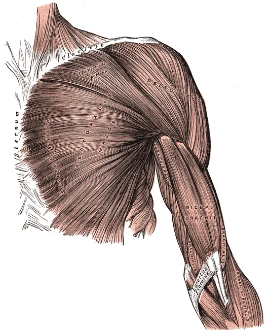
In the same 80 muscles, the attachment had a front layer and a back layer1. The clavicular part joined the front layer in every case, and the sternocostal and abdominal parts joined the back layer1. Studies disagree here, though: one study of 14 donated arms found a single-layer attachment to bone, with no true tendon layers there8.
What it moves
The muscle is important for moving the arm, especially for adduction and for turning the arm inward1. A study of 8 donated arms measured each muscle’s moment arm, meaning its leverage9. The upper fibres were among the best at flexion, which at the shoulder means raising the arm forward9. The middle and lower fibres were among the best adductors9.
One anatomist, cited by a study’s authors, describes the clavicular part as a recent addition in higher primates (anthropoids)1. It is said to help steady the arm against the chest1.
Different regions, different jobs
One study used electromyography (EMG, measuring a muscle’s activity from its electrical signals) in 29 healthy young women holding still efforts10. In some tasks, the middle sternocostal region was 12% to 108% more active than the clavicular and upper sternocostal regions10. These tasks were moving the arm backward, adduction with the arm turned outward, and turning the arm inward with it raised high10.
When the women raised the arm forward or swung it across the body, the three regions activated similarly at every force level10. The one exception was forward raising at 25% of greatest force, where the clavicular region was 21% more active than the upper sternocostal region10. The authors conclude that the regions may be specialised for different actions10.
Its nerves and blood supply
In the 80-muscle study, the lateral pectoral nerve was the main nerve to the muscle1. In specimens stained to show nerve branches, it alone supplied the clavicular part, and its branches reached about the upper half of the sternocostal part1. The medial pectoral nerve supplied the lower half and the abdominal part, when present1. Intercostal nerves, which run between two ribs, also sent branches in every stained specimen1.
A review and meta-analysis of pectoral nerve anatomy reports the split this way: upper parts from the lateral nerve, lower parts from the medial nerve11. The medial nerve also supplies the pectoralis minor, a smaller chest muscle beneath the pectoralis major11. It runs along the pectoralis minor’s lower border12. In 50% to 100% of cases, at least some of its branches pass through that muscle11.
In 11 healthy people, the zones where nerves enter the muscle lay in its inner part13. They sat about 20% to 40% of the way along the muscle-tendon unit, and moved by up to 13.3% as arm position changed13. A surgical case report describes its blood supply as mainly from one artery branch, with more from branches of two other arteries14.
Breakdown
Where injuries happen
An analysis of 365 published injury cases, from 1822 to 2010, found that 75% came from the last 20 years15. The authors of another study state that how often injuries occur differs between the clavicular, sternal and abdominal regions16. A system for sorting injuries, proposed from those 365 cases, groups them by location15:
- at the muscle’s starting point or in the muscle belly, the thick middle part15
- anywhere from the musculotendinous junction, where muscle fibres join their tendon, to the tendon’s attachment15
- a bony avulsion, where the attachment pulls away from the bone15
In a pooled analysis of 112 reported cases, all in men, most tears were complete and at the attachment on the humerus17. A later systematic review of surgical repairs agrees, with complete tears at the attachment most common (75%)18. One centre, studying consecutive patients assessed for a tear, found otherwise19. There, 60.4% of tears were at the musculotendinous junction, 34.9% at the attachment and 4.7% in the belly19.
Who tears it, and how
In a systematic review of 330 surgically repaired tears, every patient was male, with an average age of 30.4 years18. Across the published repairs in another review, the bench press was the most common cause, in 49% of cases20. In the 112 pooled cases, tears happened most often in sport, with the arm raised out to the side and turned outward17.
A review found few studies on injuries linked to the deadlift, and a pectoralis major tear was among those reported21.
When its nerves are hurt
A review reports that the pectoral nerves can be injured on their own in several ways11:
- a direct blow11
- squeezing by an enlarged muscle11
- surgery, such as breast surgery or removing lymph nodes from the armpit11
How its shape varies
The muscle’s shape varies between people1. In the 80-muscle study, the most common variation was a separate clavicular part1. The clavicular part was underdeveloped in 2.5% of muscles and fused with the deltoid in 5%1.
A rare condition present from birth
Poland’s syndrome is a rare condition, present from birth, in which the pectoralis major is missing22. Other differences of the chest wall and arm on the same side come with it22. A literature review reports that functional limits are minimal, so surgical correction is done mainly for appearance22.
Recovery
How it grows with training
Several trials measured muscle thickness, meaning how thick a muscle is from its surface to the bone beneath23. Most found the muscle grew, but one short trial did not3,6:
| People studied | What they did | What changed |
|---|---|---|
| 7 untrained young men23 | Bench press at 75% of their one-repetition maximum (1RM, the heaviest load they could lift once), 3 days a week, 24 weeks23 | Thickness rose significantly from week 1, strength from week 323 |
| 10 young men24 | Light bench press (30% of 1RM) twice a day for 2 weeks, with or without cuffs limiting blood flow to the arms24 | Thickness rose 16% with cuffs, and did not change without them24 |
| 18 young men3 | Bench press at 40% of 1RM, or push-ups set to the same load, twice a week, 8 weeks3 | Thickness rose 18.3% with push-ups and 19.4% with the bench press3 |
| 15 untrained young men4 | Pec deck, one set or three sets, 12 weeks4 | Clavicular part 17–18% thicker, sternocostal part 21%, with no difference between one and three sets4 |
| 81 active people5 | Stretching 15 minutes a day, 4 days a week, or weight training 3 days a week, 8 weeks5 | Both groups grew thicker than controls, with no difference between them5 |
| 23 moderately trained men6 | Push-ups or bench press, 3 days a week, 4 weeks6 | Both gained 1RM strength, but neither showed a significant change in thickness6 |
How growth changes over months
In the 7 men who trained for 24 weeks, thickness and strength rose significantly in each of three training phases23. The triceps grew thicker only in the first two phases23. In another 24-week study of young men, cross-sectional area (the size of a slice across the muscle) rose gradually for 6 weeks25. After that, the rate of increase gradually fell25.
That study also had a second group among its 14 men25. This group trained in three 6-week blocks, with 3-week breaks between them25. Overall gains in size and strength were similar to those of the men who trained without breaks25.
Where it grows
In 47 untrained men, flat, incline or mixed bench press once a week for 8 weeks changed thickness differently at only one of three sites26. That was the upper site, which grew most in the incline group26.
Connections
Its tendon and the deltoid
The muscle inserts on the humerus through the pectoralis major tendon7,27. In an ultrasound study of 46 volunteers, the authors concluded that the muscle ends in two separate tendons27. One came from the sternal and abdominal parts27. The other emerged between the deltoid and the clavicular fibres, and merged with a tendon inside the deltoid27.
The clavicular part may reach along the collarbone as far as the deltoid, the rounded muscle that caps the shoulder, and may fuse with it1. The authors of one study describe the two clavicular parts as working together1.
The latissimus dorsi and triceps
For pulling the arm in toward the body, this muscle and the latissimus dorsi (the lats) matter more than the triceps28.
Exercises that load it
Related exercise pages are bench press, overhead press, band external rotation and dead hang29,30,31,32.
| Exercise | What studies found |
|---|---|
| Bench press | Counted as a main mover in the traditional model, with the front deltoid and triceps29 |
| Bench press | In a review of 14 EMG studies, it showed similar activity to the triceps, and both were more active than the front deltoid29 |
| Bench press | A review reports that its activity rises with heavier loads, and with fatigue29 |
| Bench press | In 30 trained adults, upper chest activity peaked at a 30° bench, and middle and lower activity was higher on a flat bench33 |
| Bench press | In 13 men, the clavicular part was more active than the sternocostal part on the incline, and the reverse on the flat bench2 |
| Bench press | In 28 men, its activity was similar across three grip widths34 |
| Bench press and fly | In 12 college-age men and women, its activation did not differ significantly between barbell press, dumbbell press and dumbbell fly35 |
| Overhead press | In 14 trained men pressing seated, pressing in front of the head gave higher activity on the way up than pressing behind the neck30 |
| Band external rotation | In a 30-person study, the standing version with a towel roll drew its highest activity of five exercises31 |
| Dead hang | In 11 male climbers, the authors classed it as a postural muscle32 |
Pectoralis major pulls through
Pectoralis major attaches to
Pectoralis major works with to raise the arm forward
Pectoralis major works with to move the arm
Pectoralis major works with in the bench press
Pectoralis major is loaded by
Sources
- Haładaj R, Wysiadecki G, Clarke E, Polguj M, Topol M (2019). Anatomical Variations of the Pectoralis Major Muscle: Notes on Their Impact on Pectoral Nerve Innervation Patterns and Discussion on Their Clinical Relevance.. Biomed Res Int. doi:10.1155/2019/6212039 PMID 31061824
- Albarello JCDS, Cabral HV, Leitão BFM, Halmenschlager GH, Lulic-Kuryllo T, Matta TTD (2022). Non-uniform excitation of pectoralis major induced by changes in bench press inclination leads to uneven variations in the cross-sectional area measured by panoramic ultrasonography.. J Electromyogr Kinesiol. doi:10.1016/j.jelekin.2022.102722 PMID 36334406
- Kikuchi N, Nakazato K (2017). Low-load bench press and push-up induce similar muscle hypertrophy and strength gain.. J Exerc Sci Fit. doi:10.1016/j.jesf.2017.06.003 PMID 29541130
- Pinto MD, Ughini C, Nunes JP, Cadore EL, Pinto RS (2025). Pectoralis Clavicular and Sternocostal Thicknesses Increase Similarly in Response to One and Three Sets of Pec Deck Resistance Training in Untrained Young Men.. J Strength Cond Res. doi:10.1519/JSC.0000000000005045 PMID 40266636
- Wohlann T, Warneke K, Kalder V, Behm DG, Schmidt T, Schiemann S (2024). Influence of 8-weeks of supervised static stretching or resistance training of pectoral major muscles on maximal strength, muscle thickness and range of motion.. Eur J Appl Physiol. doi:10.1007/s00421-023-05413-y PMID 38240811
- Kotarsky CJ, Christensen BK, Miller JS, Hackney KJ (2018). Effect of Progressive Calisthenic Push-up Training on Muscle Strength and Thickness.. J Strength Cond Res. doi:10.1519/JSC.0000000000002345 PMID 29466268
- Solari F, Burns B (2023). Anatomy, Thorax, Pectoralis Major Major.. StatPearls. PMID 30252247
- Huang BK, Wong JH, Haghighi P, Wan L, Du J, Chang EY (2020). Pectoralis major tendon and enthesis: anatomic, magnetic resonance imaging, ultrasonographic, and histologic investigation.. Journal of Shoulder and Elbow Surgery. doi:10.1016/j.jse.2019.12.020 PMID 32169467
- Ackland DC, Pak P, Richardson M, Pandy MG (2008). Moment arms of the muscles crossing the anatomical shoulder.. Journal of Anatomy. doi:10.1111/j.1469-7580.2008.00965.x PMID 18691376
- Lulic-Kuryllo T, Negro F, Jiang N, Dickerson CR (2022). Differential regional pectoralis major activation indicates functional diversity in healthy females.. J Biomech. doi:10.1016/j.jbiomech.2022.110966 PMID 35093733
- Porzionato A, Macchi V, Stecco C, Loukas M, Tubbs RS, De Caro R (2012). Surgical anatomy of the pectoral nerves and the pectoral musculature.. Clin Anat. doi:10.1002/ca.21301 PMID 22125052
- Maldonado KA, Tadi P (2023). Anatomy, Thorax, Medial Pectoral Nerves.. StatPearls. PMID 32310519
- Mancebo FD, Cabral HV, de Souza LML, de Oliveira LF, Vieira TM (2019). Innervation zone locations distribute medially within the pectoralis major muscle during bench press exercise.. J Electromyogr Kinesiol. doi:10.1016/j.jelekin.2019.03.002 PMID 30870768
- Sio B, Zhang SL, Ng HW (2025). Rare "T-junction" aberrancy in vascular anatomy of the pectoral branch of the thoracoacromial trunk in the pectoralis major myocutaneous flap: A case report.. JPRAS Open. doi:10.1016/j.jpra.2025.06.010 PMID 40704049
- ElMaraghy AW, Devereaux MW (2012). A systematic review and comprehensive classification of pectoralis major tears.. Journal of Shoulder and Elbow Surgery. doi:10.1016/j.jse.2011.04.035 PMID 21831661
- Umehara J, Sato Y, Ikezoe T, Yagi M, Nojiri S, Nakao S, Yanase K, Hirono T, Ichihashi N (2021). Regional differential stretching of the pectoralis major muscle: An ultrasound elastography study.. J Biomech. doi:10.1016/j.jbiomech.2021.110416 PMID 33894472
- Bak K, Cameron EA, Henderson IJ (2000). Rupture of the pectoralis major: a meta-analysis of 112 cases.. Knee Surgery, Sports Traumatology, Arthroscopy. doi:10.1007/s001670050197 PMID 10795675
- Gupton M, Johnson JE (2019). Surgical Treatment of Pectoralis Major Muscle Ruptures: A Systematic Review and Meta-analysis.. Orthopaedic Journal of Sports Medicine. doi:10.1177/2325967118824551 PMID 30800687
- Carlos NBT, Drain NP, Fatora GC, Nazzal EM, Herman ZJ, Hughes JD, Rodosky MW, Lin A, Lesniak BP (2023). Myotendinous junction tears of the pectoralis major are occurring more frequently and discrepancies exist between intraoperative and radiographic assessments.. JSES International. doi:10.1016/j.jseint.2023.06.019 PMID 37969514
- Yu J, Zhang C, Horner N, Ayeni OR, Leroux T, Alolabi B, Khan M (2019). Outcomes and Return to Sport After Pectoralis Major Tendon Repair: A Systematic Review.. Sports Health. doi:10.1177/1941738118818060 PMID 30543493
- Bengtsson V, Berglund L, Aasa U (2018). Narrative review of injuries in powerlifting with special reference to their association to the squat, bench press and deadlift.. BMJ Open Sport & Exercise Medicine. doi:10.1136/bmjsem-2018-000382 PMID 30057777
- Vazirnia A, Cohen PR (2015). Poland's syndrome: a concise review of the clinical features highlighting associated dermatologic manifestations.. Am J Clin Dermatol. doi:10.1007/s40257-015-0132-x PMID 25943075
- Ogasawara R, Thiebaud RS, Loenneke JP, Loftin M, Abe T (2012). Time course for arm and chest muscle thickness changes following bench press training.. Interv Med Appl Sci. doi:10.1556/IMAS.4.2012.4.7 PMID 24265879
- Yasuda T, Fujita S, Ogasawara R, Sato Y, Abe T (2010). Effects of low-intensity bench press training with restricted arm muscle blood flow on chest muscle hypertrophy: a pilot study.. Clin Physiol Funct Imaging. doi:10.1111/j.1475-097X.2010.00949.x PMID 20618358
- Ogasawara R, Yasuda T, Ishii N, Abe T (2013). Comparison of muscle hypertrophy following 6-month of continuous and periodic strength training.. Eur J Appl Physiol. doi:10.1007/s00421-012-2511-9 PMID 23053130
- Chaves SFN, Rocha-Júnior VA, Encarnação IGA, Martins-Costa HC, Freitas EDS, Coelho DB, Franco FSC, Loenneke JP, Bottaro M, Ferreira-Júnior JB (2020). Effects of Horizontal and Incline Bench Press on Neuromuscular Adaptations in Untrained Young Men.. Int J Exerc Sci. doi:10.70252/FDNB1158 PMID 32922646
- Guiu R, Martinel V, Lapègue F, Tankeng LT, Werthel JD, Schlur C (2025). Ultrasound assessment of the distal pectoralis major: identification of two distinct tendons.. Journal of Ultrasound. doi:10.1007/s40477-025-01026-7 PMID 40402442
- Landin D, Thompson M, Jackson M (2018). Functions of the Triceps Brachii in Humans: A Review.. Journal of Clinical Medicine Research. doi:10.14740/jocmr3340w PMID 29511416
- Stastny P, Gołaś A, Blazek D, Maszczyk A, Wilk M, Pietraszewski P, Petr M, Uhlir P, Zając A (2017). A systematic review of surface electromyography analyses of the bench press movement task.. PLoS One. doi:10.1371/journal.pone.0171632 PMID 28170449
- Padovan R, Toninelli N, Longo S, Tornatore G, Esposito F, Cè E, Coratella G (2026). High-Density Surface Electromyography Excitation in Front vs. Back Overhead Press Prime Movers.. Journal of Human Kinetics. doi:10.5114/jhk/205466 PMID 42713087
- Alizadehkhaiyat O, Hawkes DH, Kemp GJ, Frostick SP (2015). Electromyographic Analysis of the Shoulder Girdle Musculature During External Rotation Exercises.. Orthopaedic Journal of Sports Medicine. doi:10.1177/2325967115613988 PMID 26740950
- Exel J, Kaufmann P, Froschauer O, Baca A, Kainz H, Mochizuki L (2026). Upper-Body Neuromechanical Coordination Strategies During Fatiguing Sustained Dead Hangs in Climbers.. European Journal of Sport Science. doi:10.1002/ejsc.70197 PMID 42168114
- Rodríguez-Ridao D, Antequera-Vique JA, Martín-Fuentes I, Muyor JM (2020). Effect of Five Bench Inclinations on the Electromyographic Activity of the Pectoralis Major, Anterior Deltoid, and Triceps Brachii during the Bench Press Exercise.. International Journal of Environmental Research and Public Health. doi:10.3390/ijerph17197339 PMID 33049982
- Saeterbakken AH, Stien N, Pedersen H, Solstad TEJ, Cumming KT, Andersen V (2021). The Effect of Grip Width on Muscle Strength and Electromyographic Activity in Bench Press among Novice- and Resistance-Trained Men.. International Journal of Environmental Research and Public Health. doi:10.3390/ijerph18126444 PMID 34198674
- Welsch EA, Bird M, Mayhew JL (2005). Electromyographic activity of the pectoralis major and anterior deltoid muscles during three upper-body lifts.. J Strength Cond Res. doi:10.1519/14513.1 PMID 15903389
Changes
- First published.