Pectoralis major tendon
Also called pec major tendon
The tendon that joins the large chest muscle to the upper arm bone. How it is built, how and in whom it tears, and what studies found after repair.
- Region
- Shoulder
- Muscles
- pectoralis major
- Updated
- September 29, 2026
- Reviewed
- September 29, 2026 · how entries are checked
Function
Where the muscle starts and ends
The pectoralis major is the biggest and highest muscle on the front of the chest wall1. It is thick and shaped like a fan, and it forms the front wall of the armpit1. It has two heads, or parts, named for where they start1. These are the clavicular head, from the clavicle (collarbone), and the sternocostal head, from the breastbone and rib cartilages1.
Its fibres start from a wide base1:
- the front of the inner half of the clavicle (collarbone)1
- the front of the sternum (breastbone)1
- the first seven costal cartilages, which link the ribs to the breastbone1
- the breastbone end of the sixth rib1
- the aponeurosis, a flat sheet of tendon, of the external oblique muscle, the outermost flat muscle on each side of the abdomen1
From this base, the muscle ends on the humerus (upper arm bone)1. It attaches to the outer lip of a groove in the humerus called the bicipital groove1. The pectoralis major is one of two axial muscles, based on the trunk, that cross the shoulder and move the humerus8. The other is the latissimus dorsi, the broad, triangular muscle of the lower back8.
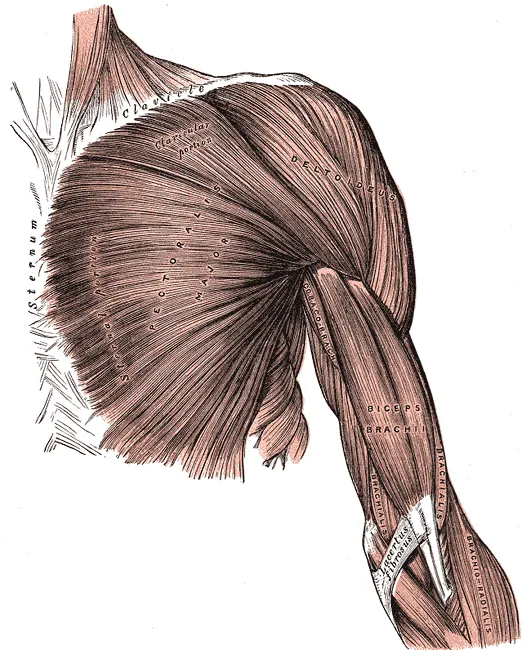
What each part of the muscle does
A study of 8 arms from body donors measured the leverage each shoulder muscle has for moving the arm9. The upper, clavicular fibres were among the best at flexion, which raises the arm forward9. The middle and lower fibres were among the best at adduction, which pulls the arm in toward the body9.
The sternocostal head is described as having 2 to 7 separate segments1. In 11 preserved donor bodies, the clavicular head was uniform, while the sternal head had 6 or 7 segments2. In each of the 11, the lowest sternal segment folded forward into a trough holding the underside of the segment above2.
How the tendon meets the bone
The footprint is the patch of the humerus that the tendon covers3. Two donor studies measured it:
| Donors studied | Footprint length, top to bottom |
|---|---|
| 14 arms, fresh-frozen | 75 mm long and about 7 mm wide3 |
| 8 male bodies, fresh-frozen | 81.4 mm in total10 |
| Same 8, sternal head alone | 42.1 mm10 |
| Same 8, clavicular head alone | 56.6 mm10 |
In the 14 arms, tendons from both the clavicular and sternal heads were found in each one3. The clavicular head’s tendon was generally the shorter of the two3.
How studies describe the tendon’s build
Studies have described the tendon’s structure near the bone in different ways2,3,4. Each finding below is what one study reported:
- Two layers. In 11 preserved donor bodies, the tendon had a longer front layer and a shorter back layer, joined along their lower edge2. This study noted no twisting of the muscle or the tendon2.
- One layer. In 14 fresh-frozen donor arms, examined with scans, dissection and a microscope, the enthesis, where tendon joins bone, was a single layer3. It was rich in fibrocartilage, a tough, fibrous kind of cartilage, and no true tendon layers were found there3.
- Two tendons. From ultrasound scans, made with sound waves, of 46 volunteers, the authors concluded that the muscle ends in two distinct tendons4. One comes from the sternal and abdominal parts; the other, the deltopectoral tendon, joins a tendon inside the deltoid4.
Breakdown
Where the tendon tears
A tear can sit at the bone, at the musculotendinous junction where muscle turns into tendon, or in the muscle belly7. Studies differ in which site was most common6,7,11:
| Study | Where tears sat |
|---|---|
| 112 published cases, pooled | Most were complete tears at the humerus11 |
| Review of surgical repairs, where type was given | Complete tears at the attachment were most common, at 75%6 |
| 102 people at one hospital (tears confirmed on MRI, a detailed scan)7 | 60.4% at the junction, 34.9% at the bone, 4.7% in the belly7 |
In the 112 pooled cases, injuries at work more often sat at the musculotendinous junction11. In the review of repairs, both heads tore together in 74% of cases that named the head6. The sternal head tore alone in 26%, and the clavicular head alone in one case6.
How the tendon tears
In the 112 pooled cases, rupture happened most often in sport: weight training, weight lifting or wrestling11. In those injuries, the arm was typically raised out to the side and rotated outward11.
A donor study copied a bench press to look for the reason12. The short lower fibres stretched out of proportion to the others during the last 30 degrees of lowering the arm12. The authors concluded that these fibres are at a mechanical disadvantage at the end of the eccentric phase12. That is the lowering phase, when the muscle works while it lengthens12. They concluded that heavy loads on these fully stretched fibres cause rupture12.
What people were doing
The bench press stands out in reviews of ruptures5,6:
- In one review of repairs, the bench press caused 49% of ruptures, contact sports 8%, and weight lifting 7%5.
- In another review of repairs, the bench press and other weight lifting caused over 70% of ruptures where the cause was given6.
- Of 365 published cases of pectoralis major injury (1822 to 2010), the authors reported 48% during weight training13.
Three-quarters of those 365 cases were from the last 20 years of that span13. In the 102 people seen at one hospital, the causes were7:
| Cause | Share of people |
|---|---|
| Weightlifting | 66.7%7 |
| Trauma | 17.6%7 |
| Sport | 10.8%7 |
| Manual labour | 4.9%7 |
One bench press article lists pectoralis major rupture among injuries reported with the lift14. It adds that high-quality data on specific bench press injuries are lacking14.
Who it happens to
Almost everyone in these studies was a man, and the average age was about 305,6,7,15:
| Group studied | Who they were |
|---|---|
| 330 surgical repairs in 63 articles | 100% men, mean age 30.4, range 18 to 746 |
| 536 repairs in 18 studies | One woman5 |
| 291 US military patients who had surgery | 100% men, mean age 30.515 |
| 102 people at one hospital | 98.1% men7 |
Among US active-duty military from 2012 to 2014, surgically repaired ruptures occurred at 60 per 100,000 person-years15. Person-years count people times the years each was followed, so 100,000 person-years could be 100,000 people for one year. Rupture was linked with serving in the Army, junior rank, and ages 25 to 3415.
What is known about anabolic steroids
Anabolic steroids are drugs related to the hormone testosterone. Some studies report how many patients used them5,7:
- In a review of 18 studies, 11% of patients (48 of 425) reported using them5.
- At one hospital, 5 of the 102 people (4.8%) confirmed current use7.
The review notes that several studies have linked steroid use with a higher chance of this rupture5. In its own pooled data, the authors judged steroid use not to look like a major factor5. They could not compare outcomes by steroid use, and they note that use may be under-reported5. In a case series of six male bodybuilders, the authors suggested steroids may play a part by pushing lifting goals too high16.
Recovery
Return to sport after surgical repair
The figures below are outcomes in the groups each study followed5,17. Going back to any sport and going back at the same level gave different numbers5:
| Group followed | What the study reported |
|---|---|
| 149 patients in 10 studies | Pooled results suggest about 90% returned to sport, a mean of 6.1 months after surgery5 |
| 128 patients in 8 studies | 74% returned to their earlier level of sport5 |
| 24 competitive athletes, mostly bodybuilders | 21 returned; 6 reached the same level with under 10% strength loss5 |
| 44 men, followed a mean of 51 months | 97.7% returned at some level; 50.0% at the same or higher intensity17 |
| 214 US military patients, 12 months or more | 95.3% returned to military duty15 |
In 44 men, 43 went back to sport at some level, but 22 went back at the same or a higher intensity17. In the same group, 17 men (38.6%) reported some apprehension, or unease, that affected their weight lifting17.
Outcomes with and without surgery
A meta-analysis, a study that pools the results of several earlier studies, combined 23 studies of 664 tears, all in men18. In this meta-analysis, tears that were surgically repaired had significantly better outcomes than tears managed without surgery18. The repaired tears did better in function, in two kinds of strength test, in satisfaction with appearance, and in deformity at rest18.
In the same meta-analysis, 14.21% of surgical repairs had a complication18. That total includes a 3.08% rate of re-rupture, where the tendon tore again18. An analysis pooling 112 reported cases, all in men, also found significantly better outcomes in people who had surgery than in those managed without it11.
A small series of 12 people with 14 ruptures gives one example12. On a strength-testing machine, those who had surgery showed peak torque and work comparable to their other side12. Peak torque is the greatest turning force a muscle produces about a joint. Those managed without surgery showed a marked shortfall in both12.
Timing of repair
The evidence on when to repair is mixed6,19. A meta-analysis of 20 studies, covering 384 injuries, all in men, compared acute and chronic repair19. Acute repair gave significantly better results for function and for satisfaction with appearance19. It also showed a trend toward more pain-free patients, but that did not reach statistical significance19.
A systematic review looked at 259 repairs whose timing was reported6. Repairs within 8 weeks of injury and later repairs showed no statistically significant difference in outcome6. About 90% of each group had outcomes rated excellent or good6.
The analysis of 112 pooled cases found that surgery, preferably within 8 weeks, did significantly better than management without surgery or delayed repair11.
Connections
The muscle and its neighbours
The tendon attaches the pectoralis major muscle to the humerus1. It works beside other muscles that move the arm at the shoulder8,9:
- Latissimus dorsi: the other axial muscle that crosses the shoulder and moves the humerus8.
- Teres major and the middle and lower latissimus dorsi: with the middle and lower pectoralis fibres, the most effective adductors in 8 donor arms9. The teres major is a thick shoulder muscle that runs from the shoulder blade to the upper arm bone.
- Anterior deltoid and supraspinatus: with the upper pectoralis fibres, among the most effective flexors in the same study9. The anterior deltoid is the front part of the deltoid (shoulder cap muscle), and the supraspinatus is a shoulder muscle.
What studies of the bench press show
The bench press is the lift most often named in these ruptures5. Its traditional model counts the pectoralis major, anterior deltoid and triceps brachii as the main movers20. The triceps brachii is the three-headed muscle on the back of the upper arm that straightens the elbow.
A review of 14 electromyography (EMG) studies, which record a muscle’s electrical activity, compared these muscles20. Pectoralis major and triceps showed similar activity, and both were higher than the anterior deltoid20. Related exercise pages: bench press, overhead press.
- In 10 elite powerlifters, the sternal part peaked as the bar began to rise and stayed about the same as it went up21.
- In 19 men, each did the lift three separate ways: full range, upper-half range and lower-half range22. Activity was significantly higher in the lower-half lift than in the full-range lift, at all four recording sites22.
- The same men showed no significant difference between the lower-half and upper-half lifts at any site22.
In 30 trained adults lifting 60% of their one-repetition maximum (1RM), each muscle’s activity was compared across five bench angles23:
| Muscle or part | Bench angle where activity was highest or higher |
|---|---|
| Upper pectoralis major | 30 degrees23 |
| Middle and lower pectoralis major | Flat, 0 degrees23 |
| Anterior deltoid | 60 degrees23 |
| Triceps brachii, medial head | Similar at each angle23 |
The same study found that inclines over 45 degrees lowered the pectoralis major’s muscular performance23. Grip width made little difference to the pectoralis major in two studies24,25. A third found the balance with triceps mostly unchanged26:
- In 28 men lifting their 6-repetition maximum, pectoralis major activity was similar across grip widths24.
- In 12 men holding a still bench press, a narrower grip raised triceps activity25. It lowered activity a little in the sternoclavicular portion of the pectoralis major25.
- In 14 men, the balance of pectoralis to triceps activity did not differ by grip, except in untrained men lowering under fatigue26.
Pectoralis major tendon works with to move the arm
Pectoralis major tendon joins a tendon inside
Pectoralis major tendon works with in the bench press
Pectoralis major tendon is loaded by
Sources
- Solari F, Burns B (2023). Anatomy, Thorax, Pectoralis Major Major.. StatPearls. PMID 30252247
- Fung L, Wong B, Ravichandiran K, Agur A, Rindlisbacher T, Elmaraghy A (2009). Three-dimensional study of pectoralis major muscle and tendon architecture.. Clinical Anatomy. doi:10.1002/ca.20784 PMID 19291757
- Huang BK, Wong JH, Haghighi P, Wan L, Du J, Chang EY (2020). Pectoralis major tendon and enthesis: anatomic, magnetic resonance imaging, ultrasonographic, and histologic investigation.. Journal of Shoulder and Elbow Surgery. doi:10.1016/j.jse.2019.12.020 PMID 32169467
- Guiu R, Martinel V, Lapègue F, Tankeng LT, Werthel JD, Schlur C (2025). Ultrasound assessment of the distal pectoralis major: identification of two distinct tendons.. Journal of Ultrasound. doi:10.1007/s40477-025-01026-7 PMID 40402442
- Yu J, Zhang C, Horner N, Ayeni OR, Leroux T, Alolabi B, Khan M (2019). Outcomes and Return to Sport After Pectoralis Major Tendon Repair: A Systematic Review.. Sports Health. doi:10.1177/1941738118818060 PMID 30543493
- Gupton M, Johnson JE (2019). Surgical Treatment of Pectoralis Major Muscle Ruptures: A Systematic Review and Meta-analysis.. Orthopaedic Journal of Sports Medicine. doi:10.1177/2325967118824551 PMID 30800687
- Carlos NBT, Drain NP, Fatora GC, Nazzal EM, Herman ZJ, Hughes JD, Rodosky MW, Lin A, Lesniak BP (2023). Myotendinous junction tears of the pectoralis major are occurring more frequently and discrepancies exist between intraoperative and radiographic assessments.. JSES International. doi:10.1016/j.jseint.2023.06.019 PMID 37969514
- Betts JG, Young KA, Wise JA, Johnson E, Poe B, Kruse DH, Korol O, Johnson JE, Womble M, DeSaix P (2022). Anatomy and Physiology 2e. OpenStax. Link
- Ackland DC, Pak P, Richardson M, Pandy MG (2008). Moment arms of the muscles crossing the anatomical shoulder.. Journal of Anatomy. doi:10.1111/j.1469-7580.2008.00965.x PMID 18691376
- Ganokroj P, Midtgaard K, Elrick BP, Dey Hazra RO, Douglass BW, Nolte PC, Peebles AM, Fossum BW, Brown JR, Millett PJ, Provencher MT (2022). Qualitative and Quantitative Anatomy of the Humeral Attachment of the Pectoralis Major Muscle and Structures at Risk: A Cadaveric Study.. Orthopaedic Journal of Sports Medicine. doi:10.1177/23259671221121333 PMID 36157089
- Bak K, Cameron EA, Henderson IJ (2000). Rupture of the pectoralis major: a meta-analysis of 112 cases.. Knee Surgery, Sports Traumatology, Arthroscopy. doi:10.1007/s001670050197 PMID 10795675
- Wolfe SW, Wickiewicz TL, Cavanaugh JT (1992). Ruptures of the pectoralis major muscle.. American Journal of Sports Medicine. doi:10.1177/036354659202000517 PMID 1443329
- ElMaraghy AW, Devereaux MW (2012). A systematic review and comprehensive classification of pectoralis major tears.. Journal of Shoulder and Elbow Surgery. doi:10.1016/j.jse.2011.04.035 PMID 21831661
- Noteboom L, Belli I, Hoozemans MJM, Seth A, Veeger HEJ, Van Der Helm FCT (2024). Effects of bench press technique variations on musculoskeletal shoulder loads and potential injury risk.. Frontiers in Physiology. doi:10.3389/fphys.2024.1393235 PMID 38974522
- Balazs GC, Brelin AM, Donohue MA, Dworak TC, Rue JP, Giuliani JR, Dickens JF (2016). Incidence Rate and Results of the Surgical Treatment of Pectoralis Major Tendon Ruptures in Active-Duty Military Personnel.. American Journal of Sports Medicine. doi:10.1177/0363546516637177 PMID 27037284
- Stefanou N, Karamanis N, Bompou E, Vasdeki D, Mellos T, Dailiana ZH (2023). Pectoralis major rupture in body builders: a case series including anabolic steroid use.. BMC Musculoskeletal Disorders. doi:10.1186/s12891-023-06382-1 PMID 37016399
- Liu JN, Gowd AK, Garcia GH, Manderle BJ, Beletsky A, Nicholson GP, Forsythe B, Romeo AA, Verma NN (2019). Analysis of Return to Sport and Weight Training After Repair of the Pectoralis Major Tendon.. American Journal of Sports Medicine. doi:10.1177/0363546519851506 PMID 31303006
- Bodendorfer BM, McCormick BP, Wang DX, Looney AM, Conroy CM, Fryar CM, Kotler JA, Ferris WJ, Postma WF, Chang ES (2020). Treatment of Pectoralis Major Tendon Tears: A Systematic Review and Meta-analysis of Operative and Nonoperative Treatment.. Orthopaedic Journal of Sports Medicine. doi:10.1177/2325967119900813 PMID 32083144
- Bodendorfer BM, Wang DX, McCormick BP, Looney AM, Conroy CM, Fryar CM, Kotler JA, Ferris WJ, Postma WF, Chang ES (2020). Treatment of Pectoralis Major Tendon Tears: A Systematic Review and Meta-analysis of Repair Timing and Fixation Methods.. The American Journal of Sports Medicine. doi:10.1177/0363546520904402 PMID 32109153
- Stastny P, Gołaś A, Blazek D, Maszczyk A, Wilk M, Pietraszewski P, Petr M, Uhlir P, Zając A (2017). A systematic review of surface electromyography analyses of the bench press movement task.. PLoS One. doi:10.1371/journal.pone.0171632 PMID 28170449
- Elliott BC, Wilson GJ, Kerr GK (1989). A biomechanical analysis of the sticking region in the bench press.. Medicine and Science in Sports and Exercise. PMID 2779404
- Fischer J, Burger C, Manieu Seguel JA, Rodoplu C, Paternoster FK, Tilp M, Konrad A (2025). Acute muscle excitation response across various bench press ranges of motion.. Scientific Reports. doi:10.1038/s41598-025-98354-9 PMID 40269203
- Rodríguez-Ridao D, Antequera-Vique JA, Martín-Fuentes I, Muyor JM (2020). Effect of Five Bench Inclinations on the Electromyographic Activity of the Pectoralis Major, Anterior Deltoid, and Triceps Brachii during the Bench Press Exercise.. International Journal of Environmental Research and Public Health. doi:10.3390/ijerph17197339 PMID 33049982
- Saeterbakken AH, Stien N, Pedersen H, Solstad TEJ, Cumming KT, Andersen V (2021). The Effect of Grip Width on Muscle Strength and Electromyographic Activity in Bench Press among Novice- and Resistance-Trained Men.. International Journal of Environmental Research and Public Health. doi:10.3390/ijerph18126444 PMID 34198674
- Lehman GJ (2005). The influence of grip width and forearm pronation/supination on upper-body myoelectric activity during the flat bench press.. Journal of Strength and Conditioning Research. doi:10.1519/R-15024.1 PMID 16095407
- Tanimoto M, Arakawa H, Sato M, Nagano A (2023). Lateral Force and EMG Activity in Wide- and Narrow-Grip Bench Press in Various Conditions.. Sports (Basel). doi:10.3390/sports11080154 PMID 37624134
Changes
- Tidy pass: minor review findings fixed, glosses aligned with the reviewed glossary.
- Added a figure showing where it sits, links to the exercise pages that load it, and tighter wording in a few places.
- First published.