Distal triceps tendon
Also called triceps tendon
The tendon that joins the triceps to the point of the elbow. How it is built, what pressing studies measure around it, how and in whom it tears, and what outcomes studies report after repair.
- Region
- Elbow
- Muscles
- triceps brachii
- Updated
- October 8, 2026
- Reviewed
- October 8, 2026 · how entries are checked
Function
What the tendon joins
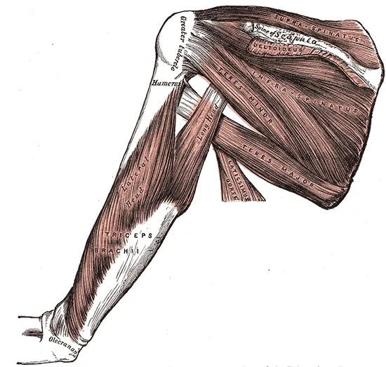
The triceps brachii is the muscle on the back of the upper arm, and it has three parts called heads7. It has long been described as a long, a lateral (outer) and a medial (inner) head that merge into one attachment on the olecranon7. The olecranon is the bony point of the elbow. Together with the small anconeus muscle, the triceps is what straightens the elbow8. A tendon joins muscle to bone, and the distal triceps tendon is where the triceps attaches at the elbow1.
The three heads start in different places1:
| Head | Where it starts |
|---|---|
| Long | The infraglenoid tubercle, a small raised area on the scapula (shoulder blade)1 |
| Lateral | The outer back of the humerus (upper arm bone), above the spiral groove, a channel in the bone1 |
| Medial | The back of the humerus, below that groove1 |
How it attaches to the elbow
A systematic review describes the attachment as a two-layered tendon spread over a wide area of the olecranon tip1. In 22 elbows from donors, the attachment was shaped like a crescent9. A central tendon sat in the middle, with muscle extending onto the bone on each side9.
A dissection of five donor arms, taken from matched pairs, found three separate attachment areas on the olecranon7:
- The back of the elbow joint’s capsule, the sleeve around the joint7.
- A deep, fleshy part formed by the medial head7.
- A surface, tendon-like part formed by the long and lateral heads7.
A review reports that the layout varies between people1. In about half of the donor arms dissected, the medial head had its own tendon part, lying under the other two heads1. In the other half, the heads shared one tendon, with the medial fibres still deepest1.
How the heads share the work
Arm position seems to change which head does most of the work10. Ten healthy people straightened the elbow with the arm held at five different angles10. With the arm at the side, a computer model estimated more force and activity from the long head than from the other two10. With the arm raised to shoulder height or higher, the medial head had the highest estimated force10.
What loading studies actually measure
The studies below did not measure the force inside the tendon itself2,3,4. Some recorded electromyography (EMG), which measures a muscle’s activity by picking up its electrical signals4,11. Others worked out joint moments, the turning effect of a load around the elbow2,3. On this page, more EMG activity or a larger moment is not read as a known tendon load.
Grip width changes the turning effect at the elbow in two pressing lifts2,3:
| Lift | Who was studied | What changed with grip width |
|---|---|---|
| Bench press | 14 trained men, at their maximum | A narrow grip gave a larger moment bending the elbow early in the lift2 |
| Seated shoulder press | 11 resistance-trained men | Wider grips lowered the net moments at the elbow3 |
In the bench press study, the authors state that straightening the elbow is one way lifters resist that bending moment2. In the shoulder press study, narrower grips also let the men lift more and move the elbow through a larger range3. A computer model of both lifts found that the elbow straighteners did little early on but took over near full extension12.
Breakdown
Partial and complete tears
A tear can go part of the way through the tendon or right through it13. Among 801 elbow MRI (magnetic resonance imaging) scans taken over 15 years, 28 people had triceps tendon tears13. Partial tears were the most common, in 18 people, and 10 had complete tears13.
In a review of athletes whose tears were repaired, 58.5% of the reported tears were complete and 41.5% partial5. Where the site of a complete tear was recorded, 25 of 26 were avulsions, where the tendon pulled away from the olecranon5. The other one tore where muscle meets tendon5.
How the tendon tears
Tears are linked to eccentric load, meaning force applied while the muscle is working but being lengthened5,14. In 21 tears among 19 American football professionals, this was the most common way the injury happened14. A review reports that tears in resistance training happen mainly while lowering the bar5. The lifts it names are the bench press and overhead pressing5.
Among athletes with repaired tears, weightlifting was the activity reported most often, at 40.7%5. American football came next, at 26.6%5.
Who tears it
At the largest university hospital in Slovakia, 23 people with the injury were seen over ten years15. The authors put the local rate at 0.46 cases per 100,000 people15. Studies of different groups agree that most people with this tear are men1,5,13,16:
| Group studied | What was found |
|---|---|
| 170 tears pooled in a review | 11 men to each woman1 |
| Pooled results in the same review | Mean age at the tear 47.5 years, from 12 to 751 |
| 277 athletes whose tears were repaired | 96.4% men5 |
| 28 people with tears on elbow MRI | 23 men and 5 women13 |
| 8,143 people with a repair in insurance claims | Mostly men, and mostly aged 40 to 5916 |
What often comes before a tear
Some people have elbow pain before the tendon tears1,5. One review reports that up to a third of people with a tear may have long-standing pain at the back of the elbow1. Tendinopathy is local pain in a tendon that comes with activities that load it. In people with earlier pain, the review reports tissue features of long-standing tendinopathy under the microscope1. Among athletes where this was recorded, 18.7% had discomfort at the back of the elbow before the injury5.
Some other conditions were recorded alongside tears5,17. Anabolic steroids are drugs related to the hormone testosterone. In 262 people who had surgery, the two most common were kidney disease, in 10%, and anabolic steroid use, in 7%17.
A corticosteroid is a drug that copies a type of hormone the body makes. Among 277 athletes, anabolic steroid use was recorded in 9, and an earlier corticosteroid injection at the back of the elbow in 85. These counts describe who was studied; they do not show cause.
Recovery
What the outcome studies describe
The figures below are outcomes in the people each study followed. Most of them had the tendon repaired by surgery5,6. They describe those groups and do not predict how any one person will recover.
Some partial tears in these studies healed without an operation14. Among 10 American football professionals with partial tears, 6 healed without surgery14.
Return to activity after repair
Reviews that pooled several studies report these rates of return after repair5,6,17:
| Group and outcome | Share who returned |
|---|---|
| 262 people, back to the pre-injury level of activity | 89%17 |
| 318 people, back to sport (where recorded) | 93.3%6 |
| 318 people, back to military duty (where recorded) | 95.3%6 |
| 318 people, back to work (where recorded) | 92.6%6 |
| 277 athletes, back to sport (where recorded), after 5.2 months on average | 93.0%5 |
Returning is not the same as returning at the same level5,18. Among athletes where this was recorded, 63.6% went back to their earlier level of competition or higher5. Among professional American football players with triceps tendon tears over 14 seasons, however they were managed, 70.8% returned to play18. Of the players with triceps tears, 41.2% got back to their pre-injury level of performance18.
Strength after repair
Strength in the repaired arm was measured with isokinetic testing, where a machine holds the arm to a fixed speed19. Across 37 published cases, the repaired arm had on average 87% of the other arm’s strength19. In one group of ten, eight were tested an average of 88 months, over seven years, after surgery20. Peak strength averaged 82% of the other arm, while endurance averaged 99%20.
Connections
The muscles it serves
The tendon carries the pull of the three triceps heads to the olecranon7. The anconeus, a small muscle, shares the job of straightening the elbow8. In the bench press, a review describes the biceps brachii (the biceps) as a steadier and as the triceps’ opposing muscle21.
What studies of the bench press show
The traditional model of the bench press names three main movers21. They are the pectoralis major (the large chest muscle), the front deltoid (the front of the shoulder muscle) and the triceps21. EMG studies show how triceps activity compares and shifts:
- Across 14 studies, triceps and pectoralis major activity were similar, and both were higher than front deltoid activity21.
- A review found lifting intensity was the biggest factor, and the triceps was the muscle whose activity changed most often21.
- In 28 men, triceps activity was lower with a wide grip than with medium or narrow grips22.
- In 14 trained men, the medial head was more active with medium and narrow grips than with a wide grip2.
- In 12 men holding the bar still, a narrower grip raised triceps activity, though the author judged the change small23.
- In 30 trained adults, medial head activity was similar at five bench angles, from flat to steeply inclined24.
What studies of the overhead press show
One EMG study calls the overhead press suited to working the triceps, because it pairs raising the arm with straightening the elbow25. In 15 men pressing standing at 80% of their maximum, triceps activity was about 39% higher with a barbell than with dumbbells4. In 14 trained men lowering a barbell, pressing behind the neck brought higher triceps activity than pressing in front26.
Push-ups and dips
Push-ups and dips also work the triceps11,27. In 20 young men, triceps activity was higher than chest activity in standard, diamond and wide push-ups27. Diamond push-ups, with the hands close together, gave the highest activity for both muscles27. In 40 people, a narrow hand position also raised triceps activity compared with a wide one28. In trained adults working to failure, triceps activity while rising in the push-up was lower than in a bench press loaded to match29.
In 13 experienced men, 11 of whom did the ring dip, mean peak triceps activity was recorded in three kinds of dip11. These are group averages, and they do not by themselves show that the dips differ:
| Dip variation | Mean peak triceps EMG |
|---|---|
| Bench dip | 0.83 millivolts11 |
| Bar dip | 1.04 millivolts11 |
| Ring dip | 1.05 millivolts11 |
Distal triceps tendon works with in the bench press
Distal triceps tendon is loaded by
Sources
- Shuttlewood K, Beazley J, Smith CD (2017). Distal triceps injuries (including snapping triceps): A systematic review of the literature.. World Journal of Orthopedics. doi:10.5312/wjo.v8.i6.507 PMID 28660143
- Larsen S, Gomo O, van den Tillaar R (2020). A Biomechanical Analysis of Wide, Medium, and Narrow Grip Width Effects on Kinematics, Horizontal Kinetics, and Muscle Activity on the Sticking Region in Recreationally Trained Males During 1-RM Bench Pressing.. Frontiers in Sports and Active Living. doi:10.3389/fspor.2020.637066 PMID 33554113
- Gundersen AH, Krosshaug T, Mausehund L, van den Tillaar R, Larsen S (2026). The impact of grip width on kinetics and kinematics in the shoulder press among resistance-trained men.. Sports Biomechanics. doi:10.1080/14763141.2025.2590028 PMID 41335596
- Saeterbakken AH, Fimland MS (2013). Effects of body position and loading modality on muscle activity and strength in shoulder presses.. Journal of Strength and Conditioning Research. doi:10.1519/JSC.0b013e318276b873 PMID 23096062
- Vandenberg CJ, Touhey DC, Brophy RH, Smith MV, Knapik DM (2026). Distal triceps tendon rupture repair results in high return to sport rates for amateur and professional athletes: a systematic review.. JSES Reviews, Reports, and Techniques. doi:10.1016/j.xrrt.2026.100694 PMID 41847163
- Johns WL, Miltenberg BH, Baumann AN, Kermanshahi N, Muchintala RR, Cohen SB (2025). Return to Work, Sport, and Sport- Related Activity After Distal Triceps Tendon Repair: A Systematic Review.. The American Journal of Sports Medicine. doi:10.1177/03635465241283970 PMID 39790046
- Barco R, Sánchez P, Morrey ME, Morrey BF, Sánchez-Sotelo J (2017). The distal triceps tendon insertional anatomy-implications for surgery.. JSES Open Access. doi:10.1016/j.jses.2017.05.002 PMID 30675548
- Betts JG, Young KA, Wise JA, Johnson E, Poe B, Kruse DH, Korol O, Johnson JE, Womble M, DeSaix P (2022). Anatomy and Physiology 2e. OpenStax. Link
- Kamineni S, Pooley J, Bachoura A, Yoshida R, Cummings J (2022). Triceps brachii insertional footprint: Under-estimated complexity.. Shoulder & Elbow. doi:10.1177/17585732221135633 PMID 38818100
- Kholinne E, Zulkarnain RF, Sun YC, Lim S, Chun JM, Jeon IH (2018). The different role of each head of the triceps brachii muscle in elbow extension.. Acta Orthopaedica et Traumatologica Turcica. PMID 29503079
- McKenzie A, Crowley-McHattan Z, Meir R, Whitting J, Volschenk W (2022). Bench, Bar, and Ring Dips: Do Kinematics and Muscle Activity Differ?. International Journal of Environmental Research and Public Health. doi:10.3390/ijerph192013211 PMID 36293792
- Evangelista P, Rum L, Picerno P, Biscarini A (2025). Decoding the Contribution of Shoulder and Elbow Mechanics to Barbell Kinematics and the Sticking Region in Bench and Overhead Press Exercises: A Link-Chain Model with Single- and Two-Joint Muscles.. Journal of Functional Morphology and Kinesiology. doi:10.3390/jfmk10030322 PMID 40843853
- Koplas MC, Schneider E, Sundaram M (2011). Prevalence of triceps tendon tears on MRI of the elbow and clinical correlation.. Skeletal Radiology. doi:10.1007/s00256-010-1043-9 PMID 20953605
- Mair SD, Isbell WM, Gill TJ, Schlegel TF, Hawkins RJ (2004). Triceps tendon ruptures in professional football players.. The American Journal of Sports Medicine. doi:10.1177/0095399703258707 PMID 14977669
- Sekáč J, Šagát P, Bartík P, Kilián M, Tohănean DI, Perez J, Vasilcova V, Durdík Š (2024). Distal Triceps Tendon Rupture-First Retrospective Study in Central Europe.. Journal of Clinical Medicine. doi:10.3390/jcm13247792 PMID 39768717
- Lee E, Stillson QA, Seidel HD, Bhattacharjee S, Koh JL, Strelzow JA, Shi LL (2023). Surgical Outcomes, Trends, and Risk Factors of Distal Triceps Repairs.. Hand (New York). doi:10.1177/15589447221095114 PMID 35658641
- Dunn JC, Kusnezov N, Fares A, Rubin S, Orr J, Friedman D, Kilcoyne K (2017). Triceps Tendon Ruptures: A Systematic Review.. Hand (New York). doi:10.1177/1558944716677338 PMID 28832209
- Garlapaty AR, Jones M, Baumann J, Gunn C, Cook JL, DeFroda SF (2025). Characterization of distal biceps tendon and triceps tendon injuries in National Football League players from 2009 to 2022.. Journal of Orthopaedics. doi:10.1016/j.jor.2025.01.033 PMID 39991508
- Tran DV, Yetter TR, Somerson JS (2022). Surgical repair of distal triceps rupture: a systematic review of outcomes and complications.. JSES Reviews, Reports, and Techniques. doi:10.1016/j.xrrt.2022.04.001 PMID 37588859
- van Riet RP, Morrey BF, Ho E, O'Driscoll SW (2003). Surgical treatment of distal triceps ruptures.. The Journal of Bone and Joint Surgery. American Volume. doi:10.2106/00004623-200310000-00015 PMID 14563805
- Stastny P, Gołaś A, Blazek D, Maszczyk A, Wilk M, Pietraszewski P, Petr M, Uhlir P, Zając A (2017). A systematic review of surface electromyography analyses of the bench press movement task.. PLoS One. doi:10.1371/journal.pone.0171632 PMID 28170449
- Saeterbakken AH, Stien N, Pedersen H, Solstad TEJ, Cumming KT, Andersen V (2021). The Effect of Grip Width on Muscle Strength and Electromyographic Activity in Bench Press among Novice- and Resistance-Trained Men.. International Journal of Environmental Research and Public Health. doi:10.3390/ijerph18126444 PMID 34198674
- Lehman GJ (2005). The influence of grip width and forearm pronation/supination on upper-body myoelectric activity during the flat bench press.. Journal of Strength and Conditioning Research. doi:10.1519/R-15024.1 PMID 16095407
- Rodríguez-Ridao D, Antequera-Vique JA, Martín-Fuentes I, Muyor JM (2020). Effect of Five Bench Inclinations on the Electromyographic Activity of the Pectoralis Major, Anterior Deltoid, and Triceps Brachii during the Bench Press Exercise.. International Journal of Environmental Research and Public Health. doi:10.3390/ijerph17197339 PMID 33049982
- Coratella G, Tornatore G, Longo S, Esposito F, Cè E (2022). Front vs Back and Barbell vs Machine Overhead Press: An Electromyographic Analysis and Implications For Resistance Training.. Frontiers in Physiology. doi:10.3389/fphys.2022.825880 PMID 35936912
- Padovan R, Toninelli N, Longo S, Tornatore G, Esposito F, Cè E, Coratella G (2026). High-Density Surface Electromyography Excitation in Front vs. Back Overhead Press Prime Movers.. Journal of Human Kinetics. doi:10.5114/jhk/205466 PMID 42713087
- Intziegianni K, Katsamis E, Michaelides M, Parpa K (2026). Electromyographic Activation of the Pectoralis Major and Triceps Brachii Muscles During Standard, Diamond, and Wide Hand Position Push-Ups.. Muscles. doi:10.3390/muscles5010018 PMID 41892990
- Cogley RM, Archambault TA, Fibeger JF, Koverman MM, Youdas JW, Hollman JH (2005). Comparison of muscle activation using various hand positions during the push-up exercise.. Journal of Strength and Conditioning Research. doi:10.1519/15094.1 PMID 16095413
- Alizadeh S, Rayner M, Mahmoud MMI, Behm DG (2020). Push-Ups vs. Bench Press Differences in Repetitions and Muscle Activation between Sexes.. Journal of Sports Science & Medicine. PMID 32390722
Changes
- Revised to report sources added after their published corrections were read
- Tidy pass: minor review findings fixed, glosses aligned with the reviewed glossary.
- Added a figure showing where it sits, links to the exercise pages that load it, and tighter wording in a few places.
- First published.