Brachialis
The thick muscle under the biceps that bends the elbow. Where it attaches, why forearm turning barely changes its job, how it tears or forms bone, how it grows with training, and what it works with.
- Region
- Upper arm
- Starts on
- the lower half of the front of the humerus2,6
- Ends on
- a rough bump (the ulnar tuberosity) and a bony lip at the front (the coronoid process) of the upper ulna2,6
- Updated
- October 22, 2026
- Reviewed
- October 22, 2026 · how entries are checked
Function
Where it sits in the arm
The brachialis lies in the lower front part of the upper arm, deeper than the biceps brachii1. It is in the arm’s front compartment, with the biceps and the coracobrachialis, a small muscle at the front of the upper arm5. Gray’s 1918 anatomy describes it covering the front of the elbow joint and the lower half of the humerus6. It forms part of the upper part of the antecubital fossa, the hollow at the front of the elbow1.
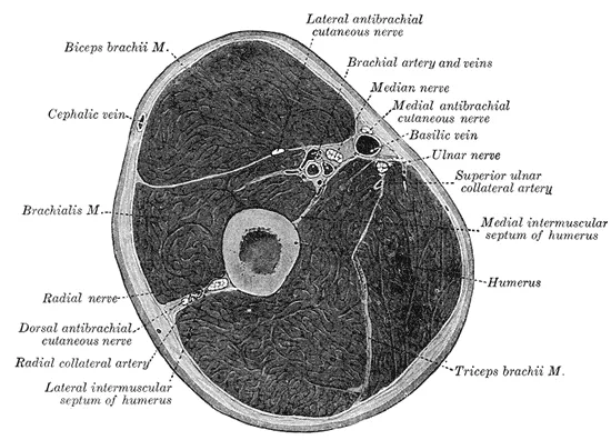
Where it starts and ends
A 2025 review describes a strong, thick muscle running from the lower front of the humerus to the upper ulna2.
In Gray’s account, the muscle starts at the deltoid’s attachment and reaches to within 2.5 cm of the elbow’s joint surface6. It also starts from the intermuscular septa, sheets of tough tissue that separate groups of muscles6. Its fibres meet in a thick tendon that attaches to the ulnar tuberosity, a bump on the ulna6. The tendon also attaches to a rough hollow on the coronoid process6. The coronoid process is a bony lip at the front of the top of the ulna, at the elbow.
Two studies of cadavers, bodies donated for study, measured the attachment on the ulna7,8. Its average length was 26.3 mm in 20 elbows and 27.9 mm in 117,8.
Two heads, described in different ways
A 2007 cadaver study notes that published accounts of brachialis anatomy have conflicted, and that its own findings differ considerably9. Three studies describe two heads, superficial and deep, but differ on where each attaches9,10,11:
- In 11 limbs, the larger superficial head ended in a thick round tendon on the ulnar tuberosity, and the deep head on the coronoid process9.
- In 240 limbs from an adult Sri Lankan population, the superficial head attached to the coronoid process and the deep head to the ulnar tuberosity10.
- In a study of 13 cadaver elbows by MRI, the one muscle that was dissected had two heads, and the superficial head attached to the ulnar tuberosity further down than the deep head11.
The 11-limb authors suggest the superficial head may give most of the muscle’s bending strength9. The smaller deep head may help start bending from a straight elbow9.
What it moves
A StatPearls chapter calls the brachialis an important flexor of the elbow, meaning a muscle that bends it1. It says the muscle bends the elbow in every natural position, and is considered a “pure flexor”1. A 2020 paper explains that it attaches only to the ulna, so it has no role in turning the forearm12. The same paper notes it makes the greatest contractile force of the elbow flexors12.
In an MRI and dynamometer study of four men, the brachialis gave the largest share of the elbow’s bending torque3. Next came the biceps and the brachioradialis, a forearm muscle that bends the elbow3. A dynamometer is a machine that measures turning force.
| Muscle | Share of elbow bending torque in four men (%) |
|---|---|
| Brachialis | 473 |
| Biceps brachii | 343 |
| Brachioradialis | 193 |
How forearm position changes its activity
Studies have used electromyography (EMG), which picks up a muscle’s electrical signals, to compare forearm positions13,14,15. Pronation is turning the palm down, and supination is turning it up. Each row is one study:
| Who was studied | What they did | What was found |
|---|---|---|
| 6 healthy men | Bent the elbow with no weight, in three positions | The brachialis activity pattern was similar palm up, neutral and palm down; the biceps patterns were not13 |
| 9 healthy people | Held the elbow bent while turning the forearm | The biceps was more active palm up; the brachialis and brachioradialis were more active palm down14 |
| 10 weight-trained men | Curled on a preacher bench, palms up and down | With either grip, the brachialis, both biceps heads and the brachioradialis were activated at similar levels to one another16 |
| 15 young healthy men | Bent the elbow as hard as they could | The three muscles were very active with the forearm neutral, fully turned down or fully turned up15 |
Its nerve and blood supply
In Gray’s account, the musculocutaneous nerve, which works the muscles at the front of the upper arm, supplies the brachialis6. It usually gets an extra filament from the radial nerve too6. A 2025 review says its blood comes from branches of the brachial artery and the recurrent radial artery2. In dissection studies, how often the radial nerve shares the supply differs9,10,19:
| Dissection study | Specimens with a radial nerve branch to the brachialis (%) |
|---|---|
| 20 cadaver arms | 6519 |
| 240 cadaver limbs | 8310 |
| 11 cadaver limbs | 1009 |
A different kind of study tested the nerves in 100 patients19. Stimulating the radial nerve drew a response from the brachialis in 90% of them19. On average, the radial nerve’s share of the muscle’s supply was 11%19.
Breakdown
How often it is injured
A 2025 review states that brachialis injuries are rare2. They can come from a contusion (a bruise from a direct blow) or from repeated overuse2. A 2020 case report counted only 10 acute tears reported up to then, all in adults12. A 2019 paper says isolated tears can look like a torn distal biceps tendon or a tumour within the muscle20. That makes them hard to identify20.
Tears in single case reports
A case report is a report on a single patient. Each row is one20,21:
| Who | How it happened | What was found |
|---|---|---|
| 31-year-old competitive male weight lifter | A 40.8 kg biceps curl | The tendon was torn fully through at its attachment on the ulna20 |
| 35-year-old woman | Possibly strenuous gym exercise | An isolated tear, confirmed by MRI and managed without surgery21 |
| 8-year-old girl | A gymnastics move forced her elbows fully straight, or perhaps beyond12 | A complete tear12 |
| 8-year-old boy | A fall on gymnastics bars | A complete rupture22 |
The authors reason that her brachialis would have been doing eccentric work when it tore, meaning muscle work done while the muscle lengthens12.
Overuse in climbers and lifters
Climbing comes up in several reports2,23,24:
- A 1990 report lists brachialis tendinitis among the soft tissue problems that replaced broken bones as climbers’ main injuries23.
- A 2025 review states that repeated palm-down pull-ups in rock climbers may strain the brachialis or cause “climber’s elbow”, a tendinopathy (tendon pain with loading)2.
- A 2023 review of indoor climbing injuries says brachialis injuries are seldom seen outside climbing24.
The 2025 review also notes case reports of brachialis injuries in football, hockey, rugby and weightlifting2.
Bone forming in the muscle
A 2021 review describes myositis ossificans as bone forming in soft tissue after injury25. It names the brachialis as one of its most common sites, with the deltoid, quadriceps and adductors25. A 2017 case report describes an 18-year-old man with no history of trauma26. He had recently started regular gym workouts without supervision26. A growing bony mass in his brachialis then limited elbow motion26.
Recovery
How it grows with training
Training studies have measured brachialis size. Each row is one study4,27:
| Who was studied | Training | What was found |
|---|---|---|
| 5 older men | 12 weeks of heavy resistance training | Brachialis volume and cross-sectional area rose 22.6%, and the biceps 13.9%4 |
| 14 older men | 12 weeks of elbow bending with one arm | The maximal cross-sectional area of the biceps and brachialis together rose 17.4% in the trained arm and did not change in the other28 |
| 21 untrained adults | 12 weeks, incline curls on one arm and preacher curls on the other | Brachialis volume rose 21.1% after preacher curls and 17.1% after incline curls27 |
Cross-sectional area is the size of a slice cut straight across a muscle. In the 21-adult study, each arm did five sets of 10 at 70% of the one-repetition maximum, twice a week27. That maximum is the heaviest load a person can lift once. The biceps grew more after incline curls27. Total elbow flexor growth did not differ between arms, and in both, the biceps and brachialis grew more in their middle region27.
What a single session shows
In a trial of twelve men, each arm was randomly assigned to a fast or a slow curl-type protocol at 60% of the one-repetition maximum29. MRI signal changes showed the biceps was used preferentially in the fast protocol29. The brachialis was used preferentially in the slow one, which spent five times as long lowering as lifting29.
In 16 well-trained young men, four sets of one-arm dumbbell curls to failure swelled the biceps more than the brachialis30. Muscle thickness rose 18.7% in the biceps and 12.3% in the brachialis30. Brachialis swelling was similar after dumbbell curls and dumbbell rows30. Europe PMC’s record of this study lists funding from Brazilian public research agencies; it does not state the funders’ role30.
After a tear
Two single case reports describe the course after a tear20,22. The 31-year-old weight lifter’s tear was managed without surgery20. Ten months after the injury he was back to weightlifting and biceps curls, and he had full strength at final follow-up20. The 8-year-old boy spent 3 weeks in a cast22. Two months after the injury, his elbow motion and daily use had no limits22.
After bone formed in the muscle
A 1992 case report, on a single patient, describes myositis ossificans of the brachialis in a 15-year-old football player31. After nine weeks of an exercise programme, he had regained full elbow motion31. At nine months, the new bone was nearly mature, and his elbow moved fully without pain31. His strength was normal, and he had returned to competition31.
Connections
The biceps above it
When a cup is lifted, the biceps brachii is the prime mover, and the brachialis helps as a synergist32. A synergist is a muscle that helps another make the same movement. The musculocutaneous nerve runs down between the two muscles and supplies both33. In 13 elbows, microscopic examination found no direct link between their tendons11. Above the elbow crease, the median nerve runs in front of the brachialis, between it and the biceps34.
The radial nerve beside it
The radial nerve, the main nerve at the back of the arm and forearm, pierces the lateral intermuscular septum35,36. It then runs between the brachioradialis and the brachialis35,36.
The humerus and the elbow joint
The brachialis starts on the humerus and covers the front of the elbow joint6. Sources differ on its link to the joint capsule, the sleeve of tough tissue that encloses a joint7,11. In 20 elbows, it attached through muscle and a tendon sheet to the capsule, the coronoid process and the upper ulna7. In 13 elbows, microscopic examination found no direct link between its tendon and the capsule11.
In nine cadaver elbows, brachialis strain rose under varus (inward-bending) forces, peaking at 30 degrees of bend with the palm down37. It rose significantly with both collateral ligaments cut, and the authors suggest the muscle may help steady the elbow against these forces37.
Exercises that load it
Related exercise pages: biceps curl and pull-up32,38. Studies of these exercises found these results:
| Exercise | What studies found |
|---|---|
| Biceps curl | In 21 untrained adults, brachialis volume grew more after 12 weeks of preacher curls (21.1%) than incline curls (17.1%)27 |
| Pull-up | In a computer model fed by motion tracking of 11 men with no history of shoulder pathology, front-grip pull-ups favoured the biceps and brachialis38 |
Brachialis lies beneath
Brachialis is supplied by branches of
Brachialis starts on
Brachialis crosses
Brachialis lies next to
Brachialis is loaded by
Sources
- Plantz MA, Bordoni B (2023). Anatomy, Shoulder and Upper Limb, Brachialis Muscle.. StatPearls. PMID 31869094
- Manske RC, Wolfe C, Page P, Voight M (2025). Diagnostic Musculoskeletal Ultrasound in the Evaluation of the Brachialis.. International journal of sports physical therapy. doi:10.26603/001c.142362 PMID 40756795
- Kawakami Y, Nakazawa K, Fujimoto T, Nozaki D, Miyashita M, Fukunaga T (1994). Specific tension of elbow flexor and extensor muscles based on magnetic resonance imaging.. European journal of applied physiology and occupational physiology. doi:10.1007/BF00244027 PMID 8194543
- Roman WJ, Fleckenstein J, Stray-Gundersen J, Alway SE, Peshock R, Gonyea WJ (1993). Adaptations in the elbow flexors of elderly males after heavy-resistance training.. Journal of applied physiology (Bethesda, Md. : 1985). doi:10.1152/jappl.1993.74.2.750 PMID 8458791
- Bounds EJ, Frane N, Jajou L, Weishuhn LJ, Kok SJ (2023). Humeral Shaft Fractures.. StatPearls. PMID 28846218
- Gray H; Lewis WH (editor) (1918). Anatomy of the Human Body. Lea & Febiger, Philadelphia (20th edition). Link
- Cage DJ, Abrams RA, Callahan JJ, Botte MJ (1995). Soft tissue attachments of the ulnar coronoid process. An anatomic study with radiographic correlation.. Clinical orthopaedics and related research. PMID 7586820
- Cucchi D, Luceri F, Menon A, Müller LP, Kabir K, Randelli PS, Arrigoni P, Wegmann K (2022). Anatomic relations of the median nerve to the ulnar insertion of the brachialis muscle: safety issues and implications for medial approaches to the elbow joint.. Archives of orthopaedic and trauma surgery. doi:10.1007/s00402-021-03753-y PMID 33484309
- Leonello DT, Galley IJ, Bain GI, Carter CD (2007). Brachialis muscle anatomy. A study in cadavers.. The Journal of bone and joint surgery. American volume. doi:10.2106/JBJS.F.00343 PMID 17545433
- Ilayperuma I, Uluwitiya SM, Nanayakkara BG, Palahepitiya KN (2019). Re-visiting the brachialis muscle: morphology, morphometry, gender diversity, and innervation.. Surgical and radiologic anatomy : SRA. doi:10.1007/s00276-019-02182-2 PMID 30820647
- Sanal HT, Chen L, Negrao P, Haghighi P, Trudell DJ, Resnick DL (2009). Distal attachment of the brachialis muscle: anatomic and MRI study in cadavers.. AJR. American journal of roentgenology. doi:10.2214/AJR.08.1150 PMID 19155412
- Forsythe CM, Burtis MT, Pfeiffer DC, Spurling GR, Ashby DB (2020). Acute brachialis muscle tear in a pediatric patient.. Radiology case reports. doi:10.1016/j.radcr.2020.01.041 PMID 32140195
- Date S, Kurumadani H, Nakashima Y, Ishii Y, Ueda A, Kurauchi K, Sunagawa T (2021). Brachialis Muscle Activity Can Be Measured With Surface Electromyography: A Comparative Study Using Surface and Fine-Wire Electrodes.. Frontiers in physiology. doi:10.3389/fphys.2021.809422 PMID 35002781
- Naito A, Sun YJ, Yajima M, Fukamachi H, Ushikoshi K (1998). Electromyographic study of the elbow flexors and extensors in a motion of forearm pronation/supination while maintaining elbow flexion in humans.. The Tohoku journal of experimental medicine. doi:10.1620/tjem.186.267 PMID 10328159
- Caufriez B, Dugailly PM, Brassinne E, Schuind F (2018). The Role of the Muscle Brachioradialis in Elbow Flexion: An Electromyographic Study.. Journal of Hand Surgery Asian-Pacific Volume. doi:10.1142/S2424835518500145 PMID 29409427
- Bankoff AD, Gushi MS, Boer NP (2007). Electromyographic study of the flexor muscles of the elbow articulation in weightlifting trained subjects.. Electromyography and clinical neurophysiology. PMID 17375882
- Coratella G, Tornatore G, Longo S, Toninelli N, Padovan R, Esposito F, Cè E (2023). Biceps Brachii and Brachioradialis Excitation in Biceps Curl Exercise: Different Handgrips, Different Synergy.. Sports (Basel). doi:10.3390/sports11030064 PMID 36976950
- Staudenmann D, Taube W (2015). Brachialis muscle activity can be assessed with surface electromyography.. Journal of electromyography and kinesiology : official journal of the International Society of Electrophysiological Kinesiology. doi:10.1016/j.jelekin.2014.11.003 PMID 25468488
- Bendersky M, Bianchi HF (2012). Double innervation of the brachialis muscle: anatomic-physiological study.. Surgical and radiologic anatomy : SRA. doi:10.1007/s00276-012-0977-0 PMID 22584807
- Curry EJ, Cusano A, Elattar O, Bogart A, Murakami A, Li X (2019). Brachialis Muscle Tendon Rupture of the Distal Ulnar Attachment in a Competitive Weight Lifter.. Orthopedics. doi:10.3928/01477447-20190221-04 PMID 30810756
- Sharma P, Mehta N, Narayan A (2017). Isolated Traumatic Brachialis Muscle Tear: A Case Report and Review of Literature.. Bulletin of emergency and trauma. doi:10.18869/acadpub.beat.5.4.476 PMID 29177180
- Yamaji A, Uesugi M, Kamada H, Ichimura H, Yamazaki M (2022). Brachialis Muscle Rupture in a Pediatric Patient Followed Up by Ultrasound Examinations: A Rare Case Report.. Case reports in orthopedics. doi:10.1155/2022/3391350 PMID 35783672
- Bollen SR (1990). Upper limb injuries in elite rock climbers.. Journal of the Royal College of Surgeons of Edinburgh. PMID 2079693
- Leung J (2023). A Guide to Indoor Rock Climbing Injuries.. Current sports medicine reports. doi:10.1249/JSR.0000000000001036 PMID 36757124
- Savvidou O, Papakonstantinou O, Lakiotaki E, Melissaridou D, Korkolopoulou P, Papagelopoulos PJ (2021). Post-traumatic myositis ossificans: a benign lesion that simulates malignant bone and soft tissue tumours.. EFORT Open Reviews. doi:10.1302/2058-5241.6.210002 PMID 34377549
- Galanis N, Stavraka C, Valavani E, Kirkos J (2017). Unsupervised Exercise-Induced Myositis Ossificans in the Brachialis Muscle of a Young Healthy Male: A Case Report.. Orthopaedic journal of sports medicine. doi:10.1177/2325967117718780 PMID 28795074
- Kobayashi Y, Maeo S, Eihara Y, Ono M, Sato M, Sugiyama T, Kanehisa H, Isaka T (2026). Exercise-specific preferential hypertrophy of individual elbow flexor muscles after incline versus preacher dumbbell curl training.. Journal of sports sciences. doi:10.1080/02640414.2026.2741751 PMID 42829982
- Brown AB, McCartney N, Sale DG (1990). Positive adaptations to weight-lifting training in the elderly.. Journal of applied physiology (Bethesda, Md. : 1985). doi:10.1152/jappl.1990.69.5.1725 PMID 2272965
- Kulig K, Powers CM, Shellock FG, Terk M (2001). The effects of eccentric velocity on activation of elbow flexors: evaluation by magnetic resonance imaging.. Medicine and science in sports and exercise. doi:10.1097/00005768-200102000-00004 PMID 11224805
- Leitão BFM, Albarello JCS, Halmenschlager GH, de Salles BF, Matta TT (2024). Exercise selection plays an important role in inducing non-uniform acute responses on elbow flexors muscle thickness.. Journal of bodywork and movement therapies. doi:10.1016/j.jbmt.2024.07.028 PMID 39593419
- De Carlo MS, Misamore GW, Carrell KR, Sell KE (1992). Rehabilitation of myositis ossificans in the brachialis muscle.. Journal of athletic training. PMID 16558137
- Betts JG, Young KA, Wise JA, Johnson E, Poe B, Kruse DH, Korol O, Johnson JE, Womble M, DeSaix P (2022). Anatomy and Physiology 2e. OpenStax. Link
- Desai SS, Arbor TC, Varacallo MA (2023). Anatomy, Shoulder and Upper Limb, Musculocutaneous Nerve.. StatPearls. PMID 30480938
- Binsaleem S (2025). Median nerve entrapment neuropathy: a review on the pronator syndrome.. JSES Reviews, Reports, and Techniques. doi:10.1016/j.xrrt.2024.10.001 PMID 39872334
- Węgiel A, Karauda P, Zielinska N, Tubbs RS, Olewnik Ł (2023). Radial nerve compression: anatomical perspective and clinical consequences.. Neurosurgical review. doi:10.1007/s10143-023-01944-2 PMID 36781706
- Latef TJ, Bilal M, Vetter M, Iwanaga J, Oskouian RJ, Tubbs RS (2018). Injury of the Radial Nerve in the Arm: A Review.. Cureus. doi:10.7759/cureus.2199 PMID 29666777
- Ott N, Harland A, Knevels M, Hackl M, Leschinger T, Lanzerath F, Scaal M, Wegmann K, Müller LP (2021). The role of the brachialis muscle in elbow stability with collateral ligament injury: A biomechanical investigation.. Clinical biomechanics (Bristol, Avon). doi:10.1016/j.clinbiomech.2021.105478 PMID 34517193
- Urbanczyk CA, Prinold JAI, Reilly P, Bull AMJ (2020). Avoiding high-risk rotator cuff loading: Muscle force during three pull-up techniques.. Scandinavian journal of medicine & science in sports. doi:10.1111/sms.13780 PMID 32715526
Changes
- First published.