Extensor carpi ulnaris tendon
Also called ECU tendon, ECU, sixth extensor compartment
The tendon on the little-finger side of the back of the wrist. How a groove and a small sleeve hold it, how it slips or wears in racquet and club sports, and what case series found afterwards.
- Region
- Wrist & hand
- Muscles
- extensor carpi ulnaris
- Updated
- October 14, 2026
- Reviewed
- October 14, 2026 · how entries are checked
Function
Where the tendon runs
The extensor carpi ulnaris is a forearm muscle on the little-finger side that pulls the wrist back and toward that side4,5. It starts from the lateral epicondyle, the outer bony bump at the elbow, and from the back border of the ulna4,5. Its tendon ends on the base of the fifth metacarpal, the long hand bone at the base of the little finger4,5.
As it leaves the forearm, the tendon passes through a tunnel of bone and fibre called the sixth extensor compartment1. The tunnel lies in a groove on the back of the ulna1. Beyond the end of the ulna, the tendon lies over part of the triangular fibrocartilage complex (TFCC) and the triquetrum, a wrist bone1.
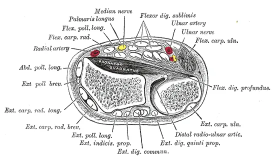
What holds it in the groove
Two layers hold the tendon1. The outer one is the extensor retinaculum1. Over this tendon it has no attachment to the ulna1. Its main job is described as stopping the tendon from lifting away from the wrist when the muscle contracts1.
The inner layer is the subsheath, which lies under the retinaculum and is anchored to the far end of the ulna1. In 12 cadaver (donated body) arms it was roughly 1 cm square6. The subsheath is listed as one part of the TFCC7. The outer edge of that complex blends with the subsheath where it attaches to the ulna7.
Cadaver studies have cut the subsheath in stages8,9:
- Far half. In 10 specimens, cutting the far half markedly increased how far the tendon moved, and cutting the near half did not8.
- Far extension. In eight arms, cutting the subsheath alone did not significantly increase subluxation, a partial slip of the tendon out of its usual place9. Also cutting an extension of it further along did, with the forearm turned palm-down9.
- Intact subsheath. In 7 of 10 specimens, the tendon left its groove in at least one forearm and wrist position with the subsheath whole8.
What the muscle does through it
The extensor carpi ulnaris pulls the wrist back and tilts the hand toward the little-finger side4,5. Supination is turning the forearm so the palm faces up, and pronation is turning it so the palm faces down. One review says the tendon adds more to pulling the wrist back with the forearm fully supinated, and less with it pronated1.
A study of muscle activity gives a different picture. Electromyography (EMG) measures a muscle’s activity by picking up its electrical signals. In 40 healthy arms producing a constant wrist extension effort, ECU activity did not change between pronation, neutral and supination10.
The muscle and tendon are also described as steadying the joint between the two forearm bones at the wrist during forearm rotation11. In 8 cadaver arms with the TFCC cut, loading the ECU tendon alone reduced back-to-front movement at that joint in supination and neutral rotation12.
How forearm position changes the load
In full pronation the tendon leaves the wrist in a straight line1. In supination it leaves the sixth compartment at an angle of about 30 degrees1. In nine cadaver arms, tunnel pressure around the tendon was 27 times higher during what the authors call the carpal supination test than in neutral13.
Breakdown
Four kinds of problem
One review groups ECU tendon problems into four kinds, which can occur alone or together1:
- Tenosynovitis, swelling of the lining that surrounds the tendon1.
- Tendinopathy, whose cause here is described as unclear1,14.
- Tendon disruption1. Rupture of a normal ECU tendon is described as very unusual1.
- Tendon instability, which ranges from a minor slip during rotation to full dislocation with the forearm locking1.
Instability follows a torn or failing subsheath1. Sudden tears usually happen in a single injury, and less often the subsheath gives way under repeated stress1. The retinaculum above it is described as often intact15. A systematic review, a study that searches for the research on a question in a set, checkable way, calls instability relatively rare3. It adds that the exact rate is unknown3.
Which sports the reports come from
ECU problems have been reported most often in tennis and golf1. Injuries to the structures that hold the tendon also occur in contact sports such as rugby league1. A review describes those structures as at particular risk of sudden injury in one position1. In it, the forearm rotates while the wrist is held bent forward and tilted toward the little finger1.
- Tennis. One paper described 28 cases of ECU-related wrist pain in elite players16. Twelve had sudden instability, 14 had tendinopathy of gradual onset and 2 had a complete rupture16. Ten of the 12 instability cases happened during a double-handed backhand16.
- Golf. Among 128 European Tour golfers studied at one 2009 championship, 38 reported 43 wrist problems17. These included 8 cases of ECU tendinosis (tendon wear) and 4 of ECU subluxation17. Subluxation was the wrist injury that cost the most absence from competition17. The European Tour paid the study’s expenses17.
- Rugby league. Unpublished data cited in a 2013 review record six sudden subsheath injuries over five years in seven English Super League clubs1.
What scans show, with and without symptoms
Several studies scanned wrists that were not painful2,18,19. In 26 symptom-free recreational tennis players, ultrasound showed structural abnormalities of the ECU tendon in 39 of 52 wrists2. Moving ultrasound found the tendon unstable in 22 of the 52 wrists2. Abnormalities meant findings such as tendon wear, sheath swelling and tears, and instability meant subluxation or dislocation2. Its criteria were set in advance, but the abstract does not give them2.
| Who was scanned | What was found |
|---|---|
| 40 symptom-free wrists of healthy volunteers | The tendon moved up to 50% beyond the edge of its groove, on the study’s own measure19 |
| 755 clinic visitors with no active wrist complaint | 50 had a snapping tendon on a provocation test; ultrasound of those 50 showed subluxation in 19 and dislocation in 318 |
| MRI reports from one institution, 2001 to 201420 | 31 of 556 patients with ECU signal changes had clinically suspected ECU tendinopathy20 |
The 705 people without snapping in the clinic study had no ultrasound18. A retrospective MRI study followed 16 people with athletic subsheath injuries21. Repeat MRI tended to show the tendon settling within its groove over time21. The authors of one paper suggest that the tendon’s displacement itself may not be the source of complaints22.
What is found alongside it
A review notes that ECU tendinosis can coexist with problems in other structures on the little-finger side of the wrist, the TFCC among them1. One study took 133 patients who had wrist MRI and keyhole surgery7. MRI showed an ECU abnormality in 84.9% of those with a tear at the outer edge of the TFCC7. The figure was 19.6% in those with no tear7.
Studies disagree on whether a shallow groove matters. Two cadaver studies, of 10 and 9 specimens, found no significant link between groove depth and how far the tendon moved out6,8. An ultrasound study of eight cadaver arms linked a shallower groove with greater subluxation9.
Recovery
Series followed without an operation
A review describes tears on the thumb side of the subsheath as less likely to heal, because the tendon lies outside the torn sheath23.
The tennis paper reported what happened to its elite players16:
- Gradual tendinopathy, 14 cases. Pain resolved within 2 to 24 weeks in most, and four were lost to follow-up16.
- Fully displaced tendon, five players. The wrist was held in plaster for two to three months, with no operation16. They were back in competition five to six months after the injury, without symptoms16.
- Subsheath detached without rupture, three players. The authors’ plan for this injury was six to eight weeks or more in plaster16. Their three players were back in competition after about four months16.
Of the 12 players with sudden instability, eight were followed to return and four were lost16. A chart review took 15 patients with ECU tenosynovitis who first had splinting and a steroid injection24. That approach failed in 7, who went on to a surgical release24. A 2013 review states that no evidence supports one type of cast over another for resting the wrist in ECU tendinosis1.
Series followed after an operation
A systematic review of operations for ECU instability after injury found eight retrospective studies covering 97 wrists3. A retrospective study looks back at past cases or records. The review classed its studies as low-level evidence with small samples, of 5 to 21 wrists each3. It concluded that the evidence does not allow one technique to be called better than another3. The review was registered in advance and reports no specific grant3.
All eight studies first rested the wrist for 2 to 8 weeks, in no set way, with anti-inflammatory medicine3. Symptoms had lasted 1.5 to 17 months before the operation, 8.6 on average3.
Across those studies, complications occurred in 9% of patients and no study reported a recurrence or a repeat rupture3. Five patients did not get back to the level of activity they had before3. Two studies reported how long return to sport or activity took: 2.6 to 8 months3. Two studies compared values before and after, and both reported significant improvement in pain, function, satisfaction and grip3.
| Series | What it reported |
|---|---|
| 2020: 15 patients, subsheath rebuilt with a retinaculum sling (pooled) | About 1 in 3 had a complication or a further operation. At a median of 8 years most were satisfied, though many had some symptoms left25 |
| 2024: 14 elite professional or college athletes, retinaculum sling | Mean time to unrestricted return to sport was 92.5 days, with no complications26 |
| 2019: 12 patients, retinaculum flap (pooled) | MRI showed an unchanged displacement pattern in five afterwards; the authors report good to excellent results wherever the tendon sat22 |
| 2008: 21 patients, sheath reattached and groove deepened (pooled) | Wrist movement, grip strength, pain and satisfaction improved significantly at 24 to 45 months27 |
The 2008 series says it compared values before and after in a prospective study, one that follows people forward in time27. The 2024 review counts the same series as a retrospective case series3. A separate 2024 paper notes the limited number of prospective studies of the options for managing ECU problems15.
Connections
The muscle and its neighbours
The tendon carries the pull of the extensor carpi ulnaris, covered with the other forearm extensors4,5. One reference text has the muscle starting from the outer elbow through the common extensor tendon4,5. A study of 10 cadaver elbows found its tendon starting lower and further back28.
At the wrist, a review names it among six forearm muscles that act on the wrist alone29. Its subsheath is counted as part of the triangular fibrocartilage complex in each of 24 anatomical studies in one scoping review30.
What exercise and stretch studies measured
Related exercise pages: wrist extension and the wrist extensor stretch10,31. In 40 healthy arms holding a wrist-extension effort, extensor carpi ulnaris activity did not change with forearm position10. In four cadaver elbows, stretching the extensor carpi ulnaris had no significant effect on force in the common extensor tendon31.
Extensor carpi ulnaris tendon carries force from
Extensor carpi ulnaris tendon is held by a sleeve counted as part of
Extensor carpi ulnaris tendon crosses
Extensor carpi ulnaris tendon belongs to a muscle that starts from
Extensor carpi ulnaris tendon is loaded by
Extensor carpi ulnaris tendon is stretched by
Sources
- Campbell D, Campbell R, O'Connor P, Hawkes R (2013). Sports-related extensor carpi ulnaris pathology: a review of functional anatomy, sports injury and management.. British journal of sports medicine. doi:10.1136/bjsports-2013-092835 PMID 24096897
- Sole JS, Wisniewski SJ, Newcomer KL, Maida E, Smith J (2015). Sonographic evaluation of the extensor carpi ulnaris in asymptomatic tennis players.. PM & R : the journal of injury, function, and rehabilitation. doi:10.1016/j.pmrj.2014.08.951 PMID 25217825
- Lari A, Burhamah W, Alherz M, Prada C (2024). Operative Management of Extensor Carpi Ulnaris Instability: A Systematic Review.. Journal of wrist surgery. doi:10.1055/s-0043-1764354 PMID 38808191
- Sawyer E, Sajjad H, Tadi P (2023). Anatomy, Shoulder and Upper Limb, Forearm Extensor Carpi Ulnaris Muscle.. StatPearls. PMID 30969582
- Gray H; Lewis WH (editor) (1918). Anatomy of the Human Body. Lea & Febiger, Philadelphia (20th edition). Link
- Shoap SC, Dennis ER, Lombardi JM, Wilkerson J, Wahood M, Rosenwasser MP (2022). An Analysis of Extensor Carpi Ulnaris Groove Morphology and Tendon Instability.. Hand (New York, N.Y.). doi:10.1177/15589447221105539 PMID 35815616
- Nevalainen MT, Zoga AC, Rivlin M, Morrison WB, Roedl JB (2023). Extensor carpi ulnaris tendon pathology and ulnar styloid bone marrow edema as diagnostic markers of peripheral triangular fibrocartilage complex tears on wrist MRI: a case-control study.. European radiology. doi:10.1007/s00330-023-09446-x PMID 36809434
- Ghatan AC, Puri SG, Morse KW, Hearns KA, von Althann C, Carlson MG (2016). Relative Contribution of the Subsheath to Extensor Carpi Ulnaris Tendon Stability: Implications for Surgical Reconstruction and Rehabilitation.. The Journal of hand surgery. doi:10.1016/j.jhsa.2015.10.024 PMID 26691954
- Inoue T, Iida A, Omokawa S, Kawamura K, Mahakkanukrauh P, Tanaka Y (2025). Ultrasonographic Evaluation of Extensor Carpi Ulnaris Tendon Subluxation at the Ulnar Groove.. The Journal of hand surgery. doi:10.1016/j.jhsa.2025.09.006 PMID 41071141
- Ikeda K, Kaneoka K, Matsunaga N, Ikumi A, Yamazaki M, Yoshii Y (2025). Effects of forearm rotation on wrist flexor and extensor muscle activities.. Journal of orthopaedic surgery and research. doi:10.1186/s13018-024-05363-x PMID 39819577
- Kalson NS, Malone PS, Bradley RS, Withers PJ, Lees VC (2012). Fibre bundles in the human extensor carpi ulnaris tendon are arranged in a spiral.. The Journal of hand surgery, European volume. doi:10.1177/1753193411433228 PMID 22190565
- Iida A, Omokawa S, Moritomo H, Aoki M, Wada T, Kataoka T, Tanaka Y (2012). Biomechanical study of the extensor carpi ulnaris as a dynamic wrist stabilizer.. The Journal of hand surgery. doi:10.1016/j.jhsa.2012.07.042 PMID 23123149
- Kataoka T, Moritomo H, Omori S, Iida A, Omokawa S, Suzuki D, Fujimiya M, Wada T, Aoki M, Yoshikawa H (2015). Pressure and tendon strain in the sixth extensor compartment of the wrist during simulated provocative maneuvers for diagnosing extensor carpi ulnaris tendinitis.. Journal of orthopaedic science : official journal of the Japanese Orthopaedic Association. PMID 26377977
- Zarro M, Goel R, Bickhart N, May CC, Abzug JM (2022). Extensor Carpi Ulnaris Tendinopathy in Athletes: A Review of the Conservative and Rehabilitative Options.. Hand (New York, N.Y.). doi:10.1177/15589447221127331 PMID 36250572
- Thirumavalavan J, Ibrahim Z, Byrne RA, Arant KR, Gil JA (2024). Extensor Carpi Ulnaris Instability: A Comprehensive Review of Pathology and Operative Techniques.. Hand (New York, N.Y.). doi:10.1177/15589447231168908 PMID 37226412
- Montalvan B, Parier J, Brasseur JL, Le Viet D, Drape JL (2006). Extensor carpi ulnaris injuries in tennis players: a study of 28 cases.. British journal of sports medicine. doi:10.1136/bjsm.2005.023275 PMID 16632573
- Hawkes R, O'Connor P, Campbell D (2013). The prevalence, variety and impact of wrist problems in elite professional golfers on the European Tour.. British journal of sports medicine. doi:10.1136/bjsports-2012-091917 PMID 24014125
- Erpala F, Ozturk T (2021). "Snapping" of the extensor carpi ulnaris tendon in asymptomatic population.. BMC musculoskeletal disorders. doi:10.1186/s12891-021-04271-z PMID 33902526
- Lee KS, Ablove RH, Singh S, De Smet AA, Haaland B, Fine JP (2009). Ultrasound imaging of normal displacement of the extensor carpi ulnaris tendon within the ulnar groove in 12 forearm-wrist positions.. AJR. American journal of roentgenology. doi:10.2214/AJR.08.1725 PMID 19696277
- Kuntz MT, Janssen SJ, Ring D (2015). Incidental signal changes in the extensor carpi ulnaris on MRI.. Hand (New York, N.Y.). doi:10.1007/s11552-015-9764-9 PMID 26568735
- Jeantroux J, Becce F, Guerini H, Montalvan B, Le Viet D, Drapé JL (2011). Athletic injuries of the extensor carpi ulnaris subsheath: MRI findings and utility of gadolinium-enhanced fat-saturated T1-weighted sequences with wrist pronation and supination.. European radiology. doi:10.1007/s00330-010-1887-3 PMID 20680292
- Peter K, Luzian H, Markus G, Ansgar R, Andrea K, Arora R (2019). Mid-term outcome (11-90 months) of the extensor retinaculum flap procedure for extensor carpi ulnaris tendon instability.. Archives of orthopaedic and trauma surgery. doi:10.1007/s00402-019-03227-2 PMID 31222435
- Byrd JN, Sasor SE, Chung KC (2021). Extensor Carpi Ulnaris Subluxation.. Hand clinics. doi:10.1016/j.hcl.2021.06.005 PMID 34602128
- Crimmins CA, Jones NF (1995). Stenosing tenosynovitis of the extensor carpi ulnaris.. Annals of plastic surgery. doi:10.1097/00000637-199507000-00020 PMID 7574276
- Verhiel SHWL, Özkan S, Chen NC, Jupiter JB (2020). Long-Term Outcomes After Extensor Carpi Ulnaris Subsheath Reconstruction With Extensor Retinaculum.. Techniques in hand & upper extremity surgery. doi:10.1097/BTH.0000000000000257 PMID 31343594
- Mastroianni MA, Leibman M, Belsky M, Vitale MA, Ruchelsman DE (2024). Radially Based Extensor Retinacular Sling Reconstruction for Extensor Carpi Ulnaris Subsheath Injuries in Elite Athletes.. Hand (New York, N.Y.). doi:10.1177/15589447231151433 PMID 36779484
- MacLennan AJ, Nemechek NM, Waitayawinyu T, Trumble TE (2008). Diagnosis and anatomic reconstruction of extensor carpi ulnaris subluxation.. The Journal of hand surgery. doi:10.1016/j.jhsa.2007.10.002 PMID 18261666
- Zoner CS, Buck FM, Cardoso FN, Gheno R, Trudell DJ, Randall TD, Resnick D (2010). Detailed MRI-anatomic study of the lateral epicondyle of the elbow and its tendinous and ligamentous attachments in cadavers.. AJR. American journal of roentgenology. doi:10.2214/AJR.09.3173 PMID 20729438
- Eschweiler J, Li J, Quack V, Rath B, Baroncini A, Hildebrand F, Migliorini F (2022). Anatomy, Biomechanics, and Loads of the Wrist Joint.. Life (Basel, Switzerland). doi:10.3390/life12020188 PMID 35207475
- van der Post AS, Jens S, Daams JG, Obdeijn MC, Maas M, Oostra RJ (2022). The triangular fibrocartilage complex in the human wrist: A scoping review toward uniform and clinically relevant terminology.. Clinical anatomy (New York, N.Y.). doi:10.1002/ca.23880 PMID 35396731
- Erak S, Day R, Wang A (2004). The role of supinator in the pathogenesis of chronic lateral elbow pain: a biomechanical study.. Journal of hand surgery (Edinburgh, Scotland). doi:10.1016/j.jhsb.2004.06.001 PMID 15336750
Changes
- First published.