Triangular fibrocartilage complex
Also called TFCC, triangular fibrocartilage, TFC, articular disc of the wrist
The disc and ligaments on the little-finger side of the wrist. What they are made of, how they share load and steady the forearm bones, how they wear and tear, and what studies found about healing.
- Region
- Wrist & hand
- Updated
- October 14, 2026
- Reviewed
- October 14, 2026 · how entries are checked
Function
Where it sits and what it is made of
The triangular fibrocartilage complex (TFCC) sits on the ulnar (little-finger) side of the wrist1. It arises from the radius (the forearm bone on the thumb side)1. It attaches to the far end of the ulna (the forearm bone on the little-finger side) and to the wrist bones on that side1. The end of the ulna does not touch any carpal bone (small bone of the wrist) directly4. The figure labels the central part “Articular disc”.
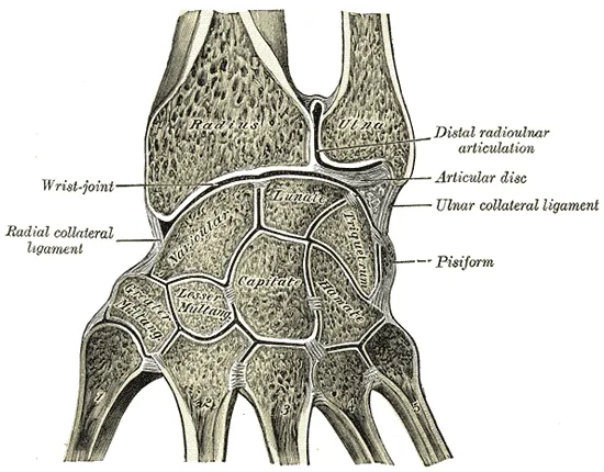
The paper that named the TFCC studied 61 specimens by dissection and biomechanical testing2,3. It described one continuous structure whose parts could not be dissected apart2,3. The parts it listed were:
- the articular disc2,3;
- the dorsal (back) and volar (palm-side) radioulnar ligaments2,3;
- the meniscus homologue2,3;
- the ulnar collateral ligament of the wrist2,3;
- the sheath of the extensor carpi ulnaris, a forearm muscle on the little-finger side2,3.
A scoping review of 24 anatomical studies found that all of them counted four of those parts2. These were the radioulnar ligaments, the disc, the extensor carpi ulnaris sheath or subsheath, and the meniscus homologue2. The greatest disagreement was over the ulnar collateral ligament2. The review’s authors concluded that this ligament is quite likely not a distinct structure2. A study of eight cadavers found the meniscus homologue to be dense connective tissue with no identity of its own under the microscope9.
The disc and its attachments
The articular disc is made of fibrocartilage, according to four anatomical studies summarised in the scoping review2. Seen from above it is triangular, and one anatomical study found its radial part thin and its ulnar part thick2.
On the radial side, sections from eight cadavers showed the disc attached to the hyaline cartilage (a smooth, glassy kind of cartilage) of the radius9. On the ulnar side, serial sections showed the radioulnar ligament arising from two places10. One is the base of the styloid process (a bony point), and the other is the fovea10. The fovea is a hollow between the head of the ulna and its styloid process2.
The subsheath of the extensor carpi ulnaris tendon is listed as one part of the TFCC11. The outer edge of the TFCC blends with the subsheath where it attaches to the ulna11.
How it shares load
A review describes the TFCC as a load-bearing structure that passes and spreads axial (lengthwise) load from the carpal bones to the ulna12. One laboratory study measured the ulna’s share of that load, and what happened when the wrist was altered13.
| Wrist in the laboratory study | Ulna’s share of the axial load |
|---|---|
| Intact | 18.4%13 |
| Articular disc removed | 6.2%13 |
| Ulna lengthened by 2.5 mm | 41.9%13 |
| Ulna shortened by 2.5 mm | 4.3%13 |
Those figures describe an ulna whose length was changed in the laboratory13. Ulnar variance is the relative length of the ulna at the wrist14. In 58 cadaver forearms, the force through the lower ulna was only weakly related to natural ulnar variance (r = 0.44)15. The authors put that down to a thicker triangular fibrocartilage in arms with a shorter ulna15.
In nine cadaver forearms, the force through the lower ulna showed no relationship to ulnar variance14. In those forearms the triangular fibrocartilage was thinner where the ulna was relatively longer14.
On radiographs (X-ray images) of 120 healthy volunteers, ulnar variance was greatest when gripping in pronation, with the palm turned down16. It was least when relaxed in supination, with the palm turned up, a mean change of 1.34 mm16. In nine cadaver forearms, the axial force through the lower ulna was greatest in pronation14.
How it steadies the forearm bones
In 16 fresh-frozen cadaver arms, the ligament parts of the TFCC were the dominant stabilisers of the distal radioulnar joint17. The palmar radioulnar ligament was the major restraint on the ulna shifting toward the back of the wrist17. The dorsal ligament was the primary restraint on it shifting toward the palm17.
Blood and nerve supply
In 10 cadaver specimens, small vessels entered the TFCC from the joint capsule (the sleeve of tough tissue that encloses a joint)18. They supplied its outer 10% to 40%18. The inner portion had no blood vessels, and no vessels crossed its attachment to the radius18. Two cadaver studies found that the outer 15% to 20% of the articular disc had blood vessels and the rest had none5,6.
Breakdown
Wear with age
A study of 100 fresh cadavers (180 wrist joints) concluded that degeneration of the triangular fibrocartilage begins in the third decade of life19. The changes ran from fewer cells and fraying through thinning to perforation, a hole through the disc19. They were always in the central part of the disc19. No disc was perforated in the first two decades of life19.
| Decade of life, 180 cadaver wrist joints | Discs with a perforation |
|---|---|
| Third | 7.6%19 |
| Fourth | 18.1%19 |
| Fifth | 40.0%19 |
| Sixth | 42.8%19 |
| Over sixty years | 53.1%19 |
A systematic review pooled 15 studies of 977 wrists7. TFCC abnormalities rose from 27% in people younger than 30 to 49% in people 70 and older7. In people without symptoms, they rose from 15% to 49% across the same ages7. A systematic review of 15 studies concluded that TFCC abnormalities are so common, with and without symptoms, that they may be incidental findings7.
Two studies using MRI (a scan that shows soft tissue in detail) in volunteers without symptoms differed over age20,21:
- On MRI of 103 volunteers, the TFCC was judged abnormal in 39 wrists, and greater age was correlated with an abnormal TFCC20.
- Of 70 volunteers aged 15 to 78, half had abnormally high signal in the triangular fibrocartilage, and age did not substantially affect that21.
A longer ulna and a hole in the disc
In a retrospective study (one that looks back at past records) of 124 patients’ MRI scans, holes in the centre of the disc were counted22. They were seen in 40% of wrists with positive ulnar variance, against 10% of those with negative variance22. Positive ulnar variance means an ulna that stands longer than the radius at the wrist23.
Tears from injury
The Palmer classification recognises traumatic and degenerative (gradual wear) lesions of the TFCC24. A review of TFCC injuries in athletes sets out its traumatic types by location25:
- 1A, a hole in the centre of the articular disc alone25;
- 1B, a tear at the ulnar edge, with or without a fracture of the ulnar styloid25;
- 1C, a disruption at the far side, from the ulnocarpal ligament25;
- 1D, a disruption at the radial side25.
The degenerative types run from 2A, wear of the TFCC, to 2E, which adds arthritis between the ulna and the wrist bones25.
One retrospective cohort study covered 500 adults with ulnar-sided wrist pain and a suspected TFCC lesion26. All had arthroscopy (keyhole surgery inside a joint, using a small camera) in one group of 28 clinics, with no standard protocol26. The arthroscopy confirmed a TFCC lesion in 368 (73.6%) and showed no clear abnormality at all in 80 (16.0%)26. The lesions most often recorded were Palmer type 1B (31.6% of all patients), 1D (15.2%) and 2C (10.4%)26. The study states that the Esser Foundation and Xpert Clinics funded it, and that one author holds stocks in Equipe Zorgbedrijven26.
Two series looked inside the wrist with a scope after a displaced fracture of the distal radius, the wrist end of that bone27,28:
- Among 60 patients with a fracture into the joint surface, 26 had a TFCC tear27.
- Among 50 fractures in adults, the TFCC was torn in 39 (78%), and its tears were statistically correlated with fractures of the ulnar styloid28.
In sport
A review of TFCC injuries in athletes reports that they make up between 3% and 9% of athletes’ hand and wrist injuries25. It says they are mainly caused by sudden or repeated axial loads on the wrist, particularly on its ulnar side with rotation or side-to-side bending25. The sports it names most often are tennis, padel, table tennis, golf and baseball25.
On MRI of 41 adolescents with no symptoms, 18 of them gymnasts, the TFCC looked no different between gymnasts and non-gymnasts29. Among ten girl gymnasts aged 12 to 16 scanned for wrist or hand pain, three had a tear of the triangular fibrocartilage30.
Recovery
Healing follows the blood supply
Two groups of authors reasoned from the vessels they saw in cadavers5,18. One suggested that tears at the outer edge may have enough blood supply to mount a repair response18. Tears in the centre or along the radial attachment have no immediate access to blood, they wrote, and are not likely to heal18. The other group judged that a repaired tear could heal in the zone with vessels, and not in the central zone without them5.
One case series (a report on a group of patients, without a comparison group) covered 35 patients with a repaired tear at the outer edge31. Nine had a second look inside the joint, and the repair had healed in seven of them31.
What outcome studies can and cannot show
The 2024 protocol of a randomised trial of TFCC surgery states that no randomised controlled trial has studied the efficacy of operating on these tears8. It notes that results reported after debridement (trimming away damaged tissue) come from studies without a comparison group8. Those results could reflect how the tears behave over time on their own, their natural course, it adds8.
A systematic review of 15 studies (478 patients) of scope-assisted repair found that an average of 84% fully returned to their previous work or sport32. The same review concluded that the literature does not say when patients can expect their function to be restored32.
A systematic review of traumatic tears managed without surgery found eight studies of 425 patients33. It described the data as mixed and scarce33. The page on wrist sprain reports the debridement, repair and non-surgical studies in more detail.
Connections
The TFCC is part of the wrist, between the head of the ulna and two wrist bones, the lunate and the triquetrum12. In 50 displaced fractures of the distal radius examined through a scope, it was torn in 3928. The page on distal radius fracture covers that injury. The page on cartilage covers the tissue in general.
In a histological (microscope) study of eight cadavers, the lower layer of the TFCC blended with the sheath of the extensor carpi ulnaris tendon9. Among 133 patients with wrist MRI and arthroscopy, an abnormal tendon on MRI was seen in 84.9% of those with a peripheral TFCC tear11.
In the 500-patient arthroscopy study, lesions of the scapholunate ligament were seen in 15.0%26. Across 155 MRI scans of acutely injured wrists with normal X-rays, 15 showed a TFCC injury34. The page on wrist sprain covers those injuries.
Related exercise pages: push-up. In 14 volunteers doing push-ups, forces travelled more toward the ulnar side on a hyperextended (bent far back) wrist than on a neutral wrist35.
Triangular fibrocartilage complex forms part of
Triangular fibrocartilage complex attaches to
Triangular fibrocartilage complex blends with the sheath of
Triangular fibrocartilage complex is injured alongside
Triangular fibrocartilage complex is injured in
Triangular fibrocartilage complex is related to
Triangular fibrocartilage complex is loaded by
Sources
- Martins T, Marappa-Ganeshan R (2023). Anatomy, Shoulder and Upper Limb, Forearm Triangular Fibrocartilage Complex.. StatPearls. PMID 32119451
- van der Post AS, Jens S, Daams JG, Obdeijn MC, Maas M, Oostra RJ (2022). The triangular fibrocartilage complex in the human wrist: A scoping review toward uniform and clinically relevant terminology.. Clinical anatomy (New York, N.Y.). doi:10.1002/ca.23880 PMID 35396731
- Palmer AK, Werner FW (1981). The triangular fibrocartilage complex of the wrist--anatomy and function.. The Journal of hand surgery. doi:10.1016/s0363-5023(81)80170-0 PMID 7229292
- Betts JG, Young KA, Wise JA, Johnson E, Poe B, Kruse DH, Korol O, Johnson JE, Womble M, DeSaix P (2022). Anatomy and Physiology 2e. OpenStax. Link
- Thiru RG, Ferlic DC, Clayton ML, McClure DC (1986). Arterial anatomy of the triangular fibrocartilage of the wrist and its surgical significance.. The Journal of hand surgery. doi:10.1016/s0363-5023(86)80065-x PMID 3958460
- Chidgey LK, Dell PC, Bittar ES, Spanier SS (1991). Histologic anatomy of the triangular fibrocartilage.. The Journal of hand surgery. doi:10.1016/s0363-5023(10)80073-5 PMID 1748755
- Chan JJ, Teunis T, Ring D (2014). Prevalence of triangular fibrocartilage complex abnormalities regardless of symptoms rise with age: systematic review and pooled analysis.. Clinical orthopaedics and related research. doi:10.1007/s11999-014-3825-1 PMID 25091224
- Kaivorinne A, Räisänen MP, Karjalainen T, Jokihaara J, Gvozdenovic R, Wilcke M, et al. (2024). tREatment of trIaNgular FibrOcaRtilage ComplEx Ruptures (REINFORCER): protocol for randomised, controlled, blinded, efficacy trial of triangular fibrocartilage complex tears.. BMJ open. doi:10.1136/bmjopen-2024-086102 PMID 39663172
- Benjamin M, Evans EJ, Pemberton DJ (1990). Histological studies on the triangular fibrocartilage complex of the wrist.. Journal of anatomy. PMID 2272909
- Nakamura T, Yabe Y (2000). Histological anatomy of the triangular fibrocartilage complex of the human wrist.. Annals of anatomy = Anatomischer Anzeiger : official organ of the Anatomische Gesellschaft. doi:10.1016/S0940-9602(00)80106-5 PMID 11125809
- Nevalainen MT, Zoga AC, Rivlin M, Morrison WB, Roedl JB (2023). Extensor carpi ulnaris tendon pathology and ulnar styloid bone marrow edema as diagnostic markers of peripheral triangular fibrocartilage complex tears on wrist MRI: a case-control study.. European radiology. doi:10.1007/s00330-023-09446-x PMID 36809434
- Eschweiler J, Li J, Quack V, Rath B, Baroncini A, Hildebrand F, Migliorini F (2022). Anatomy, Biomechanics, and Loads of the Wrist Joint.. Life (Basel, Switzerland). doi:10.3390/life12020188 PMID 35207475
- Werner FW, Glisson RR, Murphy DJ, Palmer AK (1986). Force transmission through the distal radioulnar carpal joint: effect of ulnar lengthening and shortening.. Handchirurgie, Mikrochirurgie, plastische Chirurgie : Organ der Deutschsprachigen Arbeitsgemeinschaft fur Handchirurgie : Organ der Deutschsprachigen Arbeitsgemeinschaft fur Mikrochirurgie der Peripheren Nerven und Gefasse : Organ der V.... PMID 3770570
- Harley BJ, Pereria ML, Werner FW, Kinney DA, Sutton LG (2015). Force variations in the distal radius and ulna: effect of ulnar variance and forearm motion.. The Journal of hand surgery. doi:10.1016/j.jhsa.2014.10.001 PMID 25459378
- Werner FW, Palmer AK, Fortino MD, Short WH (1992). Force transmission through the distal ulna: effect of ulnar variance, lunate fossa angulation, and radial and palmar tilt of the distal radius.. The Journal of hand surgery. doi:10.1016/0363-5023(92)90342-m PMID 1613214
- Jung JM, Baek GH, Kim JH, Lee YH, Chung MS (2001). Changes in ulnar variance in relation to forearm rotation and grip.. The Journal of bone and joint surgery. British volume. doi:10.1302/0301-620x.83b7.11062 PMID 11603517
- Stuart PR, Berger RA, Linscheid RL, An KN (2000). The dorsopalmar stability of the distal radioulnar joint.. The Journal of hand surgery. doi:10.1053/jhsu.2000.9418 PMID 10913210
- Bednar MS, Arnoczky SP, Weiland AJ (1991). The microvasculature of the triangular fibrocartilage complex: its clinical significance.. The Journal of hand surgery. doi:10.1016/s0363-5023(10)80074-7 PMID 1748756
- Mikić ZD (1978). Age changes in the triangular fibrocartilage of the wrist joint.. Journal of anatomy. PMID 670069
- Iordache SD, Rowan R, Garvin GJ, Osman S, Grewal R, Faber KJ (2012). Prevalence of triangular fibrocartilage complex abnormalities on MRI scans of asymptomatic wrists.. The Journal of hand surgery. doi:10.1016/j.jhsa.2011.10.006 PMID 22129657
- Sugimoto H, Shinozaki T, Ohsawa T (1994). Triangular fibrocartilage in asymptomatic subjects: investigation of abnormal MR signal intensity.. Radiology. doi:10.1148/radiology.191.1.7503815 PMID 7503815
- Can Y, Karaalioğlu B, Yılmaz MK, Uzel K, Kara A (2025). Does ulnar variance relate with TFCC lesions?. BMC musculoskeletal disorders. doi:10.1186/s12891-025-09265-9 PMID 41204292
- Palmer AK, Glisson RR, Werner FW (1984). Relationship between ulnar variance and triangular fibrocartilage complex thickness.. The Journal of hand surgery. doi:10.1016/s0363-5023(84)80013-1 PMID 6491211
- Palmer AK (1989). Triangular fibrocartilage complex lesions: a classification.. The Journal of hand surgery. doi:10.1016/0363-5023(89)90174-3 PMID 2666492
- Pace V, Bronzini F, Novello G, Mosillo G, Braghiroli L (2024). Review and update on the management of triangular fibrocartilage complex injuries in professional athletes.. World journal of orthopedics. doi:10.5312/wjo.v15.i2.110 PMID 38464359
- van Wijk L, Kasie S, Koeyvoets C, Souer S, Hand-Wrist Study Group, Hovius S, van der Heijden B (2025). Diagnostic wrist arthroscopy: findings in patients suspected of TFCC lesions.. Archives of orthopaedic and trauma surgery. doi:10.1007/s00402-025-06002-8 PMID 40767920
- Geissler WB, Freeland AE, Savoie FH, McIntyre LW, Whipple TL (1996). Intracarpal soft-tissue lesions associated with an intra-articular fracture of the distal end of the radius.. The Journal of bone and joint surgery. American volume. doi:10.2106/00004623-199603000-00006 PMID 8613442
- Lindau T, Arner M, Hagberg L (1997). Intraarticular lesions in distal fractures of the radius in young adults. Journal of hand surgery (Edinburgh, Scotland). doi:10.1016/s0266-7681(97)80364-6 PMID 9752922
- van der Post AS, Jens S, Kox LS, Obdeijn MC, Oostra RJ, Maas M (2026). The Influence of Early Gymnastic Exposure on the Triangular Fibrocartilage Complex in the Adolescent Wrist.. Journal of hand surgery global online. doi:10.1016/j.jhsg.2026.100969 PMID 41816629
- Dwek JR, Cardoso F, Chung CB (2009). MR imaging of overuse injuries in the skeletally immature gymnast: spectrum of soft-tissue and osseous lesions in the hand and wrist.. Pediatric radiology. doi:10.1007/s00247-009-1428-x PMID 19847413
- Estrella EP, Hung LK, Ho PC, Tse WL (2007). Arthroscopic repair of triangular fibrocartilage complex tears.. Arthroscopy : the journal of arthroscopic & related surgery : official publication of the Arthroscopy Association of North America and the International Arthroscopy Association. doi:10.1016/j.arthro.2007.01.026 PMID 17637408
- Silber ZS, Donnelly JC, Farias MJ, Lama CJ, Luther LI, Kosinski LR, et al. (2023). Return to Activity After Arthroscopically Assisted Triangular Fibrocartilage Complex Repair: A Systematic Review.. Clinical journal of sport medicine : official journal of the Canadian Academy of Sport Medicine. doi:10.1097/JSM.0000000000001114 PMID 36728770
- Choi SI, Malik S, MacLean S (2024). The Natural History of Non-operatively Treated Traumatic Triangular Fibrocartilage Complex Tears: A Systematic Review.. Journal of wrist surgery. doi:10.1055/s-0044-1786164 PMID 39619455
- Bergh TH, Lindau T, Bernardshaw SV, Behzadi M, Soldal LA, Steen K, Brudvik C (2012). A new definition of wrist sprain necessary after findings in a prospective MRI study.. Injury. doi:10.1016/j.injury.2012.06.028 PMID 22819266
- Polovinets O, Wolf A, Wollstein R (2018). Force transmission through the wrist during performance of push-ups on a hyperextended and a neutral wrist.. Journal of hand therapy. doi:10.1016/j.jht.2017.04.005 PMID 28684196
Changes
- First published.