Distal radius fracture
Also called lower radius fracture, Colles fracture, Smith fracture
Breaks of the lower end of the radius, the forearm bone on the thumb side. The patterns sources name, who they affect, the first year afterwards, return to sport and work, and what trials of wires, plates, casts and bandages found.
- Region
- Wrist & hand
- Updated
- October 14, 2026
- Reviewed
- October 14, 2026 · how entries are checked
What it is
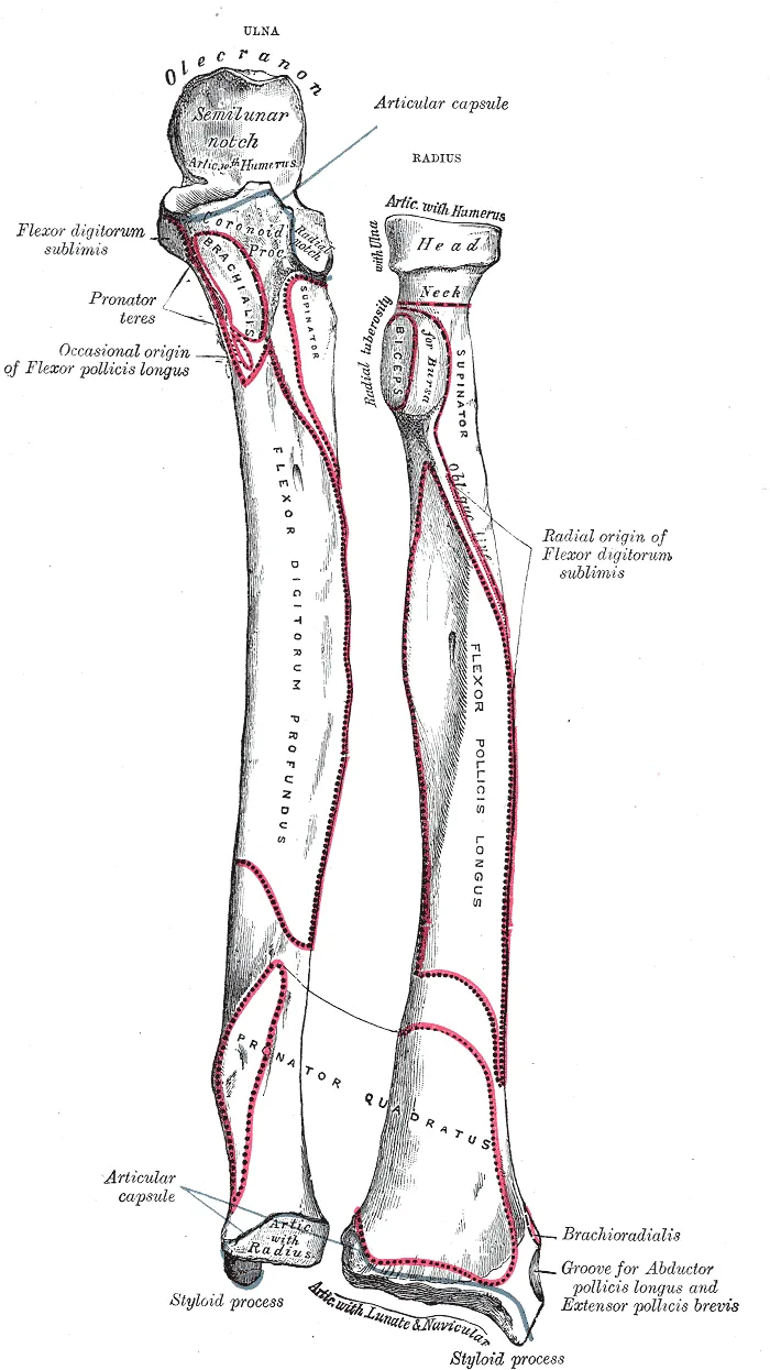
The radius runs beside the ulna (the forearm bone on the little-finger side), on the thumb side of the forearm5. Its lower end, the distal radius, meets the ulna and two wrist bones, the scaphoid and lunate6. A hard fall onto an outstretched hand sends force up the arm and may break the radius5.
A 2020 Cochrane review, a systematic review done to the Cochrane Collaboration’s published standards, gives the usual definition1. The break lies within 3 cm of the radiocarpal joint, where the radius meets the lunate and scaphoid1. The review adds that most are closed injuries, with the skin left whole1.
The patterns sources name
The sources describe three patterns of break at the lower radius5,7,8:
- Colles fracture. An anatomy textbook describes a complete break across the lower radius, with the broken-off end driven backward and upward5.
- Smith fracture. A reference chapter describes a break in which the broken-off end shifts or tilts toward the palm7. It is the less common of the two7.
- Torus (buckle) fracture. The report of a trial in children calls this the mildest form of fracture, in which the bone buckles8.
A reference chapter says the name Colles fracture is often used for any lower radius fracture angled backward9. A 2026 narrative review, a summary written without a set search method, uses a narrower meaning10. For it, the name means a break in several pieces, tilted and shifted backward, outside the joint10. On that meaning, it puts Colles fractures at about one-sixth (17.5%) of wrist fractures10.
A registry followed 1,951 children aged 4 to 18 with a lower radius fracture at four children’s hospitals11. Of their breaks, 44.0% were torus fractures and 31.3% went through both sides of the bone11. Another 21.0% involved the growth plate, the layer of cartilage near the bone’s end where it grows longer11.
Injuries found with the fracture
Two series looked inside the wrist by arthroscopy, keyhole surgery inside a joint using a small camera12,13. Both studied displaced fractures, breaks in which the bone ends have moved out of line12,13. The table counts tears of the scapholunate ligament and the triangular fibrocartilage complex, or TFCC.
| Who was examined | Scapholunate ligament torn | TFCC torn |
|---|---|---|
| 60 patients with a displaced fracture into the joint surface | 1912 | 2612 |
| 50 displaced fractures in adults (men aged 20 to 60, women aged 20 to 50) | 27, partly or fully13 | 3913 |
Who it affects
A review of who breaks this bone says children and older adults are at greatest risk2. The distal radius page gives the detail on age, sex, causes, growth and bone density. In the Swedish Fracture Register for 2015 to 2017, adults with this break had a mean age of 62.7 years14. A simple fall caused 75% of their 23,394 fractures14. In children, data the review cites put sports behind 30% of these fractures at ages 10 to 14 and 47% at 15 to 172.
What studies found
The first year and after
In the first two months after the break, most patients in one cohort had mild pain at rest and severe pain with movement3. In a cohort study of 129 patients with a lower radius fracture, most of the recovery happened within six months3. Symptoms persisted in a small minority at one year3. Over that year, personal care and household work came back sooner and more fully than work and recreation3.
A 2026 recommendations paper from a European trauma society states that recovery is most marked in the first three months15. It adds that improvement can continue beyond one year15.
Healing
Three sources give healing times and rates for this break16,17,18. The distal radius page covers how the bone mends.
- Children’s buckle fractures. A 2026 review of children’s fractures calls the buckle fracture stable, and says it heals reliably within three to four weeks16.
- 494 adults fixed with a plate on the palm side. Union, the bone ends knitting together, was recorded at a mean of about 12.3 to 12.5 weeks17. The authors caution that this reflects when X-rays were taken at set visits more than true healing time17. In 1.2% (6 of 494) the break did not unite17.
- 311 fractures in people aged 50 or older. X-rays showed no delayed union or non-union in any of them18.
Return to sport and work
Three groups of people have been measured19,20,21:
- People hurt in sport, 1995. Of 225 patients whose fracture happened in sport, 131 answered a questionnaire19. Of these, 72.5% had gone back to their original sport19. Return depended mainly on age and the standard of competition before the injury19.
- Children with a buckle fracture. A 2023 scoping review, which maps what research exists on a question, covered 22 publications20. These children commonly returned to sport by four weeks, with no refractures recorded in those back at four weeks20. The 64 surgeons it surveyed recommended a return at a mean of 6.6 weeks20.
- 227 workers seen at a hand clinic. They lost an average of 9.2 weeks from work (median 8, range 0 to 52), and 21% lost no time21. How the break looked on X-ray after care was not related to time off21. Self-rated disability and the physical demands of the job were the strongest predictors21.
Complex regional pain syndrome
A 2026 systematic review, one that searches for studies in a set, checkable way, describes this syndrome4. It calls complex regional pain syndrome a disabling pain condition that follows injury4. It says the condition may hold back recovery and arm function after this break4. The 2020 Cochrane review calls it a major complication of the fracture, with an often unclear cause1. It lists pain and tenderness, reduced joint movement, swelling, tissue changes and unsteady blood-vessel control1.
How often it follows the break depends on the study4,22:
- A 2020 systematic review of nine observational studies took its figure from the three most rigorous22. Between 3.7% and 14% of adults met the Budapest criteria, an agreed checklist of signs, within four months22. Rates were lower in breaks managed without surgery22.
- The 2026 review gives reported rates from 1% to 37%, depending on the criteria, the way the break was managed and the people studied4.
Wires compared with a plate in adults
The trials below used the patient-rated wrist score, which runs to 100, where 0 is best23. Plate fixation is surgery that holds a broken bone together with a metal plate.
DRAFFT was a randomised trial of 461 adults in 18 UK trauma centres with a backward-displaced fracture that needed surgical fixation24. Assessors did not know each patient’s group, though surgeons and patients did24. The score showed no clinically relevant difference between wires and a locking plate: 1.3 points, in favour of the plate24. The UK National Institute for Health Research funded the trial through its Health Technology Assessment scheme24. The report declares that surgeons involved had received research funding and paid educational support from implant makers, not for this study24.
At the five-year follow-up, 44% of participants gave scores, and there was no clinically relevant difference at any point25.
Wires compared with a cast in adults
DRAFFT2 was a randomised trial of 500 adults in 36 UK hospitals whose backward-displaced fractures had been manipulated23. Wires did not give a better score at 12 months than a moulded cast: 20.7 with wires and 21.2 with a cast23. In the cast group, 33 people (13%) went on to surgical fixation because the fracture lost position in the first six weeks23. The authors name as a major limitation that neither clinicians nor participants could be blind to cast or wires23. The UK NIHR Health Technology Assessment Programme funded the trial, and the report states it was not involved in the study’s design23.
The 2020 Cochrane review covered wire fixation through the skin in adults1. It judged each of its 26 trials, with 1,946 mostly older, female participants, at high risk of bias1. It concluded that randomised evidence was insufficient to show the place of wires compared with a cast alone1.
A plate compared with a cast in people aged 60 or older
CROSSFIRE randomised 166 people aged 60 or older in Australia and New Zealand with a displaced lower radius fracture26. They had either a plate on the palm side of the bone, or the bone set and a cast26. With assessors unaware of the groups, the 12-month score showed no clinically important difference: 19.8 with a plate, 21.5 with a cast26. Australia’s National Health and Medical Research Council and three other bodies funded the trial, and the report states they had no role in it26.
At 24 months, an assessor unaware of the groups followed up 151 of the 16627. There was still no clinically important difference: 13.6 with a plate, 15.8 with a cast27. The 24-month report names the same research council among its supporters, and states that the sponsors had no role27.
A 2024 meta-analysis, a study that pools earlier results, combined six randomised trials in people aged 60 or older with a displaced fracture28. On an arm disability score, a plate gave a 12-month result 3.61 points better than a set bone and a cast28. The review reports no funding and declares no conflict of interest28. The patient-rated wrist score and complications did not differ significantly28. The differences did not reach the published thresholds for a clinically important change28.
A bandage compared with rigid immobilisation in children
FORCE randomised 965 children aged 4 to 15 with a torus fracture of the lower radius, in 23 UK emergency departments8. One group was offered a soft bandage and sent home; the other had rigid immobilisation, keeping the limb still, and follow-up8. Families and clinicians knew the group, and the authors name observer bias as a concern8. Two authors sat on boards of the programme that funded it29. Britain’s National Institute for Health and Care Research funded the trial, and the report states it had no role in the study29.
Pain at three days was equivalent in the two groups, and complications were 1.0% against 0.6%8. The adjusted difference was 0.10 points on a pain scale of 0 to 10, against an equivalence margin of 1.08,29. Across six weeks, the groups showed no differences in recovery of function, quality of life or school absence8.
How long a cast stays on
Two systematic reviews compared shorter with longer cast times for fractures managed without surgery30,31.
- A 2019 review of 12 randomised trials (1,063 patients) found better grip strength and patient-reported outcome with the shorter period30. Pain, movement and X-rays did not differ, and the trials were too varied to pool30. It reports no funding and declares no conflict of interest30.
- A 2024 review of 16 studies (1,118 patients) compared under three weeks with over three weeks31. The shorter period often gave equal or better outcomes31. For displaced fractures that had been set, it concluded the period could not go below four weeks, because of the risk of complications31. Its authors declare no competing interests31.
Exercise programmes afterwards
A 2015 Cochrane review covered exercise and therapy programmes after a lower radius fracture in adults32. It included 26 trials with 1,269 mainly female and older patients32. It rated the evidence for each of its 23 comparisons as low or very low quality, and insufficient to establish how the approaches compared32.
Connections
The distal radius meets the scaphoid and lunate at the wrist joint5,6. A hard fall onto an outstretched hand may break the radius or the scaphoid, the subject of the scaphoid fracture page5.
A systematic review of 20 articles covered 1,346 patients with acute distal radius fractures examined by arthroscopy33. It found an overall rate of injury to the scapholunate ligament of 35.3%33.
One study counted emergency visits for acute wrist trauma in people aged 15 or older34. Of 69 per 10,000 inhabitants a year, distal radius fractures made up 2734. The wrist sprain page covers other wrist injuries.
Distal radius fracture involves
Distal radius fracture has been found together with tears of
Distal radius fracture can follow the same kind of fall as
Distal radius fracture has been counted alongside
Sources
- Karantana A, Handoll HH, Sabouni A (2020). Percutaneous pinning for treating distal radial fractures in adults.. The Cochrane database of systematic reviews. doi:10.1002/14651858.CD006080.pub3 PMID 32032439
- Nellans KW, Kowalski E, Chung KC (2012). The epidemiology of distal radius fractures.. Hand clinics. doi:10.1016/j.hcl.2012.02.001 PMID 22554654
- MacDermid JC, Roth JH, Richards RS (2003). Pain and disability reported in the year following a distal radius fracture: a cohort study.. BMC musculoskeletal disorders. doi:10.1186/1471-2474-4-24 PMID 14588078
- Serôdio IN, Saiz-Vázquez O, Ortiz-Huerta H, Simón-Vicente L, Santamaría-Vázquez M (2026). Preventing Complex Regional Pain Syndrome After Distal Radius Fracture: A Systematic Review of Rehabilitation and Clinical Prophylaxis Strategies.. Journal of functional morphology and kinesiology. doi:10.3390/jfmk11020158 PMID 42029526
- Betts JG, Young KA, Wise JA, Johnson E, Poe B, Kruse DH, Korol O, Johnson JE, Womble M, DeSaix P (2022). Anatomy and Physiology 2e. OpenStax. Link
- Bair MM, Zafar Gondal A (2023). Anatomy, Shoulder and Upper Limb, Forearm Radius. StatPearls. PMID 31356039
- Schroeder JD, Varacallo MA (2024). Smith Fracture Review. StatPearls. PMID 31613494
- Perry DC, Achten J, Knight R, Dutton SJ, Dritsaki M, Mason JM, Appelbe DE, Roland DT, Messahel S, Widnall J, Gibson P, Preston J, Spoors LM, Campolier M, Costa ML (2022). Offer of a bandage versus rigid immobilisation in 4- to 15-year-olds with distal radius torus fractures: the FORCE equivalence RCT.. Health technology assessment (Winchester, England). doi:10.3310/BDNS6122 PMID 35904496
- Summers K, Mabrouk A, Fowles SM (2023). Colles Fracture. StatPearls. PMID 31971712
- Kirchberg T, Nolan R, Tomberg S (2026). Emergency Medicine Management of Distal Radius and Ulna Fractures: Narrative Review.. The western journal of emergency medicine. doi:10.5811/westjem.52930 PMID 42550696
- Shah AS, Guzek RH, Miller ML, Willey MC, Mahan ST, Bae DS (2023). Descriptive Epidemiology of Isolated Distal Radius Fractures in Children: Results From a Prospective Multicenter Registry.. Journal of pediatric orthopedics. doi:10.1097/BPO.0000000000002288 PMID 36299238
- Geissler WB, Freeland AE, Savoie FH, McIntyre LW, Whipple TL (1996). Intracarpal soft-tissue lesions associated with an intra-articular fracture of the distal end of the radius.. The Journal of bone and joint surgery. American volume. doi:10.2106/00004623-199603000-00006 PMID 8613442
- Lindau T, Arner M, Hagberg L (1997). Intraarticular lesions in distal fractures of the radius in young adults. Journal of hand surgery (Edinburgh, Scotland). doi:10.1016/s0266-7681(97)80364-6 PMID 9752922
- Rundgren J, Bojan A, Mellstrand Navarro C, Enocson A (2020). Epidemiology, classification, treatment and mortality of distal radius fractures in adults: an observational study of 23,394 fractures from the national Swedish fracture register.. BMC musculoskeletal disorders. doi:10.1186/s12891-020-3097-8 PMID 32035488
- Lameijer CM, Teuben MPJ, van Delft EAK, Tomazevic M, Kastelec M, Nau C (2026). ESTES recommendations regarding distal radius fractures.. European journal of trauma and emergency surgery : official publication of the European Trauma Society. doi:10.1007/s00068-026-03203-7 PMID 42113243
- Bašković M, Buzuk J, Dujić B, Jurić D, Jurković K, Pehar K, Vuković S (2026). Conservative Treatment of Pediatric Fractures-Narrative Review of Acceptable Deformity and Remodeling Potential.. Medical sciences (Basel, Switzerland). doi:10.3390/medsci14040432 PMID 42646565
- Chang M, Kasper A, Chung J, Wright J, Pennington M, Ilyas AM (2025). The Effect of Nonsteroidal Anti-Inflammatory Drugs on Union Rates Following Operative Repair of Distal Radius Fractures.. Journal of hand surgery global online. doi:10.1016/j.jhsg.2025.03.001 PMID 40497268
- Gorter EA, Gerretsen BM, Krijnen P, Appelman-Dijkstra NM, Schipper IB (2020). Does osteoporosis affect the healing of subcapital humerus and distal radius fractures?. Journal of orthopaedics. doi:10.1016/j.jor.2020.05.004 PMID 32425424
- Lawson GM, Hajducka C, McQueen MM (1995). Sports fractures of the distal radius--epidemiology and outcome.. Injury. doi:10.1016/0020-1383(95)90549-d PMID 7868207
- Bhanushali A, Bright R, Xu L, Cundy P, Williams N (2023). Return to sport after forearm fractures in children: A scoping review and survey.. Journal of children's orthopaedics. doi:10.1177/18632521231156434 PMID 37034195
- MacDermid JC, Roth JH, McMurtry R (2007). Predictors of time lost from work following a distal radius fracture.. Journal of occupational rehabilitation. doi:10.1007/s10926-007-9069-0 PMID 17245636
- Rolls C, McCabe C, Llewellyn A, Jones GT (2020). What is the incidence of complex regional pain syndrome (CRPS) Type I within four months of a wrist fracture in the adult population? A systematic review. Hand Therapy. doi:10.1177/1758998320910179
- Costa ML, Achten J, Ooms A, Png ME, Cook JA, Lamb SE, Hedley H, Dias J, Dritsaki M (2022). Surgical fixation with K-wires versus casting in adults with fracture of distal radius: DRAFFT2 multicentre randomised clinical trial.. BMJ (Clinical research ed.). doi:10.1136/bmj-2021-068041 PMID 35045969
- Costa ML, Achten J, Parsons NR, Rangan A, Griffin D, Tubeuf S, Lamb SE, Edwards A (2014). Percutaneous fixation with Kirschner wires versus volar locking plate fixation in adults with dorsally displaced fracture of distal radius: randomised controlled trial.. BMJ (Clinical research ed.). doi:10.1136/bmj.g4807 PMID 25096595
- Costa ML, Achten J, Rangan A, Lamb SE, Parsons NR (2019). Percutaneous fixation with Kirschner wires versus volar locking-plate fixation in adults with dorsally displaced fracture of distal radius: five-year follow-up of a randomized controlled trial.. The bone & joint journal. doi:10.1302/0301-620X.101B8.BJJ-2018-1285.R1 PMID 31362548
- Lawson A, Naylor JM, Buchbinder R, Ivers R, Balogh ZJ, Smith P, Xuan W, Howard K, Vafa A, Perriman D, Mittal R, Yates P, Rieger B, Smith G, Adie S, Elkinson I, Kim W, Sungaran J, Latendresse K, Wong J, Viswanathan S, Landale K, Drobetz H, Tran P, Page R, Beattie S, Mulford J, Incoll I, Kale M, Schick B, Li T, Higgs A, Oppy A, Harris IA (2021). Surgical Plating vs Closed Reduction for Fractures in the Distal Radius in Older Patients. JAMA surgery. doi:10.1001/jamasurg.2020.5672 PMID 33439250
- Lawson A, Naylor J, Buchbinder R, Ivers R, Balogh ZJ, Smith P, Xuan W, Howard K, Vafa A, Perriman D, Mittal R, Yates P, Rieger B, Smith G, Adie S, Elkinson I, Kim W, Sungaran J, Latendresse K, Wong J, Viswanathan S, Landale K, Drobetz H, Tran P, Page R, Beattie S, Mulford J, Incoll I, Kale M, Schick B, Li T, Higgs A, Oppy A, Harris IA (2022). Plating vs Closed Reduction for Fractures in the Distal Radius in Older Patients. JAMA surgery. doi:10.1001/jamasurg.2022.0809 PMID 35476128
- Latypov N, Golubev I, Borisova A (2024). Volar Locking Plate versus Closed Reduction and Immobilization for Distal Radius Fracture in the Elderly: Systematic Review and Meta-Analysis of Randomized Controlled Trials.. Journal of wrist surgery. doi:10.1055/s-0043-1774331 PMID 39619453
- Perry DC, Achten J, Knight R, Appelbe D, Dutton SJ, Dritsaki M, Mason JM, Roland DT, Messahel S, Widnall J, Costa ML, FORCE Collaborators in collaboration with PERUKI (2022). Immobilisation of torus fractures of the wrist in children (FORCE): a randomised controlled equivalence trial in the UK.. Lancet (London, England). doi:10.1016/S0140-6736(22)01015-7 PMID 35780790
- Delft EAKV, Gelder TGV, Vries R, Vermeulen J, Bloemers FW (2019). Duration of Cast Immobilization in Distal Radial Fractures: A Systematic Review.. Journal of wrist surgery. doi:10.1055/s-0039-1683433 PMID 31579555
- de Bruijn MAN, van Ginkel LA, Boersma EZ, van Silfhout L, Tromp TN, van de Krol E, Edwards MJR, Stirler VMA, Hermans E (2024). Cast immobilization duration for distal radius fractures, a systematic review.. European journal of trauma and emergency surgery : official publication of the European Trauma Society. doi:10.1007/s00068-024-02494-y PMID 38507086
- Handoll HH, Elliott J (2015). Rehabilitation for distal radial fractures in adults.. The Cochrane database of systematic reviews. doi:10.1002/14651858.CD003324.pub3 PMID 26403335
- Begum R, Hourston GJM, Bochmann T, Chojnowski AJ (2022). A Systematic 10-Year Review of the Use of Arthroscopy in the Assessment and Treatment of Intercarpal Ligament Injuries Associated with Acute Distal Radius Fracture.. The journal of hand surgery Asian-Pacific volume. doi:10.1142/S2424835522500862 PMID 36476085
- Larsen CF, Lauritsen J (1993). Epidemiology of acute wrist trauma.. International journal of epidemiology. doi:10.1093/ije/22.5.911 PMID 8282472
Changes
- First published.