Distal radius
Also called lower radius, lower end of the radius, wrist end of the radius
The wrist end of the radius, the forearm bone on the thumb side. How it is shaped, how much load it carries, who breaks it and how, how it mends and builds up with loading, and what attaches to it.
- Region
- Wrist & hand
- Muscles
- brachioradialis, Forearm extensors
- Updated
- October 14, 2026
- Reviewed
- October 14, 2026 · how entries are checked
Function
Where the bone sits
The radius runs beside the ulna, on the thumb side of the forearm1. Its upper end is a disc-shaped head that meets the humerus (upper arm bone) at the elbow1. The interosseous membrane, a sheet of dense connective tissue, joins it to the ulna1. The distal radius is the lower end, where the bone meets the ulna and two wrist bones2. A 2020 review states that breaks of this part are usually defined as lying within 3 cm of the wrist joint7.
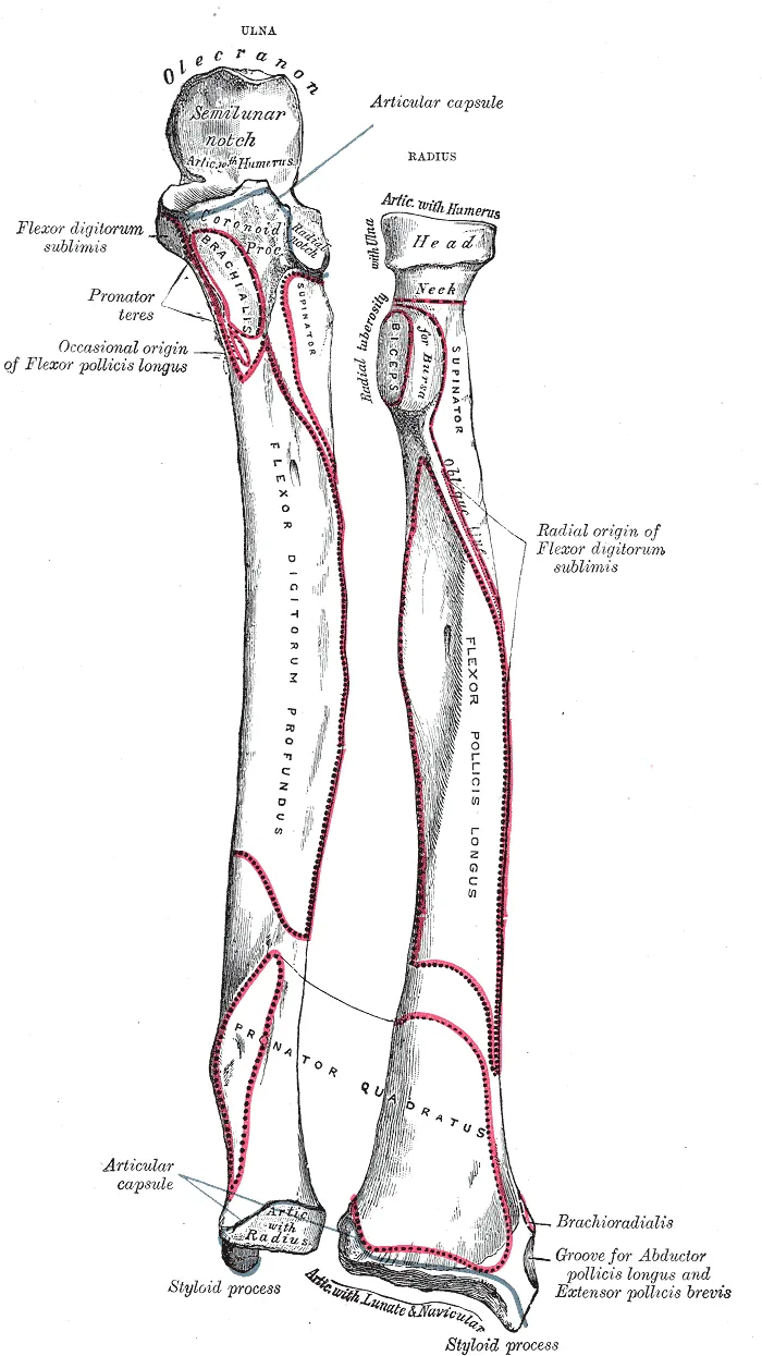
The wrist end and its joint surfaces
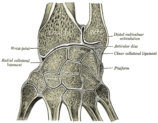
An anatomy reference names three joint surfaces at the lower end of the radius8:
- the scaphoid fossa (a fossa is a shallow hollow in a bone)8;
- the lunate fossa8;
- the ulnar notch, also called the sigmoid notch, for the lower ulna8.
The scaphoid and lunate are two of the carpal bones, the small bones of the wrist1. At the wrist joint, both meet the end of the radius directly1. The end of the ulna touches no carpal bone directly1. On the little-finger side sits the triangular fibrocartilage complex, which arises from the radius and attaches to the far end of the ulna9.
The ulnar notch is a shallow hollow on the inner side of the lower radius1. It meets the head of the ulna to form the distal radioulnar joint1. At the outer end of the radius is a point called the styloid process1. Ligaments that support the thumb side of the wrist attach to it1.
How it is built inside
A bone imaging study describes the lower end of the radius as having a thin cortex (the dense, solid outer layer of a bone)10. Trabeculae, the thin struts of spongy bone, reinforce it from the centre10. They carry load from the joint to the thicker cortex further up the bone10.
How much load it carries
Two sources give figures for how load splits across the wrist. One is a review of wrist biomechanics, for a wrist and forearm in neutral3. The other is a computer model built from 120 normal adult wrist X-rays and loaded to mimic a light grasp with the wrist in neutral11.
| Path through the wrist | Review | Computer model |
|---|---|---|
| Between radius and scaphoid | About 45%3 | 55%11 |
| Between radius and lunate | About 35%3 | 35%11 |
| Ulnar side | 20%3 | 10%, through the triangular fibrocartilage11 |
One study held eight wrists from cadavers (bodies donated for study) in a static push-up position12. A push-up force of 89 N sent an average peak of 69 N along the distal radius12. Scaled up in a straight line to a force of full body weight, that would be 663 N12.
Where the radius grows in length
A growth plate is a layer of cartilage near the end of a growing bone, where the bone grows longer. In X-rays of 200 males and females, about 80% of the radius’s growth in length came from its lower growth plate13. At age five the lower plate gave 85% of total growth, and by age eight 90%13.
Breakdown
Who breaks it
A review of who breaks this bone says children and older adults are at greatest risk4. One study read the wrist X-rays taken in 2016 in a region of southern Sweden with 182,000 residents14.
| Adults in the Swedish region, 2016 | New lower radius fractures per 10,000 people |
|---|---|
| Everyone | 2214 |
| Women | 3414 |
| Men | 1014 |
The review cites a study in which men and women aged 19 to 49 had almost identical rates4. In another study it cites, of people over 65, women’s likelihood of a distal forearm fracture was about 4.88 times men’s4.
How it breaks
A hard fall onto an outstretched hand sends force up the arm, and it may break the humerus, the radius or the scaphoid1. The review states that most of these breaks in older adults follow low-energy trauma, with a fall from standing height the leading cause4. Among 23,394 lower radius fractures in adults in the Swedish Fracture Register, a simple fall caused 75%15.
The sources name three patterns of break1,16,17:
- Colles fracture. A complete break across the lower radius, with the broken-off end driven backward and upward1. An anatomy textbook calls it the most frequent forearm fracture, common in people over 501.
- Smith fracture. A break in which the broken-off end shifts or tilts toward the palm; it is the less common of the two16.
- Torus (buckle) fracture. The report of a trial in children calls this the mildest form of fracture, in which the bone buckles17.
Children and the growth spurt
In Saskatchewan hospital and billing data, lower radius fractures peaked at 11.5 to 12.5 years in girls5. In boys the peak was at 13.5 to 14.5 years5. In Saskatchewan children, the fracture peak almost exactly matched the age of fastest growth in height5. That age was 11.9 years in girls and 14.3 in boys5.
The authors of another population study offer a possible reason18. They suggest activity rises just when fast growth’s demand for calcium leaves a brief shortfall in cortical bone mass18. The review cites data in which sports accounted for 30% of these fractures at ages 10 to 14 and 47% at ages 15 to 174.
Bone density and later fractures
Bone mineral density is how much mineral is packed into a given amount of bone. One study followed 11,392 women aged 50 to 79 for a mean of 8.5 years19. Lower density at the hip and lower spine was associated with a higher risk of wrist fracture19.
One analysis followed postmenopausal women (women after menopause) aged 50 to 79 for a mean of 11.8 years20. Of those who broke a wrist, 15.5% later had a fracture elsewhere20. A wrist fracture was associated with a higher rate of later breaks at other sites20.
Ligaments torn along with the bone
A systematic review (a study that searches for and sums up the research on a question) covered 20 articles21. Their 1,346 patients had acute distal radius fractures examined by arthroscopy, keyhole surgery inside a joint using a small camera21. The overall rate of injury to the scapholunate ligament was 35.3%21. In 50 displaced fractures in adults no older than 60, examined through a scope, the triangular fibrocartilage complex was torn in 3922.
Gymnasts and the growth plate
Eight cross-sectional studies, which look at a group once, examined gymnasts’ wrists23. Depending on the study, between 10% and 85% of the gymnasts examined had X-ray changes consistent with stress reaction of the lower radial growth plate23.
A 1997 critical review of female gymnasts looked at growth arrest, when stress stops the plate growing early24. It judged this plausible, but the evidence too weak to be conclusive24. It found no rigorous studies of whether repetitive loading inhibits growth of the radius in female gymnasts24.
Recovery
How the break mends
An anatomy textbook describes bone healing in stages1. A blood clot forms first, then calluses of cartilage and bone, which are new tissue that bridges the break1. Over several weeks spongy bone replaces the callus cartilage, and remodelling can remove outward signs of the break1. A review of fracture healing reports that about 10% of fractures do not heal normally25.
A literature review of lower radius fractures in adolescents found substantial potential for angled or overlapped fractures to remodel, even in patients older than 1226.
How long it takes at the wrist
Three studies report on union, when the bone has joined across the break27,28,29:
- Torus fractures in children. A 2026 review states that these wrist breaks heal reliably within three to four weeks27.
- Adults with a plate. One centre reviewed 494 patients whose break was fixed with a metal plate on the palm side28. Leaving out one surgeon’s 117 patients, X-rayed on a later schedule, the groups compared reached recorded union at about 12.3 to 12.5 weeks28. The authors caution that this mostly reflects when X-ray visits were scheduled, not when the bone truly joined28. Six of the 494 breaks (1.2%) did not unite28.
- Adults aged 50 or over. In another study of this age group, none of 311 lower radius fractures showed slow healing or non-union on X-ray29.
Trials of wires, plates, casts and soft bandages are covered on the distal radius fracture page17,30,31,32.
How the bone builds up with loading
A review of bone strength reports that bone adapts to the loads it usually bears33. One trial tested this at the radius, with a voluntary task that compressed the forearm6. It measured bone mineral content, the amount of mineral in a bone6.
The trial ran for 12 months in 102 healthy women aged 21 to 40, and 66 completed it6. Slips of paper drawn from an envelope set the groups, and participants were blinded to the study hypotheses6. At the far end of the radius, bone mineral content rose by 2.7% and 3.4% in the two loading groups6. It fell by 1.3% in controls6. The paper states that a United States National Institutes of Health institute fully supported it, with further support from a National Science Foundation graduate fellowship6.
The loading dose was positively related to the change in bone mineral6. Yet the authors concluded that strain size, strain rate and number of loading bouts explained only a small part of the variation6.
Four studies of sport and exercise report on bone in the arm, two of them at the lower radius34,35,36,37:
- Wrist-loading sports in youth. A 2018 systematic review covered 23 studies of young people in sports such as gymnastics, tennis and rowing34. Athletes had higher bone mineral density and content in the arms than controls34.
- Gymnastics. A meta-analysis (a study that pools the results of several earlier studies) of 17 studies compared pre-pubertal girls (girls before puberty) in gymnastics with non-gymnasts35. Gymnasts had greater bone properties, and the largest difference was in volumetric bone mineral density at the lower end of the radius35.
- Impact exercise. A 2023 meta-analysis pooled 28 randomised trials of impact exercise, with ground forces at least those of running36. It found gains in some bone structure measures elsewhere, but no effect at the distal radius36.
- Resistance training. A 2001 meta-analysis of controlled trials in women reported that this training helped radius bone density after menopause37. It did not single out the lower end37.
In ten cadaver wrists, the bone just under the cartilage was densest in two spots, one in each fossa38. The authors state that such patterns reflect the load carried over the long term38. In healthy adults scanned while bending the wrist, higher density in the lower radius went with larger joint contact areas39.
Connections
Joints it forms
The distal radius forms the wrist joint with the scaphoid and lunate, and the distal radioulnar joint with the ulna1.
Related exercise pages: push-up, in which one cadaver study measured force along the distal radius12.
Muscles and tendons attached to the radius
| Structure | Where it meets the radius |
|---|---|
| Distal biceps tendon, of the biceps brachii | Radial tuberosity, a bump near the elbow1,40 |
| Forearm flexors | Pronator teres, a forearm muscle that turns the palm down, inserts at the middle of the shaft’s outer surface41 |
| Forearm extensors | Brachioradialis, a forearm muscle that bends the elbow, inserts at the base of the styloid process41 |
| First dorsal compartment tendons | Run in a groove on the outer side of the lower end41 |
Distal radius forms part of
Distal radius meets at the wrist joint
Distal radius gives origin to
Distal radius can be injured together with
Distal radius breaks in
Distal radius is attached to by
Distal radius carries in its groove
Distal radius is loaded by
Sources
- Betts JG, Young KA, Wise JA, Johnson E, Poe B, Kruse DH, Korol O, Johnson JE, Womble M, DeSaix P (2022). Anatomy and Physiology 2e. OpenStax. Link
- Bair MM, Zafar Gondal A (2023). Anatomy, Shoulder and Upper Limb, Forearm Radius. StatPearls. PMID 31356039
- Eschweiler J, Li J, Quack V, Rath B, Baroncini A, Hildebrand F, Migliorini F (2022). Anatomy, Biomechanics, and Loads of the Wrist Joint.. Life (Basel, Switzerland). doi:10.3390/life12020188 PMID 35207475
- Nellans KW, Kowalski E, Chung KC (2012). The epidemiology of distal radius fractures.. Hand clinics. doi:10.1016/j.hcl.2012.02.001 PMID 22554654
- Bailey DA, Wedge JH, McCulloch RG, Martin AD, Bernhardson SC (1989). Epidemiology of fractures of the distal end of the radius in children as associated with growth.. The Journal of bone and joint surgery. American volume. PMID 2777851
- Troy KL, Mancuso ME, Johnson JE, Wu Z, Schnitzer TJ, Butler TA (2020). Bone Adaptation in Adult Women Is Related to Loading Dose: A 12-Month Randomized Controlled Trial.. Journal of bone and mineral research : the official journal of the American Society for Bone and Mineral Research. doi:10.1002/jbmr.3999 PMID 32154945
- Karantana A, Handoll HH, Sabouni A (2020). Percutaneous pinning for treating distal radial fractures in adults.. The Cochrane database of systematic reviews. doi:10.1002/14651858.CD006080.pub3 PMID 32032439
- Summers K, Mabrouk A, Fowles SM (2023). Colles Fracture. StatPearls. PMID 31971712
- Martins T, Marappa-Ganeshan R (2023). Anatomy, Shoulder and Upper Limb, Forearm Triangular Fibrocartilage Complex.. StatPearls. PMID 32119451
- Bala Y, Bui QM, Wang XF, Iuliano S, Wang Q, Ghasem-Zadeh A, Rozental TD, Bouxsein ML, Zebaze RM, Seeman E (2015). Trabecular and cortical microstructure and fragility of the distal radius in women.. Journal of bone and mineral research : the official journal of the American Society for Bone and Mineral Research. doi:10.1002/jbmr.2388 PMID 25327362
- Schuind F, Cooney WP, Linscheid RL, An KN, Chao EY (1995). Force and pressure transmission through the normal wrist. A theoretical two-dimensional study in the posteroanterior plane.. Journal of biomechanics. doi:10.1016/0021-9290(94)00093-j PMID 7775494
- Smith JM, Werner FW, Harley BJ (2018). Forces in the Distal Radius During a Pushup or Active Wrist Motions.. The Journal of hand surgery. doi:10.1016/j.jhsa.2018.05.020 PMID 29945841
- Pritchett JW (1991). Growth plate activity in the upper extremity.. Clinical orthopaedics and related research. PMID 2060215
- Ali M, Eiriksdottir A, Murtadha M, Åkesson A, Atroshi I (2020). Incidence of distal radius fracture in a general population in southern Sweden in 2016 compared with 2001.. Osteoporosis international : a journal established as result of cooperation between the European Foundation for Osteoporosis and the National Osteoporosis Foundation of the USA. doi:10.1007/s00198-020-05282-7 PMID 31930451
- Rundgren J, Bojan A, Mellstrand Navarro C, Enocson A (2020). Epidemiology, classification, treatment and mortality of distal radius fractures in adults: an observational study of 23,394 fractures from the national Swedish fracture register.. BMC musculoskeletal disorders. doi:10.1186/s12891-020-3097-8 PMID 32035488
- Schroeder JD, Varacallo MA (2024). Smith Fracture Review. StatPearls. PMID 31613494
- Perry DC, Achten J, Knight R, Dutton SJ, Dritsaki M, Mason JM, Appelbe DE, Roland DT, Messahel S, Widnall J, Gibson P, Preston J, Spoors LM, Campolier M, Costa ML (2022). Offer of a bandage versus rigid immobilisation in 4- to 15-year-olds with distal radius torus fractures: the FORCE equivalence RCT.. Health technology assessment (Winchester, England). doi:10.3310/BDNS6122 PMID 35904496
- Khosla S, Melton LJ, Dekutoski MB, Achenbach SJ, Oberg AL, Riggs BL (2003). Incidence of Childhood Distal Forearm Fractures Over 30 Years. JAMA. doi:10.1001/jama.290.11.1479 PMID 13129988
- Crandall CJ, Hovey KM, Andrews CA, Cauley JA, Manson JE, Wactawski-Wende J, Wright NC, Li W, Beavers K, Curtis JR, LeBoff MS (2015). Bone Mineral Density as a Predictor of Subsequent Wrist Fractures: Findings From the Women's Health Initiative Study.. The Journal of clinical endocrinology and metabolism. doi:10.1210/jc.2015-2568 PMID 26367200
- Crandall CJ, Hovey KM, Cauley JA, Andrews CA, Curtis JR, Wactawski-Wende J, Wright NC, Li W, LeBoff MS (2015). Wrist Fracture and Risk of Subsequent Fracture: Findings from the Women's Health Initiative Study.. Journal of bone and mineral research : the official journal of the American Society for Bone and Mineral Research. doi:10.1002/jbmr.2559 PMID 25990562
- Begum R, Hourston GJM, Bochmann T, Chojnowski AJ (2022). A Systematic 10-Year Review of the Use of Arthroscopy in the Assessment and Treatment of Intercarpal Ligament Injuries Associated with Acute Distal Radius Fracture.. The journal of hand surgery Asian-Pacific volume. doi:10.1142/S2424835522500862 PMID 36476085
- Lindau T, Arner M, Hagberg L (1997). Intraarticular lesions in distal fractures of the radius in young adults. Journal of hand surgery (Edinburgh, Scotland). doi:10.1016/s0266-7681(97)80364-6 PMID 9752922
- Caine D, DiFiori J, Maffulli N (2006). Physeal injuries in children's and youth sports: reasons for concern?. British journal of sports medicine. doi:10.1136/bjsm.2005.017822 PMID 16807307
- Caine D, Howe W, Ross W, Bergman G (1997). Does repetitive physical loading inhibit radial growth in female gymnasts?. Clinical journal of sport medicine : official journal of the Canadian Academy of Sport Medicine. doi:10.1097/00042752-199710000-00007 PMID 9397328
- Einhorn TA, Gerstenfeld LC (2015). Fracture healing: mechanisms and interventions.. Nature reviews. Rheumatology. doi:10.1038/nrrheum.2014.164 PMID 25266456
- Greig D, Silva M (2021). Management of Distal Radius Fractures in Adolescent Patients.. Journal of pediatric orthopedics. doi:10.1097/BPO.0000000000001778 PMID 34096530
- Bašković M, Buzuk J, Dujić B, Jurić D, Jurković K, Pehar K, Vuković S (2026). Conservative Treatment of Pediatric Fractures-Narrative Review of Acceptable Deformity and Remodeling Potential.. Medical sciences (Basel, Switzerland). doi:10.3390/medsci14040432 PMID 42646565
- Chang M, Kasper A, Chung J, Wright J, Pennington M, Ilyas AM (2025). The Effect of Nonsteroidal Anti-Inflammatory Drugs on Union Rates Following Operative Repair of Distal Radius Fractures.. Journal of hand surgery global online. doi:10.1016/j.jhsg.2025.03.001 PMID 40497268
- Gorter EA, Gerretsen BM, Krijnen P, Appelman-Dijkstra NM, Schipper IB (2020). Does osteoporosis affect the healing of subcapital humerus and distal radius fractures?. Journal of orthopaedics. doi:10.1016/j.jor.2020.05.004 PMID 32425424
- Costa ML, Achten J, Parsons NR, Rangan A, Griffin D, Tubeuf S, Lamb SE, Edwards A (2014). Percutaneous fixation with Kirschner wires versus volar locking plate fixation in adults with dorsally displaced fracture of distal radius: randomised controlled trial.. BMJ (Clinical research ed.). doi:10.1136/bmj.g4807 PMID 25096595
- Costa ML, Achten J, Ooms A, Png ME, Cook JA, Lamb SE, Hedley H, Dias J, Dritsaki M (2022). Surgical fixation with K-wires versus casting in adults with fracture of distal radius: DRAFFT2 multicentre randomised clinical trial.. BMJ (Clinical research ed.). doi:10.1136/bmj-2021-068041 PMID 35045969
- Lawson A, Naylor JM, Buchbinder R, Ivers R, Balogh ZJ, Smith P, Xuan W, Howard K, Vafa A, Perriman D, Mittal R, Yates P, Rieger B, Smith G, Adie S, Elkinson I, Kim W, Sungaran J, Latendresse K, Wong J, Viswanathan S, Landale K, Drobetz H, Tran P, Page R, Beattie S, Mulford J, Incoll I, Kale M, Schick B, Li T, Higgs A, Oppy A, Harris IA (2021). Surgical Plating vs Closed Reduction for Fractures in the Distal Radius in Older Patients. JAMA surgery. doi:10.1001/jamasurg.2020.5672 PMID 33439250
- Hart NH, Nimphius S, Rantalainen T, Ireland A, Siafarikas A, Newton RU (2017). Mechanical basis of bone strength: influence of bone material, bone structure and muscle action.. Journal of musculoskeletal & neuronal interactions. PMID 28860414
- Kox LS, Kuijer PPFM, Thijssen DAJ, Kerkhoffs GMMJ, van Rijn RR, Frings-Dresen MHW, Maas M (2018). Health Effects of Wrist-Loading Sports During Youth: A Systematic Literature Review.. Journal of physical activity & health. doi:10.1123/jpah.2017-0328 PMID 29741447
- Burt LA, Greene DA, Ducher G, Naughton GA (2013). Skeletal adaptations associated with pre-pubertal gymnastics participation as determined by DXA and pQCT: a systematic review and meta-analysis.. Journal of science and medicine in sport. doi:10.1016/j.jsams.2012.07.006 PMID 22951266
- Ng CA, Gandham A, Mesinovic J, Owen PJ, Ebeling PR, Scott D (2023). Effects of Moderate- to High-Impact Exercise Training on Bone Structure Across the Lifespan: A Systematic Review and Meta-Analysis of Randomized Controlled Trials.. Journal of bone and mineral research. doi:10.1002/jbmr.4899 PMID 37555459
- Kelley GA, Kelley KS, Tran ZV (2001). Resistance Training and Bone Mineral Density in Women. American journal of physical medicine & rehabilitation. doi:10.1097/00002060-200101000-00017 PMID 11138958
- Hoogbergen MM, Niessen WJ, Schuurman AH, Spauwen PH, Kauer JM (2002). Subchondral bone mineral density patterns representing the loading history of the wrist joint.. Journal of hand surgery (Edinburgh, Scotland). doi:10.1054/jhsb.2001.0714 PMID 12027489
- Straatman L, Norman E, Knowles N, Walton D, Suh N, Lalone E (2023). Use it or lose it: The relationship between two image-based biomarkers in better understanding osteoarthritis progression in the wrist.. Journal of biomechanics. doi:10.1016/j.jbiomech.2023.111849 PMID 37931557
- Hsu D, Anand P, Mabrouk A, Chang KV (2023). Biceps Tendon Rupture.. StatPearls. PMID 30020607
- Gray H; Lewis WH (editor) (1918). Anatomy of the Human Body. Lea & Febiger, Philadelphia (20th edition). Link
Changes
- First published.