First dorsal compartment tendons
Also called first extensor compartment, first dorsal compartment, abductor pollicis longus tendon, extensor pollicis brevis tendon, APL tendon, EPB tendon
The two thumb tendons that share a tunnel on the thumb side of the wrist. How the tunnel is built, how wrist position changes gliding inside it, and what studies of De Quervain's found there.
- Region
- Wrist & hand
- Muscles
- abductor pollicis longus, extensor pollicis brevis
- Updated
- October 14, 2026
- Reviewed
- October 14, 2026 · how entries are checked
Function
Where the two tendons run
A retinaculum is a band of tissue that holds tendons in place where they cross a joint. An anatomical study of the extensor retinaculum describes six tendon compartments on the back of the wrist12. Six lengthwise septa (dividing walls) separate them12. The septa run from the retinaculum’s upper layer down to the radius, the forearm bone on the thumb side12.
The first compartment holds two tendons. One belongs to abductor pollicis longus (APL) and the other to extensor pollicis brevis (EPB)1,2. They pass through a tunnel of bone and fibrous tissue3,4. The tunnel lies along the radial styloid, the bony point on the thumb side of the wrist3,4.
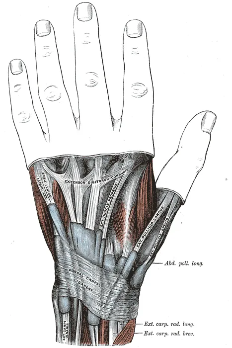
Both muscles belong to the deep group of the forearm extensors13. Before the two tendons reach the retinaculum, they cross over the two radial wrist extensors of the second compartment1. Further down, they form the thumb-side border of the anatomical snuffbox, the hollow at the base of the thumb14.
Where they attach and what they do
In a dissection of 50 adult cadaver (donated body) upper limbs, the main APL tendon attached to the base of the first metacarpal15. That is the bone at the root of the thumb15. In the standard textbook description, the EPB tendon attaches to the thumb’s first bone beyond the knuckle, at the back of its base16. A literature review of four cadaver studies covering 268 hands found that this textbook attachment appears uncommon in practice16.
The APL moves the thumb away from the hand at the joint at its base17. It is also described as steadying the wrist while it bends and straightens17. It is one of the main muscles that tilt the wrist toward the thumb side17. The main job given for the EPB is straightening the thumb at its knuckle16.
How many strands each tendon has
Studies have counted the strands, called slips, of each tendon15,18:
- 50 adult cadaver upper limbs. The APL never had a single tendon, and every limb had between two and six15.
- 40 dissected cadaver hands. The mean number of slips was 2.6 for the APL and 1.1 for the EPB18.
- 130 people having the compartment opened for De Quervain’s. In this group, 78% had more than one APL slip and 92% had a single EPB tendon19.
A meta-analysis of 68 studies and 5,229 wrists found two APL tendons and one EPB tendon to be the commonest arrangement2. That held in cadavers and in people with De Quervain’s, with no significant difference between the two groups2.
How often a septum divides the compartment
In 66 dissected cadaver hands, a septum divided the first compartment in 3120. Three reports compared cadavers with people operated on for De Quervain’s5,21,22:
| Report | Cadavers with a septum | De Quervain’s surgery cases with a septum |
|---|---|---|
| Literature review of 21 studies | 43.7%21 | 62.2%21 |
| Meta-analysis | 35%5 | 67%5 |
| One study of 96 cadaver wrists and 85 wrists at surgery | 72.9%22 | 61.2%22 |
The single study found no significant difference between its two groups22. A meta-analysis of 68 studies and 5,229 wrists found a septum much more often in people with De Quervain’s than in cadavers2.
The nerve branches beside the compartment
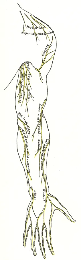
In 52 wrists from 28 cadavers, a mean of 4.2 skin nerve branches lay in the area around the first compartment23. Of these, 3.3 came from the superficial radial nerve23. That nerve is one of the two branches the radial nerve divides into24. Other studies measured how close the branches lie19,25,26:
- 20 fresh cadaver arms. The nearest branch lay a mean of 0.4 cm from the centre of the compartment, at the level of the retinaculum25.
- 20 forearms from 11 cadavers. The branch to the back of the thumb lay on average 2 mm from the compartment’s midline26. In 8 forearms it passed directly over the compartment along its whole length26.
- 130 people having the compartment opened for De Quervain’s. Surgeons met at least one branch of the nerve in 61% of operations19.
How wrist position changes gliding and pressure
One study tested 15 cadaver wrists in seven fixed positions6. The wrist was bent forward or back by 30 or 60 degrees, held straight, or tilted to either side6. In 15 cadaver wrists, wrist position significantly changed the gliding resistance of both tendons6. Resistance was measured in newtons (N), a unit of force.
| Tendon | Positions with significantly higher resistance | Other positions tested |
|---|---|---|
| APL, 15 wrists | 0.33 N, bent forward 60 degrees6 | The rest under 0.26 N6 |
| EPB, 8 wrists with a septum | 0.51 N, bent forward 60 degrees6 | The rest under 0.26 N6 |
| EPB, 7 wrists with no septum | 0.20 N, bent forward 60 degrees, and 0.22 N, bent back 60 degrees6 | The rest under 0.15 N6 |
Overall, gliding resistance did not differ between wrists with and without a septum6.
Another study measured pressure inside the first compartment in 14 arms from seven fresh frozen cadavers27. Pressure is given in kilopascals (kPa), a unit of force per area. The change in pressure was significantly greater with the wrist bent forward than straight or bent back27.
| Wrist position | Mean pressure change in the compartment |
|---|---|
| Bent forward 45 degrees | 79.5 kPa27 |
| Straight | 36.3 kPa27 |
| Bent back 45 degrees | 50.4 kPa27 |
Seven of those wrists had a septum27. The pressure change on the EPB side of it averaged 81.7 kPa27. That was significantly greater than on the APL side, at 32.8 kPa, or in wrists with no septum, at 54.6 kPa27.
Breakdown
De Quervain’s and the first compartment
De Quervain’s tenosynovitis is painful swelling around two thumb tendons at the wrist. The De Quervain’s tenosynovitis page covers how it is defined, its course and the trials. The authors of a cadaver study wrote that its cause is unknown6. They wrote that repetitive motion with an awkward wrist position, and a divided compartment, are considered causes6. As a hypothesis, they proposed that these might raise the resistance to tendon gliding and so cause small-scale damage6.
What tissue studies found, and where they disagree
Studies of tissue removed at surgery for De Quervain’s disagree on whether inflammation is present7,9. Inflammation is the body’s response to injury or irritation. Two studies found the tissue was not marked by inflammation, and a third found only minimal round cell infiltration7,8,28:
- 35 people with De Quervain’s. The thickening of the retinaculum and tendon lining was due to fibrosis (scarring) in every layer28. Round cell infiltration was minimal28.
- 23 people operated on, against 24 controls. The tendon sheath and synovium, its lining tissue, were not marked by inflammation7. The authors wrote that they believe the disease results from degenerative processes within the tissue, rather than from inflammation7.
- 6 patients whose De Quervain’s began in pregnancy or within 12 months of childbirth. The thickened sheath showed no acute or chronic inflammatory changes8.
Two studies of the retinaculum reported signs of inflammation9,10:
- 13 people operated on. Neutrophil elastase and cyclooxygenase were found in the tissue, more of them where the collagen was more disrupted9. The study concluded that inflammation is present9.
- 16 people operated on. Cells staining for three inflammatory factors were more frequent in areas with more disrupted collagen10. So were markers of new blood vessels10.
What scans show
Ultrasound is a scan made with sound waves. A systematic review of ultrasound in De Quervain’s listed the commonest features5. They were a thickened extensor retinaculum, a swollen tendon sheath, fluid around the tendons and enlarged tendons5.
One study scanned both wrists of 51 people whose De Quervain’s had not responded to non-surgical care29. The tendon sheath was thicker on the affected side, at 1.5 mm against 0.95 mm29. Both tendons also had a significantly larger cross-sectional area on the affected side29. The septum between the tendons was thicker too, at 1.1 mm against 0.72 mm29.
Who is affected, by work and sport
A systematic review searches for the research on a question in a set, checkable way. One such review pooled controlled cohort studies, which follow groups of people over time11. It gave an odds ratio of 2.89 for De Quervain’s with repetitive, forceful or ergonomically stressful manual work11. An odds ratio shows how many times higher the odds of an outcome are in one group than in another. The 95% confidence interval, a range that shows how precise the result is, ran from 1.4 to 5.9711.
The same review covered 80 articles on the cause of De Quervain’s11. It found no evidence that met the Bradford Hill criteria for a causal link with risk factors at work11. It concluded that the evidence was not sufficient to confirm one11.
A case series reports on a group of patients with the same condition, without a comparison group. One took 45 volleyball players with De Quervain’s, 27 of them professional30. Total training quantity was about twice as high in those with severe signs as in those with mild signs, a significant difference30.
By 2019, three case reports of injury attributed to kettlebell training had been published31. One described De Quervain’s that was attributed to repeated trauma to the EPB tendon31.
Recovery
What happens to the tendons once the compartment is opened
Several studies looked at the two tendons after a release, an operation that cuts open the first compartment4,19,32:
- Six fresh frozen cadaver forearms. Opening the compartment increased how far the APL tendon travelled by 2.5 mm, and the EPB tendon by 2.18 mm4. The two tendons shifted out of place by a mean of 12.2 mm4.
- 130 people having a release for De Quervain’s. Tendon stability was checked during the operation19. In 9% the tendons perched on the edge when the wrist was passively bent forward19. In one person they dislocated toward the palm during active bending19.
- 46 patients in a randomised trial, which assigns people to groups by chance. Ultrasound at 24 weeks measured tendon shift after one of two incisions32. The shift showed no significant correlation with disability and pain scores or with grip and pinch strength32.
The authors of that trial conclude that tendon subluxation, a partial slip out of place, does not affect clinical outcomes after the release32.
What is known about adaptation
The tendon page covers how tendons respond to training. A systematic review of tendon training studies notes which tendons its findings rest on33. They are the Achilles, patellar and quadriceps tendons and one thigh aponeurosis, a flat sheet of tendon33. The two tendons on this page are not on that list33.
Isometric exercise is work in which the muscle contracts but the joint does not move. The authors of a randomised feasibility trial in people with De Quervain’s wrote that it had not been investigated34. They wrote that further research on exercise in this group is warranted34.
Connections
Muscles, nerve and bone
The two tendons carry the pull of two muscles of the forearm extensors, the APL and the EPB1,2,13. They share a groove on the outer side of the radius at its lower end35. The distal radius page covers that part of the bone. Skin branches of the radial nerve run beside the compartment23,24. The De Quervain’s tenosynovitis page covers the condition.
What one study of forearm twisting measured
Electromyography (EMG) measures a muscle’s activity by picking up its electrical signals. One EMG study recorded 15 forearm muscles in 11 subjects making maximal isometric twisting efforts with a power grip36. The APL was significantly more active when turning the forearm palm-up36.
First dorsal compartment tendons carries force from
First dorsal compartment tendons is affected in
First dorsal compartment tendons runs beside skin branches of
First dorsal compartment tendons runs along the lower end of
First dorsal compartment tendons crosses
First dorsal compartment tendons has a case report in
Sources
- Beutel BG, Taylor K, Michols NJ, Taqi M (2025). Intersection Syndrome.. StatPearls. PMID 28613649
- Bonczar M, Walocha J, Pasternak A, Depukat P, Dziedzic M, Ostrowski P, Bonczar T, Warchoł Ł, Koziej M (2023). Anatomical variations in the first dorsal compartment of the wrist: meta-analysis.. Folia morphologica. doi:10.5603/FM.a2022.0081 PMID 36165900
- Satteson E, Tannan SC (2023). De Quervain Tenosynovitis.. StatPearls. PMID 28723034
- Gupta A, Mirzaee F, Farahanchi Baradaran A, Aslani MA, Aslani H (2021). Biomechanical study of the abductor pollicis longus & extensor pollicis brevis after release and reconstruction of the first dorsal compartment of the wrist.. Annals of medicine and surgery (2012). doi:10.1016/j.amsu.2021.102863 PMID 34584688
- Abi-Rafeh J, Mojtahed Jaberi M, Kazan R, Alabdulkarim A, Boily M, Thibaudeau S (2022). Utility of Ultrasonography and Significance of Surgical Anatomy in the Management of de Quervain Disease: A Systematic Review and Meta-Analysis.. Plastic and reconstructive surgery. doi:10.1097/PRS.0000000000008792 PMID 35077418
- Kutsumi K, Amadio PC, Zhao C, Zobitz ME, An KN (2005). Gliding resistance of the extensor pollicis brevis tendon and abductor pollicis longus tendon within the first dorsal compartment in fixed wrist positions.. Journal of orthopaedic research : official publication of the Orthopaedic Research Society. doi:10.1016/j.orthres.2004.06.014 PMID 15734232
- Clarke MT, Lyall HA, Grant JW, Matthewson MH (1998). The histopathology of de Quervain's disease.. Journal of hand surgery (Edinburgh, Scotland). doi:10.1016/s0266-7681(98)80085-5 PMID 9888670
- Read HS, Hooper G, Davie R (2000). Histological appearances in post-partum de Quervain's disease.. Journal of hand surgery (Edinburgh, Scotland). doi:10.1054/jhsb.1999.0308 PMID 10763729
- Kuo YL, Hsu CC, Kuo LC, Wu PT, Shao CJ, Wu KC, Wu TT, Jou IM (2015). Inflammation is present in de Quervain Disease--correlation study between biochemical and histopathological evaluation.. Annals of plastic surgery. doi:10.1097/SAP.0000000000000459 PMID 25650747
- Shen PC, Wang PH, Wu PT, Wu KC, Hsieh JL, Jou IM (2015). The Estrogen Receptor-β Expression in De Quervain's Disease.. International journal of molecular sciences. doi:10.3390/ijms161125968 PMID 26556342
- Stahl S, Vida D, Meisner C, Lotter O, Rothenberger J, Schaller HE, Stahl AS (2013). Systematic review and meta-analysis on the work-related cause of de Quervain tenosynovitis. Plastic and reconstructive surgery. doi:10.1097/01.prs.0000434409.32594.1b PMID 24005369
- Taleisnik J, Gelberman RH, Miller BW, Szabo RM (1984). The extensor retinaculum of the wrist.. The Journal of hand surgery. doi:10.1016/s0363-5023(84)80100-8 PMID 6747233
- Fazal ZL, Hafeez AM, Sinkler MA, Arain A (2023). Anatomy, Shoulder and Upper Limb, Forearm Compartments.. StatPearls. PMID 30969606
- Hallett Reid S, Jozsa F, Ashurst JV (2025). Anatomy, Shoulder and Upper Limb: Hand Anatomical Snuff Box.. StatPearls. PMID 29489241
- El-Beshbishy RA, Abdel-Hamid GA (2013). Variations of the abductor pollicis longus tendon: an anatomic study.. Folia morphologica. doi:10.5603/fm.2013.0027 PMID 23740505
- Jabir S, Lyall H, Iwuagwu FC (2013). The extensor pollicis brevis: a review of its anatomy and variations.. Eplasty. PMID 23882301
- Shah DS, Middleton C, Gurdezi S, Horwitz MD, Kedgley AE (2018). The importance of abductor pollicis longus in wrist motions: A physiological wrist simulator study.. Journal of biomechanics. doi:10.1016/j.jbiomech.2018.07.011 PMID 30054091
- Nam YS, Doh G, Hong KY, Lim S, Eo S (2018). Anatomical study of the first dorsal extensor compartment for the treatment of de Quervain's disease.. Annals of anatomy = Anatomischer Anzeiger : official organ of the Anatomische Gesellschaft. doi:10.1016/j.aanat.2018.04.007 PMID 29746921
- Matzon JL, Graham JG, Lutsky KF, Takei TR, Gallant GG, Beredjiklian PK (2019). A Prospective Evaluation of the Anatomy of the First Dorsal Compartment in Patients Requiring Surgery for De Quervain's Tenosynovitis.. Journal of wrist surgery. doi:10.1055/s-0039-1688700 PMID 31579546
- Gonzalez MH, Sohlberg R, Brown A, Weinzweig N (1995). The first dorsal extensor compartment: an anatomic study.. The Journal of hand surgery. doi:10.1016/S0363-5023(05)80286-2 PMID 7594297
- Lee ZH, Stranix JT, Anzai L, Sharma S (2017). Surgical anatomy of the first extensor compartment: A systematic review and comparison of normal cadavers vs. De Quervain syndrome patients.. Journal of plastic, reconstructive & aesthetic surgery : JPRAS. doi:10.1016/j.bjps.2016.08.020 PMID 27693273
- Bernstein DT, Gonzalez MA, Hendrick RG, Petersen NJ, Nolla JM, Netscher DT (2019). Impact of Septated First Dorsal Compartments on Symptomatic de Quervain Disease.. Plastic and reconstructive surgery. doi:10.1097/PRS.0000000000005827 PMID 31348348
- Lee HI, Lee HI, Lee S, Song WC (2024). Cutaneous nerve distribution around first extensor compartment of the wrist: Clinical implications for ultrasound-guided injections.. JPRAS open. doi:10.1016/j.jpra.2024.01.013 PMID 38380184
- Wheeler R, DeCastro A (2023). Posterior Interosseous Nerve Syndrome.. StatPearls. PMID 31082090
- Abrams RA, Brown RA, Botte MJ (1992). The superficial branch of the radial nerve: an anatomic study with surgical implications.. The Journal of hand surgery. doi:10.1016/s0363-5023(09)91056-5 PMID 1430933
- Gurses IA, Coskun O, Gayretli O, Kale A, Ozturk A (2014). The relationship of the superficial radial nerve and its branch to the thumb to the first extensor compartment.. The Journal of hand surgery. doi:10.1016/j.jhsa.2013.12.004 PMID 24495622
- Sugiura S, Matsuura Y, Suzuki T, Nishikawa S, Toyooka T, Ohtori S (2022). Biomechanical assessment of the first dorsal compartment of the wrist: A fresh cadaver study with relevance to de Quervain's disease.. Clinical anatomy (New York, N.Y.). doi:10.1002/ca.23872 PMID 35434856
- Takahashi Y, Hashizume H, Inoue H, Ogura T (1994). Clinicopathological analysis of de Quervain's disease.. Acta medica Okayama. PMID 8191920
- Morisaki S, Tsuchida S, Oda R, Takahashi K (2023). Quantitative Analysis of Ultrasonography for de Quervain's Disease: Comparison of the Affected Side With the Asymptomatic Side.. Journal of ultrasound in medicine : official journal of the American Institute of Ultrasound in Medicine. doi:10.1002/jum.16152 PMID 36514245
- Rossi C, Cellocco P, Margaritondo E, Bizzarri F, Costanzo G (2005). De Quervain disease in volleyball players.. The American journal of sports medicine. doi:10.1177/0363546504268134 PMID 15716259
- Meigh NJ, Keogh JWL, Schram B, Hing WA (2019). Kettlebell training in clinical practice: a scoping review.. BMC sports science, medicine & rehabilitation. doi:10.1186/s13102-019-0130-z PMID 31497302
- Kim JH, Yang SW, Ham HJ, Kim JP (2019). Tendon Subluxation After Surgical Release of the First Dorsal Compartment in De Quervain Disease.. Annals of plastic surgery. doi:10.1097/SAP.0000000000001860 PMID 31082847
- Lazarczuk SL, Maniar N, Opar DA, Duhig SJ, Shield A, Barrett RS, Bourne MN (2022). Mechanical, Material and Morphological Adaptations of Healthy Lower Limb Tendons to Mechanical Loading: A Systematic Review and Meta-Analysis.. Sports Medicine. doi:10.1007/s40279-022-01695-y PMID 35657492
- McBain B, Rio E, Cook J, Sanderson J, Docking S (2023). Isometric thumb extension exercise as part of a multimodal intervention for de Quervain's syndrome: A randomised feasibility trial.. Hand therapy. doi:10.1177/17589983231158499 PMID 37904860
- Gray H; Lewis WH (editor) (1918). Anatomy of the Human Body. Lea & Febiger, Philadelphia (20th edition). Link
- Bader J, Boland MR, Greybe D, Nitz A, Uhl T, Pienkowski D (2018). Muscle activity during maximal isometric forearm rotation using a power grip.. Journal of biomechanics. doi:10.1016/j.jbiomech.2017.12.011 PMID 29305049
Changes
- First published.