Ulnar collateral ligament of the elbow
Also called elbow UCL, UCL of the elbow, medial ulnar collateral ligament, medial collateral ligament of the elbow, anterior bundle
The thick band on the inner side of the elbow that holds the joint against sideways opening. How it is built, how throwing and contact load it, and what scans show of how it changes.
- Region
- Elbow
- Muscles
- flexor carpi ulnaris, flexor digitorum superficialis, triceps brachii
- Updated
- October 8, 2026
- Reviewed
- October 8, 2026 · how entries are checked
Function
One ligament, several names
The same structure at the elbow goes by four names2. They are the medial ulnar collateral ligament, the ulnar collateral ligament, the medial collateral ligament and the anterior bundle2. This page calls it the ligament. A ligament is a tough band of tissue that holds bones together at a joint.
It is a thick triangular band with a front and a back portion, joined by a thinner middle portion1. Each portion has its own path:
- The front portion starts on the front of the medial epicondyle of the humerus1. It runs obliquely forward to the inner margin of the coronoid process of the ulna1. The medial epicondyle is the inner bony bump at the elbow end of the upper arm bone. The ulna is the forearm bone on the little-finger side, and the coronoid process is a bony lip at its front.
- The back portion is also triangular1. It runs from the lower back part of the medial epicondyle to the inner margin of the olecranon, the bony point of the elbow1.
- A transverse band spans the notch that separates the coronoid process from the olecranon1.
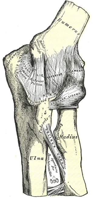
A 2018 review describes a complex of three ligaments: the medial ulnar collateral ligament proper, the posterior bundle and the transverse ligament2. In 28 cadaver elbows, the ligament had anterior and posterior bundles and only occasionally a transverse bundle5. A cadaver is a body donated for study.
How the front bundle is built
The anterior bundle starts on the front and underside of the medial epicondyle2. It ends on the sublime tubercle of the ulna, a tubercle being a small, rounded bony bump2. Cadaver studies have measured it:
| What was measured | What was found |
|---|---|
| Length of the ligament | Averaged 53.9 mm in a study of 10 cadaver specimens and 100 bone specimens6 |
| Attachment on the ulna | Ran 29.2 mm along a ridge of bone extending down from the sublime tubercle, in the same study6 |
| Area of its two ends | Averaged 45.5 mm² at its start and 127.8 mm² at its end in 13 cadaver elbows, widening toward the ulna7 |
| Change in length with bend | Lengthened by 2.2 mm between 0° and 90° of flexion (bending) in 10 cadaver elbows8 |
Bundles from 34 cadaver specimens were pulled in tension in one study9. The anterior bundle was stiffer and stronger than the posterior bundle, and its fibres of collagen (a strong, rope-like protein) were more strongly aligned9.
What it holds steady
A 2018 review says the ligament has been shown to be the elbow’s main stabiliser against valgus stress2. Valgus is an angle at a joint where the lower part points away from the body’s midline. Dynamic stabilisers such as the flexor-pronator muscles come later in that account2. These are the forearm muscles that start at the inner side of the elbow.
A cutting study of 28 cadaver elbows reported which part does this work at each angle of flexion5:
- The anterior bundle was made of a front band and a back band that tightened in turn as the elbow bent and straightened5.
- At 30°, 60° and 90° of flexion the front band was the main restraint to valgus rotation, and at 120° it shared that role5.
- The back band shared the main restraint at 120°, and was a secondary restraint at 30° and 90°5.
- The posterior bundle was a secondary restraint to valgus rotation at 30° of flexion only5.
The transverse ligament is generally thought not to add significantly to elbow stability2. Its direct attachment onto the anterior bundle may give it a role2. In 12 embalmed cadaver elbows held at 30° of flexion, cutting it made the inner joint space significantly wider under valgus stress10. The authors took this to suggest that the transverse bundle may be involved in valgus stability10.
Breakdown
The load of a throw
The ligament is commonly injured in overhead throwing athletes2. This happens when a valgus load is placed on the elbow in the late cocking and early acceleration phases of the throw2. Studies have put numbers on that load and on the ligament’s strength11,12. Torque, a turning force, is given in newton-metres (N·m) and force in newtons (N).
| What was measured | What was found |
|---|---|
| Varus torque the elbow generated in a pitch | 64 N·m in 26 highly skilled, healthy adult pitchers, by motion analysis11 |
| Valgus torque across 81 professional pitchers | A mean of 60 N·m, with individual pitchers’ means from 41 to 94 N·m2 |
| Force at which the anterior ligament failed | An average of 260 N, tested with bone left at both ends12 |
Varus is an angle at a joint where the lower part points toward the body’s midline. One account puts the load on the inner elbow in overhead throwing at 290 N of pulling force2. That is more than the 260.9 N at which the anterior bundle failed in a cadaver model2. Earlier studies indicated that throwing asks more of the ligament than its failure strength, which suggests that muscle forces must contribute13.
Throwers and contact athletes
A 2022 review separates two patterns of injury3. In overhead throwers it is typically a chronic wearing-thin of the ligament from repeated valgus stress3. In contact athletes it is usually an acute tear or avulsion of otherwise normal ligament3. An avulsion is a tear in which a tendon or ligament pulls away from the bone it attaches to.
NCAA surveillance of 25 college sports recorded 109 of these injuries from 2009-10 to 2013-1414. Of those, 76.1% came from contact and 23.9% from throwing14. Men’s wrestling and men’s baseball had the highest rates14. Throwing athletes had a significantly higher share of severe injuries than contact athletes14.
Reports from outside throwing describe other loads15,16:
- Gymnastics. One report says gymnasts load the arm along its length again and again with the elbow nearly straight15. Among five female collegiate gymnasts injured over three years, MRI showed four avulsions at the lower attachment and one at the upper15. MRI is a scan that shows soft tissue in detail.
- Dislocation. In 31 patients with an elbow dislocation and no fracture, surgery found the medial ligaments of every elbow completely ruptured or avulsed from the epicondyle16. A dislocation is when a bone is forced fully out of its normal place at a joint.
The UCL tear page covers how the injury is defined, who it affects and what studies of surgery found. A systematic review of 18 studies in baseball found the injuries mostly affected professional or collegiate pitchers17.
Changes seen in elbows without symptoms
On MRI of 426 skeletally mature baseball players, the grade of ligament abnormality did not differ between elbows with and without symptoms18. About 30% of elbows in both groups showed high-grade injury18.
Ultrasound, a scan made with sound waves, was used on 26 major league pitchers without symptoms19. Dark spots in the ligament were seen in 69% of pitching arms against 12% of the other arms19.
Recovery
How the ligament changes with throwing
Ultrasound studies have compared pitchers’ throwing arms with their other arms, and one season with the next4,20.
- Thicker in the throwing arm. Preseason scans over 10 years covered 368 professional pitchers without symptoms20. The ligament averaged 6.15 mm thick in the throwing elbow and 4.82 mm in the other20.
- By high school. Among 71 uninjured school baseball players without symptoms, it was thicker in the throwing arm in the high school players only21.
- Through the season. In 185 professional pitchers in one club, the ligament became thicker and the elbow more lax to valgus stress over the season4. Both changes reversed over the off-season4. Laxity is looseness in a joint or ligament.
- Within one outing. In 25 high school baseball players who each threw 100 pitches, the inner joint space under valgus stress had widened significantly after 60 pitches22. In 15 adult male pitchers without symptoms, neither the joint gap nor the ligament’s thickness or length differed after up to 110 fastballs23.
Among the 368 professional pitchers scanned without symptoms, 12 later injured the ligament20. Their baseline thickness and joint opening were higher than the others’, but not significantly so20.
Blood supply along the ligament
In 18 cadaver elbows injected with ink, the upper part of the ligament had a dense blood supply and the lower part had little24. The upper part appeared to get blood from an artery of the flexor-pronator muscles, and possibly from the bone of the medial epicondyle24. The authors suggested this difference may be a factor in how well injuries at each site heal24.
The graft as tissue
In reconstruction, surgery rebuilds a torn ligament using a graft, a piece of tissue moved from one place to rebuild a damaged part. The palmaris longus tendon, a thin tendon of the forearm, is the graft used most often to rebuild this ligament12. In one laboratory study it was similar to the anterior ligament, at 357 N against 260 N12.
One ultrasound study scanned eight professional pitchers at least two years after reconstruction, and eight matched healthy pitchers25. Joint space at rest, joint space under stress and laxity did not differ significantly between the groups25. The rebuilt ligament was thicker, as grafts typically are25. The UCL tear page covers outcomes of surgery and return to sport.
Connections
The ligament joins the humerus to the ulna on the inner side of the elbow joint1. It lies against the triceps brachii, the flexor carpi ulnaris and the ulnar nerve1. Part of the flexor digitorum superficialis, a forearm muscle that bends the fingers, arises from it1.
A 2020 review names the flexor carpi ulnaris and flexor digitorum superficialis as the dynamic stabilisers of the inner elbow26. The flexor carpi ulnaris is a forearm muscle that bends the wrist toward the little finger. In eight elbow specimens, loading either muscle significantly relieved strain on the ligament, the flexor carpi ulnaris most27. The common flexor tendon page covers these muscles and their shared attachment.
Related exercise pages: push-up. In nine healthy men doing push-ups, the valgus torque at the elbow, which the medial ligaments oppose, rose significantly with the hands placed further up28. It rose by 42% in a one-handed push-up that simulated a fall28.
Ulnar collateral ligament of the elbow steadies
Ulnar collateral ligament of the elbow joins
Ulnar collateral ligament of the elbow is injured in
Ulnar collateral ligament of the elbow shares valgus restraint with
Ulnar collateral ligament of the elbow lies against
Ulnar collateral ligament of the elbow is loaded by
Sources
- Gray H; Lewis WH (editor) (1918). Anatomy of the Human Body. Lea & Febiger, Philadelphia (20th edition). Link
- Labott JR, Aibinder WR, Dines JS, Camp CL (2018). Understanding the medial ulnar collateral ligament of the elbow: Review of native ligament anatomy and function.. World journal of orthopedics. doi:10.5312/wjo.v9.i6.78 PMID 29984194
- Vaswani R, White A, Dines J (2022). Medial Ulnar Collateral Ligament Injuries in Contact Athletes.. Current reviews in musculoskeletal medicine. doi:10.1007/s12178-022-09785-0 PMID 35917095
- Chalmers PN, English J, Cushman DM, Zhang C, Presson AP, Yoon S, Schulz B, Li B (2021). The ulnar collateral ligament responds to stress in professional pitchers.. Journal of shoulder and elbow surgery. doi:10.1016/j.jse.2020.06.027 PMID 32650069
- Callaway GH, Field LD, Deng XH, Torzilli PA, O'Brien SJ, Altchek DW, Warren RF (1997). Biomechanical evaluation of the medial collateral ligament of the elbow.. The Journal of bone and joint surgery. American volume. doi:10.2106/00004623-199708000-00015 PMID 9278083
- Farrow LD, Mahoney AJ, Stefancin JJ, Taljanovic MS, Sheppard JE, Schickendantz MS (2011). Quantitative analysis of the medial ulnar collateral ligament ulnar footprint and its relationship to the ulnar sublime tubercle.. The American journal of sports medicine. doi:10.1177/0363546511406220 PMID 21555509
- Dugas JR, Ostrander RV, Cain EL, Kingsley D, Andrews JR (2007). Anatomy of the anterior bundle of the ulnar collateral ligament.. Journal of shoulder and elbow surgery. doi:10.1016/j.jse.2006.11.009 PMID 17583541
- Camp CL, Jahandar H, Sinatro AM, Imhauser CW, Altchek DW, Dines JS (2018). Quantitative Anatomic Analysis of the Medial Ulnar Collateral Ligament Complex of the Elbow.. Orthopaedic journal of sports medicine. doi:10.1177/2325967118762751 PMID 29637082
- Smith MV, Castile RM, Brophy RH, Dewan A, Bernholt D, Lake SP (2019). Mechanical Properties and Microstructural Collagen Alignment of the Ulnar Collateral Ligament During Dynamic Loading.. The American journal of sports medicine. doi:10.1177/0363546518812416 PMID 30495972
- Edama M, Matsuzawa K, Yokota H, Hirabayashi R, Sekine C, Maruyama S, Sato N (2021). Elbow valgus stability of the transverse bundle of the ulnar collateral ligament.. BMC musculoskeletal disorders. doi:10.1186/s12891-021-04760-1 PMID 34641828
- Fleisig GS, Andrews JR, Dillman CJ, Escamilla RF (1995). Kinetics of baseball pitching with implications about injury mechanisms.. The American journal of sports medicine. doi:10.1177/036354659502300218 PMID 7778711
- Regan WD, Korinek SL, Morrey BF, An KN (1991). Biomechanical study of ligaments around the elbow joint.. Clinical orthopaedics and related research. doi:10.1097/00003086-199110000-00023 PMID 1914292
- Park MC, Ahmad CS (2004). Dynamic contributions of the flexor-pronator mass to elbow valgus stability.. The Journal of bone and joint surgery. American volume. doi:10.2106/00004623-200410000-00020 PMID 15466738
- Li NY, Goodman AD, Lemme NJ, Owens BD (2019). Epidemiology of Elbow Ulnar Collateral Ligament Injuries in Throwing Versus Contact Athletes of the National Collegiate Athletic Association: Analysis of the 2009-2010 to 2013-2014 Seasons.. Orthopaedic journal of sports medicine. doi:10.1177/2325967119836428 PMID 31008136
- Nicolette GW, Gravlee JR (2018). Ulnar collateral ligament injuries of the elbow in female division I collegiate gymnasts: a report of five cases.. Open access journal of sports medicine. doi:10.2147/oajsm.s159624 PMID 30233260
- Josefsson PO, Johnell O, Wendeberg B (1987). Ligamentous injuries in dislocations of the elbow joint.. Clinical orthopaedics and related research. doi:10.1097/00003086-198708000-00027 PMID 3301144
- Huang D, Lew WJ, Shimoda B, Snyder EM, Khor KS, Cifu DX, Lew HL (2025). Mechanisms of ulnar collateral ligament injury in baseball: criteria and rationale for return to play - a systematic review.. BMC sports science, medicine & rehabilitation. doi:10.1186/s13102-025-01499-3 PMID 41456039
- Hoshika S, Matsuki K, Takeuchi Y, Takahashi N, Sugaya H (2024). Microscopic Magnetic Resonance Imaging Comparing Asymptomatic and Symptomatic Ulnar Collateral Ligament Injuries in Baseball Players.. The American journal of sports medicine. doi:10.1177/03635465241259472 PMID 39101734
- Nazarian LN, McShane JM, Ciccotti MG, O'Kane PL, Harwood MI (2003). Dynamic US of the anterior band of the ulnar collateral ligament of the elbow in asymptomatic major league baseball pitchers.. Radiology. doi:10.1148/radiol.2271020288 PMID 12616000
- Ciccotti MG, Atanda A, Nazarian LN, Dodson CC, Holmes L, Cohen SB (2014). Stress sonography of the ulnar collateral ligament of the elbow in professional baseball pitchers: a 10-year study.. The American journal of sports medicine. doi:10.1177/0363546513516592 PMID 24473498
- Nagamoto H, Yamamoto N, Kurokawa D, Takahashi H, Muraki T, Tanaka M, Koike Y, Sano H, Itoi E (2015). Evaluation of the thickness of the medial ulnar collateral ligament in junior high and high school baseball players.. Journal of medical ultrasonics (2001). doi:10.1007/s10396-014-0605-1 PMID 26576792
- Hattori H, Akasaka K, Otsudo T, Hall T, Amemiya K, Mori Y (2018). The effect of repetitive baseball pitching on medial elbow joint space gapping associated with 2 elbow valgus stressors in high school baseball players.. Journal of shoulder and elbow surgery. doi:10.1016/j.jse.2017.10.031 PMID 29289491
- van Trigt B, Goethem JV, van den Bekerom MMPJ, Veeger DHEJ, Hoozemans MMJM (2024). The ulnar collateral ligament response to valgus stress, repetitive pitching, and elbow muscle contraction in asymptomatic baseball pitchers.. JSES reviews, reports, and techniques. doi:10.1016/j.xrrt.2023.11.005 PMID 38706678
- Buckley PS, Morris ER, Robbins CM, Kemler BR, Frangiamore SJ, Ciccotti MG, Huard J, LaPrade RF, Hackett TR (2019). Variations in Blood Supply From Proximal to Distal in the Ulnar Collateral Ligament of the Elbow: A Qualitative Descriptive Cadaveric Study.. The American journal of sports medicine. doi:10.1177/0363546519831693 PMID 30896969
- Hanna AJ, Sonnier JH, Fliegel BE, Erickson BJ, Jack RA, Cohen SB (2024). Dynamic Stress Ultrasound Assessment of Professional Baseball Players' Elbows After Ulnar Collateral Ligament Reconstruction.. The American journal of sports medicine. doi:10.1177/03635465241227436 PMID 38353117
- Cinque ME, Schickendantz M, Frangiamore S (2020). Review of Anatomy of the Medial Ulnar Collateral Ligament Complex of the Elbow.. Current reviews in musculoskeletal medicine. doi:10.1007/s12178-020-09609-z PMID 31989529
- Lin F, Kohli N, Perlmutter S, Lim D, Nuber GW, Makhsous M (2007). Muscle contribution to elbow joint valgus stability.. Journal of shoulder and elbow surgery. doi:10.1016/j.jse.2007.03.024 PMID 17936028
- Donkers MJ, An KN, Chao EY, Morrey BF (1993). Hand position affects elbow joint load during push-up exercise. Journal of biomechanics. doi:10.1016/0021-9290(93)90026-b PMID 8514808
Changes
- Revised to report sources added after their published corrections were read