Osgood-Schlatter disease
Also called Osgood-Schlatter, Osgood-Schlatter syndrome, Osgood-Schlatter's disease, traction apophysitis of the tibial tubercle, tibial tubercle apophysitis
Pain at the bony bump below the knee where the patellar ligament attaches, in growing children and teenagers. How reviews define it, who studies found it in, how it changes over the years, and what trials and reviews found.
- Region
- Knee
- Muscles
- quadriceps
- Updated
- October 15, 2026
- Reviewed
- October 15, 2026 · how entries are checked
What it is
How reviews and guidelines describe it
A Cochrane review names the condition traction apophysitis of the tibial tubercle1. An apophysis is a bony bump where a muscle attaches that grows from its own growth plate, and apophysitis is soreness and irritation there. Osgood-Schlatter’s disease is the condition’s eponym, a name taken from a person1. It was first described in 1903, separately by Osgood in the United States and Schlatter in Switzerland4.
A 2021 systematic review, a study that searches for all the research on a question, calls it a knee condition linked to sport and growth7. It places painful changes around the apophysis of the tibial tuberosity, and says up to 10% of adolescents are affected7. A 2022 systematic review also links it with growth and sport4.
A 2022 Dutch general-practice study cites its 2016 guideline on knee complaints that did not follow an injury8. Under it, the condition is identified from the history and a physical examination8. It is managed conservatively, without surgery, according to how severe the symptoms are8.
Where the pull falls
The tibial tuberosity (a bump on the front of the shinbone, just below the knee) is where the tendon from the kneecap attaches2. The four quadriceps muscles share one tendon that inserts into the kneecap and carries on below it as the patellar ligament2. Through the kneecap and this ligament, the quadriceps straighten the knee2.
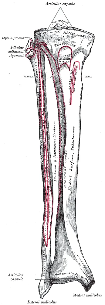
It is thought of as a traction apophysitis of the tibial tubercle6. It is put down to high forces from the patellar tendon pulling on its attachment while the growth plate is still open6. A growth plate is a layer of cartilage near the end of a growing bone. Traction growth plates like this one sit where major tendons attach, and they mainly take tension9.
The Cochrane review states that this apophysis is commonly present from 8 to 12 in girls and 12 to 15 in boys1. It says rapid growth and weight change in these years are considered the main reason these injuries appear at these ages1.
Who it affects
How often studies found it
| Group studied | What was found |
|---|---|
| 956 school students aged 12 to 15 in Natal, Brazil | 9.8% met the study’s criteria, with boys at 11.0% and girls at 8.3%3 |
| 389 students answering a questionnaire in 1985 | 12.9% had had it: 21.2% of those active in sport at 13, against 4.5% of those who were not10 |
| Dutch general-practice records covering over 200,000 patients, 2012 to 2017 | 3.8 new cases per 1,000 person-years at ages 8 to 18: 4.9 in boys and 2.7 in girls8 |
| 127 youth elite male football players at a pre-season health check | 17% had it, and 80% of those had lost no time from training or matches11 |
Prevalence is the share of people in a group who have a condition at a given time. Incidence is how many new cases appear in a group over a set time. Person-years are a count of people multiplied by the years each was followed. The Cochrane review notes that general-population figures exist only for the knee and heel forms of apophyseal injury1.
Boys, girls and sport
The 2022 review reports a male-to-female ratio of 3 to 1, and both knees affected in 20 to 30% of patients4. It lists soccer, gymnastics, basketball, football, volleyball and athletics as sports commonly linked with it4. In a series of 126 consecutive patients, 101 were boys, 64 played football and 18 basketball5.
Where studies disagree on risk
The 2022 review covered 16 studies and found the causes and risk factors controversial4. Its main studies looked at muscle factors, chiefly a tight rectus femoris, the quadriceps muscle that also bends the hip4,12. They also looked at the patellar tendon, repeated loading and sport, and the shape of the shinbone4. Single studies point different ways3,5,11:
- Muscle length: in the Brazilian study, regular sport and a shortened rectus femoris were the factors linked with it3. In a one-year study of 37 adolescent male football players, quadriceps tightness was among the risk factors13.
- Growth and build: in the 126-patient series, onset age, growth rate, body mass index (weight for height) and muscle imbalance did not reach significance5. In a case-control study of 30 youth elite male football players, growth speed, bone maturity, flexibility and strength were not linked with it11. A case-control study compares people who have a condition with similar people who do not.
- Earlier heel pain: in those 30 players, earlier Sever’s disease was strongly linked with it11. The odds ratio, how many times higher the odds are in one group, was 16.8, with a wide confidence interval (a range showing precision)11.
What studies found
How it changes over time
A prospective study follows people forward in time, and a cohort is a group followed over time. Studies that followed patients for months or years report how many still had pain5,6,14.
| Patients followed | What was found |
|---|---|
| 126 patients in a prospective series, 105 followed a median of 3.6 years | 78.8% still had pain on kneeling or pressing the bump, which did not hold them back5 |
| 51 adolescents aged 10 to 14 in a prospective cohort, all after a 12-week programme | 37% of the 46 followed up still had knee pain from it at 24 months14 |
| 43 responders from one orthopaedic department, a median of 3.75 years later | 26 (60.5%) reported knee pain from it, and 42.9% of those had pain daily6 |
| 400 of 1,281 Danish adults seen in hospital for it as teenagers, who answered a survey | 85% reported a bony bump at the tubercle, and 73% ongoing pain or problems there15 |
In the series of 126, half were free of symptoms in or after sport by 16 months, and three-quarters by 25 months5. Its authors concluded that the condition settles by itself without specific measures5. The authors of the 43-responder study concluded it may not always be self-limiting6. The Cochrane review notes increasing evidence that traction apophysitis of the tibial tubercle can bring chronic pain and limits on activity into adulthood1.
In the series of 126, 28.3% had switched sport because of the condition5. In the cohort of 51, more than 1 in 5 had stopped sport because of knee pain at 24 months14. The Danish adults scored below healthy population estimates on all knee-health scales15.
In the cohort of 51, ultrasound (a scan made with sound waves) at 24 months showed mainly tendon changes, such as thickening14. An ununited ossicle, a small separate bone, showed in 32% of those scanned14.
What the Cochrane review compared
The review searched up to January 2025 and found 10 randomised controlled trials on apophyseal injuries, in 654 children1. Such trials assign people to groups by chance. Three were on this condition1. Certainty of evidence is how sure reviewers are that a finding is right.
| Comparison | Evidence | What the review judged |
|---|---|---|
| Dexamethasone (a drug) against placebo (a dummy version), in children | One study of 23 participants, very low certainty | Very uncertain whether it changes short-term pain, physical function or sport participation1 |
| Dexamethasone against usual care, in children | One study, very low certainty | Very uncertain for the same outcomes1 |
| 12.5% dextrose (a simple sugar) with lidocaine (a local anaesthetic), injected, against usual care | 43 participants medium term, 35 long term, very low certainty | Very uncertain whether pain during activity changes1 |
| Adverse events, dexamethasone or dextrose against placebo | Two studies of 74 participants, very low certainty | Very uncertain; withdrawals because of adverse events were not measured1 |
The review concludes that trials give too little evidence to support most approaches commonly used for this condition or the heel form1. In many outcomes, the comparison groups got better too1. It states that no trial of any design has rigorously tested ice, anti-inflammatory drugs, painkillers or stretching1.
A randomised trial at Hvidovre University Hospital, called SOGOOD, compared a new approach with usual care in 130 adolescents aged 10 to 1616. Its ClinicalTrials.gov record lists it as completed in March 2024, with no results posted16. The Cochrane review’s declarations say physiotherapy associations and six charitable foundations funded it1. Two of the review’s authors are named investigators in it1.
Other reviews and studies
A 2025 meta-analysis, which pools the results of several earlier studies, looked at dextrose injections17. It included four studies of 166 patients, 162 of them male: three randomised trials and one case series17. At three months, it found no significant difference in patient-reported improvement between dextrose and placebo injections17. Pooling two trials, it found greater improvement at one year with dextrose (standardised mean difference, a gap on a common scale: 1.09)17. In two trials of athletic young patients, pain-free return to sport at three months was about twice as likely after dextrose (risk ratio 2.11)17.
The authors say dextrose may help, and that more high-quality trials are needed17. One trial enrolled adult military personnel and the others young athletes17. The authors judged every trial to have some concerns about risk of bias, mainly because the protocols were not registered17. The same meta-analysis was supported by a Korea University grant, and its authors declared no conflicts of interest17.
A 2021 systematic review of non-surgical options included 13 studies, only two of them randomised trials, of poor to moderate quality7. It found no randomised trial comparing specific exercises with sham or usual care7. A 2026 review followed PRISMA, a reporting standard for reviews, but calls itself a scoping review18. Such a review maps what research exists on a topic18. It included 15 studies of 712 patients, most of them retrospective, which look back at past records18.
The 2026 review concluded that non-surgical approaches are effective18. In the non-surgical studies it included, full recovery of daily activities ranged from 14.3% to 100%18. That conclusion differs from the Cochrane review’s, which found too little trial evidence for most common approaches1.
One study followed 51 adolescents aged 10 to 14 through 12 weeks of activity changes and knee strengthening, with no comparison group19. A successful outcome was reported by 80% at 12 weeks and 90% at 12 months19. These are not two separate groups: the 24-month cohort above followed the same 51 adolescents after this programme14.
Surgery
The 2026 review concluded that surgery is rare and seems reserved for people with closed growth plates and lasting symptoms18. It describes removing or smoothing the raised tubercle, removing the bursa (a small fluid-filled sac) and nearby irritated tissue, or removing loose ossicles18. Its three ossicle-removal studies were retrospective: one cohort study of 11 knees and two case series of 18 and 23 knees18. A case series reports on a group of patients with no comparison group.
Across six surgical studies, 79% to 100% of patients returned to daily activities18. Five of the six reported complications, at rates from 0% to 75.8%, including infections, thickened scars and leftover pain18.
Among Finnish military recruits over 13 years, surgery for unresolved Osgood-Schlatter disease occurred at 42 per 100,000 recruits20. Of 107 of these men followed a median of ten years, 87% reported no limits in everyday life or work20. Of them, 38% reported no pain at all when kneeling20. Six had minor complications and two had a further operation20.
Connections
The condition sits where the patellar ligament attaches to the tibia, at the tibial tuberosity2.
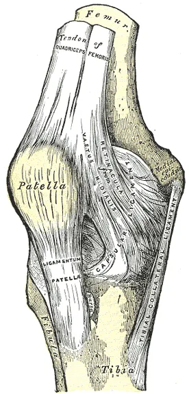
Sever’s disease is the heel form, and the Cochrane review counts the two as the most common lower-limb apophyseal injuries1. The quadriceps include the rectus femoris, the only one of the four that crosses two joints12.
Traction growth plates of cartilage shape a bone but do not add to its length, unlike the growth plates that lengthen it9. Overuse conditions of them, including this one and Sever’s disease, are common in young athletes9.
Osgood-Schlatter disease involves the attachment of
Osgood-Schlatter disease involves the growth plate of
Osgood-Schlatter disease has studies of tightness in
Osgood-Schlatter disease is studied alongside
Osgood-Schlatter disease involves a traction growth plate, described in
Osgood-Schlatter disease is related to
Sources
- Williams CM, Krommes K, Paterson KL, Haines T, Caserta A, Thorborg K (2026). Non-surgical treatment for lower limb apophyseal injuries.. Cochrane Database of Systematic Reviews. doi:10.1002/14651858.CD015156.pub2 PMID 42454655
- Betts JG, Young KA, Wise JA, Johnson E, Poe B, Kruse DH, Korol O, Johnson JE, Womble M, DeSaix P (2022). Anatomy and Physiology 2e. OpenStax. Link
- de Lucena GL, dos Santos Gomes C, Guerra RO (2011). Prevalence and associated factors of Osgood-Schlatter syndrome in a population-based sample of Brazilian adolescents.. The American journal of sports medicine. doi:10.1177/0363546510383835 PMID 21076014
- Lucenti L, Sapienza M, Caldaci A, Cristo C, Testa G, Pavone V (2022). The Etiology and Risk Factors of Osgood-Schlatter Disease: A Systematic Review.. Children (Basel, Switzerland). doi:10.3390/children9060826 PMID 35740763
- Gaulrapp H, Nührenbörger C (2022). The Osgood-Schlatter disease: a large clinical series with evaluation of risk factors, natural course, and outcomes.. International orthopaedics. doi:10.1007/s00264-021-05178-z PMID 34427770
- Guldhammer C, Rathleff MS, Jensen HP, Holden S (2019). Long-term Prognosis and Impact of Osgood-Schlatter Disease 4 Years After Diagnosis: A Retrospective Study.. Orthopaedic journal of sports medicine. doi:10.1177/2325967119878136 PMID 31700938
- Neuhaus C, Appenzeller-Herzog C, Faude O (2021). A systematic review on conservative treatment options for OSGOOD-Schlatter disease.. Physical therapy in sport : official journal of the Association of Chartered Physiotherapists in Sports Medicine. doi:10.1016/j.ptsp.2021.03.002 PMID 33744766
- van Leeuwen GJ, de Schepper EI, Rathleff MS, Bindels PJ, Bierma-Zeinstra SM, van Middelkoop M (2022). Incidence and management of Osgood-Schlatter disease in general practice: retrospective cohort study.. The British journal of general practice : the journal of the Royal College of General Practitioners. doi:10.3399/BJGP.2021.0386 PMID 34990396
- Caine D, DiFiori J, Maffulli N (2006). Physeal injuries in children's and youth sports: reasons for concern?. British journal of sports medicine. doi:10.1136/bjsm.2005.017822 PMID 16807307
- Kujala UM, Kvist M, Heinonen O (1985). Osgood-Schlatter's disease in adolescent athletes. Retrospective study of incidence and duration.. The American journal of sports medicine. doi:10.1177/036354658501300404 PMID 4025675
- Schultz M, Tol JL, Veltman L, Kaaden van der L, Reurink G (2022). Osgood-Schlatter Disease in youth elite football: Minimal time-loss and no association with clinical and ultrasonographic factors.. Physical therapy in sport : official journal of the Association of Chartered Physiotherapists in Sports Medicine. doi:10.1016/j.ptsp.2022.02.024 PMID 35305497
- Begum FA, Kayani B, Chang JS, Tansey RJ, Haddad FS (2020). The management of proximal rectus femoris avulsion injuries.. EFORT Open Reviews. doi:10.1302/2058-5241.5.200055 PMID 33312709
- Nakase J, Goshima K, Numata H, Oshima T, Takata Y, Tsuchiya H (2015). Precise risk factors for Osgood-Schlatter disease.. Archives of orthopaedic and trauma surgery. doi:10.1007/s00402-015-2270-2 PMID 26133498
- Holden S, Olesen JL, Winiarski LM, Krommes K, Thorborg K, Hölmich P, Rathleff MS (2021). Is the Prognosis of Osgood-Schlatter Poorer Than Anticipated? A Prospective Cohort Study With 24-Month Follow-up.. Orthopaedic journal of sports medicine. doi:10.1177/23259671211022239 PMID 34435066
- Krommes K, Bjerre A, Thorborg K, Nielsen MF, Hölmich P (2025). Long-Term Knee Health in Adults with a History of Adolescent Osgood-Schlatter: A National Cohort Study of Patients in Secondary Care in Denmark 1977-2020.. Sports medicine (Auckland, N.Z.). doi:10.1007/s40279-025-02214-5 PMID 40439870
- Krommes K (principal investigator); Hvidovre University Hospital (sponsor) (2021). A Comparison of Two Different Treatment Approaches for Adolescents With Osgood Schlatter (ClinicalTrials.gov NCT05174182). ClinicalTrials.gov. Link
- Rhim HC, Bjork LB, Shin J, Park J, DeLuca SE, McCarron KC, Jang KM, Ha C (2025). Efficacy of Hyperosmolar Dextrose Injection for Osgood-Schlatter Disease: A Systematic Review with Meta-Analysis.. Diagnostics (Basel, Switzerland). doi:10.3390/diagnostics15101282 PMID 40428275
- Ndjonko LCM, Klein JH, Chakraborty Y, Kata S, Alinda A, Abuelenein I, Kalluvila AT, Green DW, Fowowe O, Simpson S, Wooldridge T (2026). Treatments for Osgood Schlatter Disease: A Systematic Review of the Literature.. Orthopaedic journal of sports medicine. doi:10.1177/23259671251387354 PMID 41788553
- Rathleff MS, Winiarski L, Krommes K, Graven-Nielsen T, Hölmich P, Olesen JL, Holden S, Thorborg K (2020). Activity Modification and Knee Strengthening for Osgood-Schlatter Disease: A Prospective Cohort Study.. Orthopaedic journal of sports medicine. doi:10.1177/2325967120911106 PMID 32284945
- Pihlajamäki HK, Mattila VM, Parviainen M, Kiuru MJ, Visuri TI (2009). Long-term outcome after surgical treatment of unresolved Osgood-Schlatter disease in young men.. The Journal of bone and joint surgery. American volume. doi:10.2106/JBJS.H.01796 PMID 19797569
Changes
- First published.