Hip labrum
Also called acetabular labrum, labrum of the hip, cotyloid ligament
The rim of fibrocartilage around the edge of the hip socket. What it is made of, how it seals the joint, where and in whom it tears, how often scans show tears in hips without pain, and what studies found about healing.
- Region
- Hip
- Updated
- October 8, 2026
- Reviewed
- October 8, 2026 · how entries are checked
Function
A rim around the socket
The labrum of the hip joint is a fibrocartilage structure that runs around the edge of the hip socket1. It is continuous, usually triangular, and attached to the bony rim of the socket1. At the bottom, the transverse acetabular ligament completes the ring across a notch in the socket1. The figure labels the labrum “Cotyloid lig.”, for cotyloid ligament, its name in 1918.
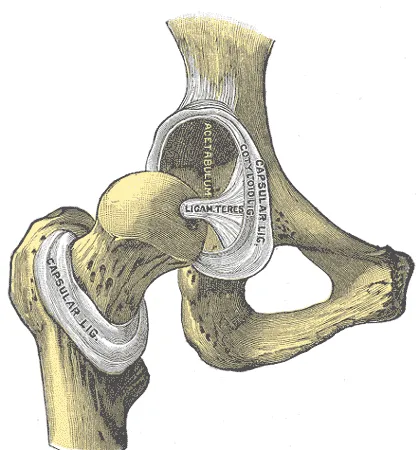
In 55 embalmed (preserved) hips, the labrum averaged 5.3 mm wide, and was wider at the front and top than at the back6. In the same hips, the calculated surface area of the socket was 28.8 square centimetres without the labrum and 36.8 with it6.
The labrum runs into the socket’s articular cartilage (the thin layer of cartilage covering the ends of bones in a joint)7. In a study of 55 embalmed and 12 fresh-frozen adult hips, the two tissues joined through a transition zone 1 to 2 mm wide7.
What it is made of
Most of the labrum is thick bundles of type I collagen (a thick, strong form of collagen)1. The bundles lie mainly parallel to the socket rim1. One study of its structure found dense connective tissue in the outer part and fibrocartilage in the inner layer8.
In that study, blood vessels entered the labrum from the neighbouring joint capsule (the sleeve of tough tissue that encloses a joint)8. They were found in its outer third, and the inner part had none8. A silicone-injection study of 35 hips in 28 fresh cadavers reported a different source9. The supplying vessels ran in a loose layer on the labrum’s capsular surface, and no contribution from the capsule could be shown9. In a study of 12 hips from 6 cadavers, the labrum had a relatively poor blood supply overall10.
The labrum has nerve endings too: in 23 labra from fresh cadavers, free nerve endings were seen in every specimen11. The authors suggested that these endings may take part in sensing pain and joint position11.
How it seals the joint
One review article describes the labrum as a seal1. It helps hold synovial fluid (the slippery fluid that oils a joint) and fluid pressure in the joint1. In one laboratory study, eight cadaver hips were compressed with 2.7 times body weight2. The table gives the average fluid pressure inside the joint, as a share of its value with the labrum intact2.
| State of the labrum | Fluid pressure, % of intact value | Source |
|---|---|---|
| Torn | 75% | 2 |
| Partly removed | 53% | 2 |
| Fully removed | 24% | 2 |
| Partly removed, then rebuilt with a graft | 110% | 2 |
The graft (a piece of tissue moved to rebuild a damaged part) was taken from the iliotibial band2. In a separate laboratory study of six human hip joints, the cartilage layers compressed 22% faster at first after the whole labrum was cut away12. The femoral head is the ball at the top of the thigh bone. The authors concluded that the sealing depends on how well the labrum fits against it12.
In eight cadaver hips pulled apart at the joint, the labrum gave 77% of the resistance at 1 mm of displacement13. It gave 70% at 2 mm, more than the capsule13. The capsule’s share grew with displacement, reaching 52% at 5 mm13.
Breakdown
Where and how it tears
Histology (tissue seen under a microscope) of 55 embalmed and 12 fresh-frozen adult hips showed two types of tear7. In one, the labrum had come away from the articular cartilage at the transition zone7. In the other, one or more splits of varying depth ran within the labrum itself7.
Of 436 consecutive patients having hip arthroscopy (keyhole surgery inside a joint), 241 had labral tears15. Every tear was at the edge facing the joint surface, not the edge facing the capsule15. In that study, 73% of the patients whose labrum was frayed or torn also had cartilage change15.
One review article states that most tears reported in the United States were in the front of the labrum1. In Japan, most were at the back1. In seven cadaver hips loaded with twisting movements, the highest strain (stretch) was concentrated where the labrum joins the bone16.
What a tear changed in cadaver hips
Three studies tested cadaver hips with labral tears13,17,18. Two pulled the cadaver joint apart, and one pressed it together13,17,18.
- Pulled apart, six hips. In six cadaver hips, the thigh bone was pulled 3 mm out of the socket17. That took 43% less force after the capsule was vented (opened), and 60% less after a 15-mm labral tear was made17.
- The seal pulled apart, eight hips. In eight cadaver hips, the peak pulling force the seal withstood averaged 76% of its intact value with a labral tear13. In those cadaver hips, it averaged 29% after part of the labrum was removed and 27% after all of it was removed13.
- Pressed together, 11 hips. Eleven cadaver hips without degeneration or dysplasia were pressed together in a neutral (natural resting) position, with an average force of 25 N18. Circumferential (around the rim) tears of 1, 2 and 3 cm did not significantly change the stability ratio of these cadaver hips18. That ratio is the peak force at dislocation of the cadaver hip, divided by the compressive load (squeezing force)18.
The third study’s authors concluded that in this cadaver set-up the labrum still resists dislocation of the femoral head once detached from the socket rim18. In 11 cadaver hips tested the same way, a radial (across the rim) tear did not significantly change the stability ratio18. Removing 1 cm of labrum from those cadaver hips did not either, while removing 2 cm significantly lowered it18. The authors concluded from the cadaver tests that 2 cm or more of labrum must be removed before hip stability falls18. Having tested cadaver hips in a neutral position, they said further studies are needed in other positions, naming abduction, extension and external rotation18.
In 22 cadaver hips tested the same way, strain in the front and front-upper labrum was unchanged or higher after a circumferential tear18. After a radial tear, strain there fell significantly in the cadaver hips18.
What tears are linked to
One review article lists at least five proposed causes of labral tears1:
- trauma1
- femoroacetabular impingement, in which the upper thigh bone and the hip socket make early contact1,19
- a loose joint capsule or a very mobile hip1
- dysplasia1
- degeneration1
The same article states that labral tears in athletes can follow a single traumatic event or repeated trauma1. Up to 74.1%, it reports, are tied to no known event and come on gradually1. It also states that sports with repeated pivoting on a loaded thigh bone have been associated with labral damage1.
A systematic review covered hip dislocation and subluxation (partial dislocation) in sport20. Its studies that looked inside the joint by arthroscopy found labral tears in 100% of hips20.
A systematic review of 43 articles found labral tears in an average of 78.8% of hips with dysplasia21. The average was 93.8% in hips with femoroacetabular impingement21. The ranges were wide: 17 to 100% and 33 to 100%21. Finite element models compared ten people with normal sockets and ten with dysplasia22. The dysplastic labrum carried 2.8 to 4.0 times more of the joint load22.
Three studies report how often tears were seen at different ages7,23,24:
- In pelvic MRI scans of 108 children aged 2 to 18 without hip pain or disease, 3 of 216 hips had a labral tear23.
- In 45 volunteers without hip symptoms aged 15 to 66, tears on MRI were not associated with age or sex24.
- In a study of embalmed and fresh-frozen adult hips, mean age 78, 53 of 55 hips had labral tears7.
Tears in hips without pain
A systematic review of imaging covered 26 studies and 2,114 hips of volunteers without symptoms, mean age 253. The 7 studies that reported on the labrum saw labral injury on MRI in 68.1% of hips3.
Two meta-analyses report labral tears in people with and without symptoms4,25.
| Who was scanned | With symptoms | Without symptoms | Source |
|---|---|---|---|
| People in 29 imaging studies | 62% (limited evidence) | 54% (moderate evidence) | 25 |
| Athletes in 20 imaging studies | 20% of hips (moderate evidence) | 33% of hips (moderate evidence); 54% of athletes (limited evidence) | 4 |
The authors of the first meta-analysis concluded that findings inside the hip are often seen in people without symptoms25. They wrote that this points to a possible mismatch between imaging findings and pain25. An international consensus group separated two things26. It stated that labral conditions causing hip-related pain should not be confused with incidental imaging findings in people without symptoms26.
Recovery
Whether a tear heals
One review article states that whether labral tears can heal is controversial1. Few vessels reach into the labrum, and for that reason some authors concluded that no part of it can repair itself1.
Two cadaver studies looked at blood vessels in torn labra7,9. In the study of 55 embalmed and 12 fresh-frozen hips, both types of tear were associated with more small blood vessels forming within the tear7. In the silicone-injection study, the blood supply was intact in all seven hips that had a labral tear9.
One study took biopsies from 6 patients whose tear had been stitched by arthroscopy and who later had a hip replacement5. All the repaired labra more or less kept their triangular shape and appeared to have healed5. The tissue looked like typical fibrocartilage with a limited blood supply, and small clefts were present in all cases5.
Two other reports describe tears that had not healed1,27. The review article reports one person whose repeat arthroscopy several months later showed no healing of the torn tissue1. That person had returned to sport without the pain coming back1. In a case series, 23 patients had an osteotomy (surgery that cuts and reshapes a bone) for hip dysplasia27. On MRI at 1 year, the labral tears persisted27.
What happened to painless tears over time
Two studies followed people who had MRI scans of hips without symptoms28,29:
- Of 21 professional hockey players with no previous hip or groin pain, 15 had labral tears on MRI in one or both hips28. Over the next 4 years, 3 players developed any hip or pelvis symptoms and 19 were still playing professionally28.
- A second study covered 100 patients with femoroacetabular impingement symptoms in one hip29. Two surgeons reading MRI found a tear in 41% and 43% of the symptom-free opposite hips29. Over 2 years, 9 patients went on to have a symptomatic labral tear in that previously symptom-free hip29.
After surgery on the labrum
A systematic review and meta-analysis pooled 33 cadaver studies of hip mechanics, covering 322 hips30. A repaired or rebuilt labrum resisted being pulled apart more than a debrided (trimmed) one, but less than a torn labrum left as it was30. Trials in living patients, including arthroscopy against exercise programmes, are on the femoroacetabular impingement page.
Connections
The labrum is attached to the bony rim of the hip socket1. The socket forms on the outer side of the hip bone, part of the pelvis31. The authors of one laboratory study concluded that its seal depends on its fit against the femoral head, the ball of the femur (thigh bone)12.
In eight cadaver hips pulled apart at the joint, the capsule of the hip joint gave 41% of the resistance at 3 mm of displacement13.
The labrum runs into the socket’s articular cartilage7. In tests of cattle labrum, its stiffness in compression was comparable to that of meniscus (a pad of cartilage in the knee)32. The page on cartilage covers the tissue in general.
Related exercise pages: lunge. One study measured the lunge in 21 patients with symptomatic one-sided labral tears and 17 controls without symptoms33. On the way up, average activity of the gluteus maximus (the large muscle of the buttock) was lower in the patients33.
Hip labrum forms part of
Hip labrum attaches to
Hip labrum seals against
Hip labrum merges with
Hip labrum is injured in
Hip labrum is compared with
Hip labrum is related to
Hip labrum has been studied in
Sources
- Groh MM, Herrera J (2009). A comprehensive review of hip labral tears.. Current Reviews in Musculoskeletal Medicine. doi:10.1007/s12178-009-9052-9 PMID 19468871
- Philippon MJ, Nepple JJ, Campbell KJ, Dornan GJ, Jansson KS, LaPrade RF, Wijdicks CA (2014). The hip fluid seal--Part I: the effect of an acetabular labral tear, repair, resection, and reconstruction on hip fluid pressurization.. Knee Surgery, Sports Traumatology, Arthroscopy. doi:10.1007/s00167-014-2874-z PMID 24519614
- Frank JM, Harris JD, Erickson BJ, Slikker W, Bush-Joseph CA, Salata MJ, Nho SJ (2015). Prevalence of Femoroacetabular Impingement Imaging Findings in Asymptomatic Volunteers: A Systematic Review.. Arthroscopy. doi:10.1016/j.arthro.2014.11.042 PMID 25636988
- Heerey JJ, Kemp JL, Mosler AB, Jones DM, Pizzari T, Scholes MJ, Agricola R, Crossley KM (2019). What is the Prevalence of Hip Intra-Articular Pathologies and Osteoarthritis in Active Athletes with Hip and Groin Pain Compared with Those Without? A Systematic Review and Meta-Analysis.. Sports Medicine. doi:10.1007/s40279-019-01092-y PMID 30972659
- Audenaert EA, Dhollander AA, Forsyth RG, Corten K, Verbruggen G, Pattyn C (2012). Histologic assessment of acetabular labrum healing.. Arthroscopy. doi:10.1016/j.arthro.2012.06.012 PMID 23084152
- Tan V, Seldes RM, Katz MA, Freedhand AM, Klimkiewicz JJ, Fitzgerald RH (2001). Contribution of acetabular labrum to articulating surface area and femoral head coverage in adult hip joints: an anatomic study in cadavera.. American Journal of Orthopedics. PMID 11757858
- Seldes RM, Tan V, Hunt J, Katz M, Winiarsky R, Fitzgerald RH (2001). Anatomy, histologic features, and vascularity of the adult acetabular labrum.. Clinical Orthopaedics and Related Research. doi:10.1097/00003086-200101000-00031 PMID 11153993
- Petersen W, Petersen F, Tillmann B (2003). Structure and vascularization of the acetabular labrum with regard to the pathogenesis and healing of labral lesions.. Archives of Orthopaedic and Trauma Surgery. doi:10.1007/s00402-003-0527-7 PMID 12802599
- Kalhor M, Horowitz K, Beck M, Nazparvar B, Ganz R (2010). Vascular supply to the acetabular labrum.. The Journal of Bone and Joint Surgery. American Volume. doi:10.2106/JBJS.I.01719 PMID 21048175
- Kelly BT, Shapiro GS, Digiovanni CW, Buly RL, Potter HG, Hannafin JA (2005). Vascularity of the hip labrum: a cadaveric investigation.. Arthroscopy. doi:10.1016/j.arthro.2004.09.016 PMID 15650660
- Kim YT, Azuma H (1995). The nerve endings of the acetabular labrum.. Clinical Orthopaedics and Related Research. doi:10.1097/00003086-199511000-00029 PMID 7586824
- Ferguson SJ, Bryant JT, Ganz R, Ito K (2003). An in vitro investigation of the acetabular labral seal in hip joint mechanics.. Journal of Biomechanics. doi:10.1016/s0021-9290(02)00365-2 PMID 12547354
- Nepple JJ, Philippon MJ, Campbell KJ, Dornan GJ, Jansson KS, LaPrade RF, Wijdicks CA (2014). The hip fluid seal--Part II: The effect of an acetabular labral tear, repair, resection, and reconstruction on hip stability to distraction.. Knee Surgery, Sports Traumatology, Arthroscopy. doi:10.1007/s00167-014-2875-y PMID 24509878
- Henak CR, Ellis BJ, Harris MD, Anderson AE, Peters CL, Weiss JA (2011). Role of the acetabular labrum in load support across the hip joint.. Journal of Biomechanics. doi:10.1016/j.jbiomech.2011.06.011 PMID 21757198
- McCarthy JC, Noble PC, Schuck MR, Wright J, Lee J (2001). The Role of Labral Lesions to Development of Early Degenerative Hip Disease. Clinical Orthopaedics and Related Research. doi:10.1097/00003086-200112000-00004 PMID 11764355
- Dy CJ, Thompson MT, Crawford MJ, Alexander JW, McCarthy JC, Noble PC (2008). Tensile strain in the anterior part of the acetabular labrum during provocative maneuvering of the normal hip.. The Journal of Bone and Joint Surgery. American Volume. doi:10.2106/JBJS.G.00467 PMID 18594094
- Crawford MJ, Dy CJ, Alexander JW, Thompson M, Schroder SJ, Vega CE, Patel RV, Miller AR, McCarthy JC, Lowe WR, Noble PC (2007). The 2007 Frank Stinchfield Award. The biomechanics of the hip labrum and the stability of the hip.. Clinical Orthopaedics and Related Research. doi:10.1097/BLO.0b013e31815b181f PMID 17906586
- Smith MV, Panchal HB, Ruberte Thiele RA, Sekiya JK (2011). Effect of acetabular labrum tears on hip stability and labral strain in a joint compression model.. The American journal of sports medicine. doi:10.1177/0363546511400981 PMID 21709039
- Hunter DJ, Eyles J, Murphy NJ, Spiers L, Burns A, Davidson E, Dickenson E, Fary C, Foster NE, Fripp J, Griffin DR, Hall M, Kim YJ, Linklater JM, Molnar R, Neubert A, O'Connell RL, O'Donnell J, O'Sullivan M, Randhawa S, Reichenbach S, Schmaranzer F, Singh P, Tran P, Wilson D, Zhang H, Bennell KL (2021). Multi-centre randomised controlled trial comparing arthroscopic hip surgery to physiotherapist-led care for femoroacetabular impingement (FAI) syndrome on hip cartilage metabolism: the Australian FASHIoN trial.. BMC musculoskeletal disorders. doi:10.1186/s12891-021-04576-z PMID 34399702
- Chona DV, Minetos PD, LaPrade CM, Cinque ME, Abrams GD, Sherman SL, Safran MR (2022). Hip Dislocation and Subluxation in Athletes: A Systematic Review.. The American journal of sports medicine. doi:10.1177/03635465211036104 PMID 34623933
- Savoye-Laurens T, Verdier N, Wettstein M, Baulot E, Gédouin JE, Martz P (2023). Labral tears in hip dysplasia and femoroacetabular impingement: A systematic review.. Orthopaedics & Traumatology: Surgery & Research. doi:10.1016/j.otsr.2022.103539 PMID 36587762
- Henak CR, Abraham CL, Anderson AE, Maas SA, Ellis BJ, Peters CL, Weiss JA (2014). Patient-specific analysis of cartilage and labrum mechanics in human hips with acetabular dysplasia.. Osteoarthritis and Cartilage. doi:10.1016/j.joca.2013.11.003 PMID 24269633
- Georgiadis AG, Seeley MA, Chauvin NA, Sankar WN (2016). Prevalence of acetabular labral tears in asymptomatic children.. Journal of Children's Orthopaedics. doi:10.1007/s11832-016-0717-9 PMID 26907862
- Register B, Pennock AT, Ho CP, Strickland CD, Lawand A, Philippon MJ (2012). Prevalence of Abnormal Hip Findings in Asymptomatic Participants. The American Journal of Sports Medicine. doi:10.1177/0363546512462124 PMID 23104610
- Heerey JJ, Kemp JL, Mosler AB, Jones DM, Pizzari T, Souza RB, Crossley KM (2018). What is the prevalence of imaging-defined intra-articular hip pathologies in people with and without pain? A systematic review and meta-analysis.. British Journal of Sports Medicine. doi:10.1136/bjsports-2017-098264 PMID 29540366
- Reiman MP, Agricola R, Kemp JL, Heerey JJ, Weir A, van Klij P, Kassarjian A, Mosler AB, Ageberg E, Hölmich P, Warholm KM, Griffin D, Mayes S, Khan KM, Crossley KM, Bizzini M, Bloom N, Casartelli NC, Diamond LE, Di Stasi S, Drew M, Friedman DJ, Freke M, Gojanovic B, Glyn-Jones S, Harris-Hayes M, Hunt MA, Impellizzeri FM, Ishøi L, Jones DM, King MG, Lawrenson PR, Leunig M, Lewis CL, Mathieu N, Moksnes H, Risberg MA, Scholes MJ, Semciw AI, Serner A, Thorborg K, Wörner T, Dijkstra HP (2020). Consensus recommendations on the classification, definition and diagnostic criteria of hip-related pain in young and middle-aged active adults from the International Hip-related Pain Research Network, Zurich 2018.. British Journal of Sports Medicine. doi:10.1136/bjsports-2019-101453 PMID 31959678
- Yang F, Shao J, Cui N, Xu Y, Wang J, Ju X (2025). Persistence of Labral Tears and Resolution of Paralabral Cysts With 2-Year Magnetic Resonance Imaging Analysis After Periacetabular Osteotomy for Hip Dysplasia.. Arthroscopy. doi:10.1016/j.arthro.2024.10.036 PMID 39510205
- Gallo RA, Silvis ML, Smetana B, Stuck D, Lynch SA, Mosher TJ, Black KP (2014). Asymptomatic hip/groin pathology identified on magnetic resonance imaging of professional hockey players: outcomes and playing status at 4 years' follow-up.. Arthroscopy. doi:10.1016/j.arthro.2014.04.100 PMID 24996873
- Vahedi H, Aalirezaie A, Azboy I, Daryoush T, Shahi A, Parvizi J (2019). Acetabular Labral Tears Are Common in Asymptomatic Contralateral Hips With Femoroacetabular Impingement.. Clinical Orthopaedics and Related Research. doi:10.1097/CORR.0000000000000567 PMID 30444756
- Hoffer AJ, Beel W, Ng KCG, Degen RM (2024). The Contribution of Soft Tissue and Bony Stabilizers to the Hip Suction Seal: A Systematic Review of Biomechanical Studies.. The American Journal of Sports Medicine. doi:10.1177/03635465231208193 PMID 38318815
- Betts JG, Young KA, Wise JA, Johnson E, Poe B, Kruse DH, Korol O, Johnson JE, Womble M, DeSaix P (2022). Anatomy and Physiology 2e. OpenStax. Link
- Ferguson SJ, Bryant JT, Ito K (2001). The material properties of the bovine acetabular labrum.. Journal of Orthopaedic Research. doi:10.1016/S0736-0266(01)00007-9 PMID 11562138
- Dwyer MK, Lewis CL, Hanmer AW, McCarthy JC (2016). Do Neuromuscular Alterations Exist for Patients With Acetabular Labral Tears During Function?. Arthroscopy : the journal of arthroscopic & related surgery : official publication of the Arthroscopy Association of North America and the International Arthroscopy Association. doi:10.1016/j.arthro.2016.03.016 PMID 27129378
Changes
- Revised to report sources added after their published corrections were read
- First published.