Internal oblique
Also called internal abdominal oblique, obliquus internus abdominis
The middle of the three flat muscles on each side of the abdomen. How it twists and steadies the trunk, where side strains tear it, how it thickens or thins, and what it works with.
- Region
- Trunk
- Starts on
- the iliac crest (top rim of the hip bone), the groin (inguinal) ligament and the thoracolumbar fascia (Pelvis)2,4
- Ends on
- the lower ribs, the sheath around the rectus abdominis and, with transversus abdominis, the pubic crest via the conjoint tendon (Pelvis)2,4,9
- Updated
- September 30, 2026
- Reviewed
- September 30, 2026 · how entries are checked
Function
Where it lies and how its fibres run
In the classic account, the side and front abdominal wall lies in layers1. The internal oblique sits between the external oblique outside and the transversus abdominis inside1. Its fibres run at right angles to those of the external oblique, up and toward the midline2.
A study of 26 embalmed cadavers (bodies donated for study) looked more closely7. There, the upper and middle fascicles (bundles of muscle fibres lying side by side) ran up and toward the midline7. The lower fascicles ran down and toward the midline7. In its lower and middle regions, the muscle could be split into two distinct layers7.
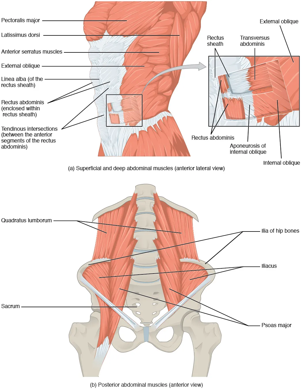
What it attaches to
A baseball injury study describes the internal oblique as starting from the iliac crest, the curved top rim of the hip bone4. It also starts from the inguinal ligament, a band across the groin4. A third start is the thoracolumbar fascia, a sheet of tough connective tissue over the lower and middle back4. It ends on the lower ribs and nearby tissue, including the rectus sheath, the casing of flat tendon sheets around the rectus abdominis4. Both obliques and the transversus abdominis attach to the ilium, the wide upper part of the hip bone in the pelvis2.
Toward the front, the muscle helps build casings and walls8,9:
- The rectus sheath around the rectus abdominis is made from the coverings of the three flat side muscles8.
- Its aponeurosis (a broad, flat sheet of tendon), as usually described, splits to pass in front of and behind the rectus muscles10.
- The conjoint tendon forms from the sheet the internal oblique shares with the transversus abdominis9. Its fibres attach to the pubic crest, the ridge along the top of the pubic bone9. It makes up most of the inner part of the back wall of the inguinal canal, a passage through the groin9.
Its nerves
An expert anatomy consensus describes the side abdominal wall muscles as supplied by the 7th to 11th intercostal nerves, which run between the ribs11. The subcostal nerve, which runs beneath the lowest rib, supplies them too11. In 40 dissected half-walls of the abdomen, the 10th intercostal nerve was consistently found between these two muscles12. It sent a median (middle value) of nine branches to the internal oblique12.
How it twists the trunk
OpenStax, an anatomy textbook, names both obliques as prime movers, the main muscles, for twisting at the waist2. It names them for bending the spine to the side too2. One trunk-rotation study cites earlier work: turning toward the same side, activity reached 58% of maximum voluntary contraction, the hardest possible effort13. Turning the other way, it was 16%13.
In 25 volunteers twisting as hard as they could, the internal oblique reached 55% of the maximum it showed in non-twisting efforts14. Opposing trunk muscles worked together to a large degree, known as co-contraction14. The author took this to suggest that holding the lower-back joints steady matters more in twisting than making a large turning force14.
Pressure and timing
Six men did repeated, varied breath-hold strains15. Peak pressure inside the abdomen tracked the activity of the internal oblique and the other abdominal muscles measured, though not the rectus abdominis15. The same men also pushed the trunk forward while holding a maximal breath-hold strain15. Internal oblique activity rose markedly15. Pushing backward, it fell, unlike the transversus abdominis15.
Studies of when the muscle switches on give different pictures for different tasks16,17:
- In 15 healthy people moving the hip, trunk muscles switched on before the leg’s main mover16. The obliques’ reaction time was consistent whichever way the hip moved16.
- In 15 people making fast arm movements while standing, the obliques’ start time varied with the direction of movement17.
- In 11 people without disabling low back pain making fast arm movements, the middle and lower regions switched on at similar times18. In the six who also moved the arm while sitting, the abdominal muscles switched on later sitting than standing18.
Breakdown
Where side strains tear it
An MRI study of athletes with side strain found the internal oblique torn away from the underside of one of the lower four ribs3. The tear could also be from one of their costal cartilages, the strips of cartilage that join ribs to the breastbone3. A narrative review of side strain in sport found that imaging typically shows the internal oblique torn from the lower ribs19. A study of one professional baseball team also found most strains at the lower ribs4. MRI, a scan that shows soft tissue in detail, can show where the tear is and how severe it is3.
In a case series (a report on several patients) of four cricketers, the authors conclude side strain comes from eccentric loading of the torso20. That is load while the muscle lengthens20. They describe it as rare where these muscles attach to the pelvis20. In those four cricketers, the injuries involved only the internal oblique’s attachment to the iliac crest20. In one, MRI showed a tear right through the front half of that attachment, 4 cm long20.
A case report (a report on a single patient) describes a professional tennis player with an internal oblique strain21. It came from an unbalanced twist of the trunk while the muscle lengthened, and MRI confirmed it21.
Strains in baseball
Across the major and minor leagues from 2011 to 2021, there were 1,928 oblique injuries that cost playing time22. The number per season rose significantly22. These counts are for oblique injuries, not the internal oblique alone22.
One Japanese professional team of 188 players was followed for a decade4. Of these, 23 players (12.2%) had an internal oblique strain confirmed by MRI4. Fielders had it significantly more often than pitchers4:
| Group | Share with an internal oblique strain |
|---|---|
| Fielders | 24.7%4 |
| Pitchers | 7.5%4 |
In that team, most strains were where the muscle attaches to the lower ribs4. They were more common on the side opposite the batting or throwing side, but happened on either side4. The authors state that swinging a bat turns the trunk toward that opposite side, which asks a lot of the internal oblique there4. Five of the 23 players (21.7%) had another strain, on average 36.5 months after the first4. Most second strains were on the side opposite the first4.
Thinning and weak spots
A systematic review, which gathers and sums up the research on a question, looked at trunk muscles in adults aged 50 and over23. The internal oblique, rectus abdominis and external oblique were 36% to 48% thinner in older than in younger adults23.
Recovery
Time away from sport
Average time out after a strain was measured in two baseball studies4,22.
| Group | Average days out |
|---|---|
| Major and minor leagues, batting injuries | 22 days22 |
| One Japanese team, internal oblique strains | 27.7 days4 |
| Major and minor leagues, pitching injuries | 29 days22 |
How it adapts to training
In a randomised trial of 73 people aged 18 to 45, 40 did 16 sessions of rotation exercises over four weeks25. Their internal oblique grew thicker on ultrasound, a scan made with sound waves, compared with their own first scan25. The published summary gives no comparison with the untrained group25. In a one-time comparison, 16 female rugby players had a thicker right internal oblique than 16 women who did not play sport5.
An MRI study looked at professional tennis and soccer players26. The tennis players’ extra oblique and transversus abdominis muscle was confined to the upper segments of the trunk26. In the soccer players, it was similar along the trunk26.
A meta-analysis looked at people with long-lasting low back pain27. It found no difference in resting thickness between motor control training and other approaches27. Motor control training trains how the deep trunk muscles are switched on and coordinated.
What happens without load
Five astronauts spent six months on the International Space Station, across seven missions6. Between the scans before and after flight, their internal oblique thinned by 15.4% on ultrasound6.
Connections
Muscles that share its work
The internal oblique is described as working with the transversus abdominis and latissimus dorsi on the side the trunk turns toward13. The external oblique and rectus abdominis are described as working on the opposite side13. In 24 healthy young men turning the trunk on a machine, the external oblique showed more activity than the internal oblique or latissimus dorsi28.
Different ways of tightening the belly have been studied29,30,31:
- Bracing is described as tightening the rectus abdominis, both obliques and the transversus abdominis together, pushing the waist out29.
- Hollowing, drawing the belly button in toward the spine, thickened the internal oblique more than bracing in 44 healthy young adults30.
- Posterior pelvic tilt, tucking the tailbone under, made the middle internal oblique the most active muscle in one study; bracing mainly used the external oblique31.
In the bird dog, 29 healthy volunteers showed the most activity in the internal oblique on the side of the raised arm32. The erector spinae, the long muscles beside the spine that straighten the back, peaked on the other side32.
Exercises that load it
A systematic review of healthy adults found the highest internal oblique activity in core stability exercises33. Many of the results below use EMG (electromyography, which picks up a muscle’s electrical signals)33; others measure thickness on ultrasound30. They compare activity with MVIC, a muscle’s hardest effort while the joint stays still33. Each row below comes from one study or one review33,34:
| Task | What was found |
|---|---|
| Front plank, shoulder blades drawn together and pelvis tucked (review) | About 120% of MVIC, the highest of the core stability exercises33 |
| Plank with the belly drawn in, 20 people | More activity than a plain plank34 |
| Plank with feet raised, 24 young adults | More activity than feet on the ground35 |
| Side plank (one study, in a review) | About 26% of maximum33 |
| Side plank with a hard breath out (one study, in a review) | About 55% of maximum, against about 18% in the ordinary side plank33 |
| Side plank, bird dog and beast crawl, 44 young adults | Largest thickness gain in the side plank, then the bird dog, then the beast crawl30 |
| Full bird dog against one arm raised, 30 women | More activity in the full position, except the right internal oblique36 |
| Bridge with one leg lifted, 15 adults | More activity on both sides than the standard bridge37 |
| Kettlebell swing with a forceful finish (review of three studies) | About 81% of MVIC, the highest of the free-weight exercises33 |
The hollow hold page covers related holds such as the V-sit38. In 30 adults, internal oblique activity did not differ detectably between a V-sit and a plank38.
Internal oblique lies beneath
Internal oblique forms the conjoint tendon with
Internal oblique forms part of the sheath of
Internal oblique attaches to
Internal oblique works with in the bird dog
Internal oblique is loaded by
Sources
- Seeras K, Qasawa RN, Ju R, Prakash S (2023). Anatomy, Abdomen and Pelvis: Anterolateral Abdominal Wall.. StatPearls. PMID 30247850
- Betts JG, Young KA, Wise JA, Johnson E, Poe B, Kruse DH, Korol O, Johnson JE, Womble M, DeSaix P (2022). Anatomy and Physiology 2e. OpenStax. Link
- Connell DA, Jhamb A, James T (2003). Side strain: a tear of internal oblique musculature.. AJR American Journal of Roentgenology. doi:10.2214/ajr.181.6.1811511 PMID 14627566
- Komatsu S, Kaneko H, Nagashima M (2022). Characteristics of internal oblique muscle strain in professional baseball players: a case series.. BMC Sports Science, Medicine and Rehabilitation. doi:10.1186/s13102-022-00510-5 PMID 35752868
- Abuín-Porras V, de la Cueva-Reguera M, Benavides-Morales P, Ávila-Pérez R, Cruz-Torres B, Pareja-Galeano H, Blanco-Morales M, Romero-Morales C (2019). Comparison of the Abdominal Wall Muscle Thickness in Female Rugby Players Versus Non-Athletic Women: A Cross-Sectional Study.. Medicina (Kaunas). doi:10.3390/medicina56010008 PMID 31881780
- Hides JA, Lambrecht G, Sexton CT, Pruett C, Petersen N, Jaekel P, Rosenberger A, Weerts G (2021). The effects of exposure to microgravity and reconditioning of the lumbar multifidus and anterolateral abdominal muscles: implications for people with LBP.. The Spine Journal. doi:10.1016/j.spinee.2020.09.006 PMID 32966906
- Urquhart DM, Barker PJ, Hodges PW, Story IH, Briggs CA (2005). Regional morphology of the transversus abdominis and obliquus internus and externus abdominis muscles.. Clinical Biomechanics. doi:10.1016/j.clinbiomech.2004.11.007 PMID 15698694
- Sevensma KE, Leavitt L, Pihl KD (2023). Anatomy, Abdomen and Pelvis, Rectus Sheath.. StatPearls. PMID 30725838
- Jiang J, Koay J (2023). Anatomy, Abdomen and Pelvis: Conjoint Tendon (Inguinal Aponeurotic Falx).. StatPearls. PMID 31747179
- Cavalli M, Aiolfi A, Bruni PG, Manfredini L, Lombardo F, Bonfanti MT, Bona D, Campanelli G (2021). Prevalence and risk factors for diastasis recti abdominis: a review and proposal of a new anatomical variation.. Hernia. doi:10.1007/s10029-021-02468-8 PMID 34363190
- Nip L, Parker SG, Scala A, Halligan S, Rockall TA, Thomas R, Giordano P, Simpson G, Chitsabesan P, Boermeester M, Zhao S, Windsor ACJ, Garcia-Urena MA (2025). Essential Anatomy for the Abdominal Wall Surgeon: Expert Consensus of Anatomical Concepts and Operative Steps for Posterior Component Separation.. World Journal of Surgery. doi:10.1002/wjs.70155 PMID 41266107
- Solé Cruz E, Rabattu PY, Todesco A, Bellier A, Chaffanjon PC, Faguet R, Piolat C, Robert Y (2020). Study of abdominal wall muscle innervation applied to large-defect closure in congenital diaphragmatic hernia.. Clinical Anatomy. doi:10.1002/ca.23503 PMID 31625184
- Sugaya T, Sakamoto M, Nakazawa R, Wada N (2016). Relationship between spinal range of motion and trunk muscle activity during trunk rotation.. Journal of Physical Therapy Science. doi:10.1589/jpts.28.589 PMID 27065549
- McGill SM (1991). Electromyographic activity of the abdominal and low back musculature during the generation of isometric and dynamic axial trunk torque: implications for lumbar mechanics.. Journal of Orthopaedic Research. doi:10.1002/jor.1100090112 PMID 1824571
- Cresswell AG, Grundström H, Thorstensson A (1992). Observations on intra-abdominal pressure and patterns of abdominal intra-muscular activity in man.. Acta Physiologica Scandinavica. doi:10.1111/j.1748-1716.1992.tb09314.x PMID 1534959
- Hodges PW, Richardson CA (1997). Contraction of the abdominal muscles associated with movement of the lower limb.. Physical Therapy. doi:10.1093/ptj/77.2.132 PMID 9037214
- Hodges PW, Richardson CA (1997). Feedforward contraction of transversus abdominis is not influenced by the direction of arm movement.. Experimental Brain Research. doi:10.1007/pl00005644 PMID 9166925
- Urquhart DM, Hodges PW, Story IH (2005). Postural activity of the abdominal muscles varies between regions of these muscles and between body positions.. Gait & Posture. doi:10.1016/j.gaitpost.2004.09.012 PMID 16274910
- Nealon AR, Kountouris A, Cook JL (2017). Side strain in sport: a narrative review of pathomechanics, diagnosis, imaging and management for the clinician.. Journal of Science and Medicine in Sport. doi:10.1016/j.jsams.2016.08.016 PMID 27616432
- Mehta CP, Daftary A, Lawande M (2021). Side Strain and Impact Injuries at the Iliac Crest Attachment of Lateral Abdominal Wall Musculature in Cricketers: An Uncommon Injury Pattern.. Indian Journal of Radiology and Imaging. doi:10.1055/s-0041-1734345 PMID 34556921
- Maquirriain J, Ghisi JP (2006). Uncommon abdominal muscle injury in a tennis player: internal oblique strain.. British Journal of Sports Medicine. doi:10.1136/bjsm.2005.023457 PMID 16632581
- Tenner ZM, Lamba A, Camp CL, Griffith TB, Conte S (2024). Abdominal Oblique and Rectus Muscle Injuries in Major and Minor League Baseball Players: An Updated Epidemiological Review.. Orthopaedic Journal of Sports Medicine. doi:10.1177/23259671241293454 PMID 39555318
- Cuellar WA, Wilson A, Blizzard CL, Otahal P, Callisaya ML, Jones G, Hides JA, Winzenberg TM (2017). The assessment of abdominal and multifidus muscles and their role in physical function in older adults: a systematic review.. Physiotherapy. doi:10.1016/j.physio.2016.06.001 PMID 27667760
- Kei N, Takahiko T (2025). Pseudohernia Following Herpes Zoster Presenting With Transient Abdominal Wall Bulging: A Case Report.. Cureus. doi:10.7759/cureus.91076 PMID 41018410
- Niewiadomy P, Szuścik-Niewiadomy K, Kuszewski M, Kurpas A, Kochan M (2021). The influence of rotational movement exercise on the abdominal muscle thickness and trunk mobility - Randomized control trial.. Journal of Bodywork and Movement Therapies. doi:10.1016/j.jbmt.2021.05.008 PMID 34391272
- Sanchis-Moysi J, Idoate F, Izquierdo M, Calbet JA, Dorado C (2013). The hypertrophy of the lateral abdominal wall and quadratus lumborum is sport-specific: an MRI segmental study in professional tennis and soccer players.. Sports Biomechanics. doi:10.1080/14763141.2012.725087 PMID 23724609
- Shanbehzadeh S, ShahAli S, Hides J, Ebrahimi-Takamjani I, Rasouli O (2022). Effect of Motor Control Training on Trunk Muscle Morphometry, Pain, and Disability in People With Chronic Low Back Pain: A Systematic Review and Meta-Analysis.. Journal of Manipulative and Physiological Therapeutics. doi:10.1016/j.jmpt.2022.06.003 PMID 35879124
- Fan JZ, Liu X, Ni GX (2014). Angular velocity affects trunk muscle strength and EMG activation during isokinetic axial rotation.. BioMed Research International. doi:10.1155/2014/623191 PMID 24804227
- Golob I, Opara Zupančič M, Kozinc Ž (2024). Abdominal Hollowing vs. Abdominal Bracing: A Scoping Review of Clinical Trials on Effectiveness for Trunk Stability and Rehabilitation.. Journal of Functional Morphology and Kinesiology. doi:10.3390/jfmk9040193 PMID 39449487
- Tsartsapakis I, Bagioka I, Fountoukidou F, Kellis E (2024). A Comparison between Core Stability Exercises and Muscle Thickness Using Two Different Activation Maneuvers.. Journal of Functional Morphology and Kinesiology. doi:10.3390/jfmk9020070 PMID 38651428
- Urquhart DM, Hodges PW, Allen TJ, Story IH (2005). Abdominal muscle recruitment during a range of voluntary exercises.. Manual Therapy. doi:10.1016/j.math.2004.08.011 PMID 15922235
- García-Vaquero MP, Moreside JM, Brontons-Gil E, Peco-González N, Vera-Garcia FJ (2012). Trunk muscle activation during stabilization exercises with single and double leg support.. Journal of Electromyography and Kinesiology. doi:10.1016/j.jelekin.2012.02.017 PMID 22436839
- Oliva-Lozano JM, Muyor JM (2020). Core Muscle Activity During Physical Fitness Exercises: A Systematic Review.. International Journal of Environmental Research and Public Health. doi:10.3390/ijerph17124306 PMID 32560185
- García-Jaén M, Cortell-Tormo JM, Hernández-Sánchez S, Tortosa-Martínez J (2020). Influence of Abdominal Hollowing Maneuver on the Core Musculature Activation during the Prone Plank Exercise.. International Journal of Environmental Research and Public Health. doi:10.3390/ijerph17207410 PMID 33053717
- Moreno-Navarro P, Sabido R, Barbado D, Prat-Luri A, Vera-Garcia FJ (2024). Trunk muscle activation in prone plank exercises with different body tilts.. Journal of Back and Musculoskeletal Rehabilitation. doi:10.3233/BMR-230209 PMID 38217576
- Pirouzi S, Emami F, Taghizadeh S, Ghanbari A (2013). Is Abdominal Muscle Activity Different from Lumbar Muscle Activity during Four-Point Kneeling?. Iranian Journal of Medical Sciences. PMID 24293787
- Yoon JO, Kang MH, Kim JS, Oh JS (2018). Effect of modified bridge exercise on trunk muscle activity in healthy adults: a cross sectional study.. Brazilian Journal of Physical Therapy. doi:10.1016/j.bjpt.2017.09.005 PMID 28943402
- Musalem LL, Stankovic T, Glisic D, Cook GE, Beach TA (2015). Biomechanical and Electromyographic Comparisons of Isometric Trunk Flexor Endurance Test Postures: Prone Plank Versus V-Sit.. Journal of Applied Biomechanics. doi:10.1123/jab.2014-0197 PMID 26252077
Changes
- First published.