Proximal hamstring tendon
Also called hamstring origin, common hamstring tendon
The tendons that anchor the hamstring muscles to the sitting bone. How they are built and loaded, how they wear or pull away from the bone, what studies found afterwards, and what they work with.
- Region
- Hip
- Muscles
- biceps femoris, semitendinosus, semimembranosus
- Updated
- September 29, 2026
- Reviewed
- September 29, 2026 · how entries are checked
Function
Where the hamstrings start
The hamstrings are the three long muscles at the back of the thigh: the biceps femoris, the semitendinosus and the semimembranosus7,8. Of the three, only the biceps femoris has two heads, a long head and a short head1. All of them except the short head start from the ischial tuberosity, a round knob of bone at the lower back of the hip bone1,2. The short head starts lower, from the linea aspera, a ridge down the back of the thigh bone1.
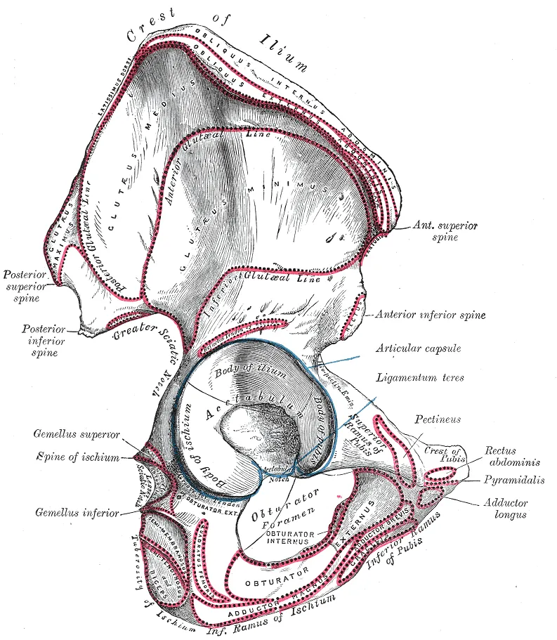
Three of them cross both the hip and the knee: the biceps femoris long head, the semimembranosus and the semitendinosus1. The hamstrings extend the hip, swinging the thigh back, and flex the knee7,9. Their origin lies deep to the gluteus maximus, the large muscle of the buttock3.
How the tendons share the bone
In 14 cadaver specimens, the semitendinosus and biceps femoris long head shared one origin through a conjoined (joined) tendon10. The semimembranosus attached by its own crescent-shaped area, in front of and to the outer side of the conjoined tendon10. A cadaver study found the conjoined tendon to be the largest of these attachments10.
Another study, of 6 cadaver pelvises, found one common hamstring tendon at the origin in 5 of them11. The sixth had a separate semimembranosus attachment11. In that study, the semimembranosus joined the common tendon about 2.3 centimetres below the attachment11.
The studies give these sizes3,10,11:
| Part | Size reported |
|---|---|
| Whole hamstring attachment, 6 pelvises | About 10 square centimetres11 |
| Conjoined tendon attachment, 14 specimens | About 567 square millimetres, oval10 |
| Semimembranosus attachment, 14 specimens | About 412 square millimetres10 |
| Attachment widths, from a review | About 3.4 cm for the conjoined tendon and 4.2 cm for the semimembranosus3 |
In the 6 pelvises, the centre of the attachment lay about 3.7 centimetres from the tip of the ischial tuberosity11.
How load reaches the tendons
In a modelling study of 20 recreationally active adults, peak musculotendon force (the force in muscle and tendon together) came during acceleration for two muscles12. It reached about 2.7 times body weight in the semimembranosus and 1.5 times in the biceps femoris long head12. In the same study, slowing down stretched all the two-joint hamstrings the most, about 11 to 14 percent, more than cutting or speeding up12.
A computer-model study of seven people sprinting found that each muscle was loaded in its own way13:
- The biceps femoris long head was stretched the most13.
- The semitendinosus lengthened the fastest13.
- The semimembranosus produced the highest peak force13.
All three reached their peak stretch and force in terminal swing, the end of the leg’s forward swing13.
The tendons also take squeezing force3. A review states that they press against the ischium (sitting bone) when the hip bends, as in sitting or lunging3. The same review states that high pulling and compressive load together are the most harmful mix for tendons, especially when repeated3. A review states that the semimembranosus tendon is thought to be the most exposed to compression, because of its deep, outer origin3.
Breakdown
What tendinopathy is like here
A review defines tendinopathy as lasting tendon pain and loss of function linked to mechanical load3. A narrative review describes proximal hamstring tendinopathy as usually causing deep buttock pain at the common hamstring origin14. A systematic review describes the pain as coming on with running, sitting and lunging15.
A review describes the main problem as tendinopathy where the tendons attach, mostly in the semimembranosus3. The same review describes tendinopathic tendon as having disordered fibres of collagen (a strong, rope-like protein), extra small blood vessels and new nerve growth3. A review reports that symptoms often follow a rise in heavy hamstring work with the hip bent, such as hill walking or running3. It also reports that symptoms are worse at faster running speeds3.
Who it affects
A systematic review reports that it mostly affects active people of both sexes, in rugby, soccer, distance running, and track and field15. It also affects people who do not play sport15. Another review reports that it affects less active people, usually women around menopause, as well as runners3. That review cites a prevalence of 12% in distance runners, meaning 12% had it at the time3.
What scans show, with and without symptoms
In MRI of 253 consecutive patients with no proximal hamstring symptoms, the scans showed4:
- normal tendons on both sides in 35%4;
- changes on both sides in 52%4;
- partial tears on both sides in 15%, and complete ruptures on both sides in 2%4.
Among those with a tear in only one of the tendons, the semimembranosus was the one torn 74% of the time4.
In MRI of 118 consecutive patients, more than 90% of these tendons showed raised signal inside them16. That signal was not linked to symptoms of tendinopathy16. A larger tendon, fluid around the tendon and bone oedema in the ischial tuberosity were linked to symptoms16. Oedema is swelling from fluid build-up, and these features could also be seen in people without symptoms16. In 31 people with symptoms, tearing-type findings appeared only in those over 4517.
When the tendon pulls away from the bone
Avulsion is where the tendon pulls away from the bone. A systematic review reports that avulsion of the hamstring origin is 3% to 11% of hamstring injuries5. A review reports that most hamstring injuries in athletes are strains where muscle joins tendon, most often at the upper end1. A review reports that partial or complete ruptures here usually follow sudden injury with the hip fully bent and the knee straight3. A cohort study states that avulsion is more common in football, rugby and other sports that demand acceleration18.
In 263 people who had surgery for a complete avulsion19:
- 53% were men, the mean age was 49, and 56% were aged 45 to 5919;
- 52% were injured in sport19;
- two thirds had pulled back from the bone by more than 2 centimetres19;
- 5% had loss of feeling put down to the sciatic nerve (the large nerve from the lower back down the back of the thigh)19.
In the same 263 people, women were older and mostly injured in daily life, while men were younger and mostly injured in sport19. Those whose injury pulled off a piece of bone were significantly younger than those whose tendon had pulled fully off the bone19.
A series of 72 surgical repairs had a mean age of 4020. Waterskiing was the most common cause20. Most were complete avulsions, and the tendon had pulled back 7 centimetres on average20.
Recovery
How the tendon responds to training
In a randomised study of 30 recreationally active men, 10 weeks of Nordic hamstring training raised semimembranosus tendon volume more than no training6. Otherwise, that study gave limited evidence of hamstring tendon or aponeurosis (a broad, flat sheet of tendon) growth6. A meta-analysis found that tendons respond to many kinds of loading21. Its data suggested that load size mattered more than the type of muscle contraction21. Its 27 studies were on the Achilles and patellar tendons, not the hamstring tendon21.
How long tendinopathy lasts
A narrative review reports that pain and loss of function in this tendinopathy are often long-standing and limit sport and daily life14. A review reports that some improvement over weeks and months is common3. Sitting pain is often slow to settle and may last up to a year3. A systematic review of 12 studies in 424 people found no high- or moderate-quality evidence for any intervention15.
What trials in tendinopathy found
One randomised trial studied 40 professional athletes with long-standing proximal hamstring tendinopathy22. At 3 months, 85% of the shockwave therapy group had at least halved their pain22. In the group given a conventional programme, 10% had done so22. The trial sits beside the review above, which found no high- or moderate-quality evidence for any intervention15.
The same review included case series of surgery for this tendinopathy15. In them, 77% to 100% of people got back to their earlier level of sport15. The two studies that gave timing reported a mean of 4.4 to 5 months15. Complications, mostly minor, came in 10% of surgical cases15. Though reporting was incomplete, surgery had the most unwanted events of the interventions studied15.
What studies found after avulsion
Reviews that combined studies of surgical repair after avulsion reported these results5,23,24:
| Studies pooled | What was reported |
|---|---|
| 35 studies, 1,530 repairs | 92.6% satisfied; strength 87% of the other side; 84.5% back in sport at 6.5 months on average5 |
| 21 studies, 846 patients, mean age 41 | 87% back in some sport and 77% at their earlier level, at a mean of 5.8 months23 |
| 13 studies, 387 people | 76% to 100% back in sport; 55% to 100% at their earlier activity level24 |
In the series of 72 repairs, strength averaged 84% of the uninjured side, and endurance 89%20. People varied widely20.
In the review of 13 studies, some pain remained in 8% to 61% of patients, and all 13 studies were of low method quality24.
A meta-analysis of 24 studies found higher satisfaction and strength after repair, though the group without surgery was small25. Satisfaction was 91% against 53%, and strength 85% against 64%25. A meta-analysis of seven studies found no significant difference in patient-reported outcomes with and without surgery26. In that analysis, more people got back to their earlier sport level after surgery, 65.2% against 50.7%26.
Complications and re-rupture
The 24-study meta-analysis reported complications in about 23% of repairs, and found most of its studies to be of low method quality25. The 35-study meta-analysis found re-rupture, the tendon tearing again, in 1.2%5. It found sciatic nerve problems in 3.5%, and both rates were lower with acute (early) repair5. In a cohort of 740 patients followed for 4.9 years on average, 4.59% re-ruptured18. Of those, 75% happened in the first 6 months, and 74% without a new injury18.
In that cohort, surgery more than 32 days after injury and a complete first rupture were significant risk factors for re-rupture18. A systematic review of 18 studies found better satisfaction, strength and return to sport with repair within 4 weeks27. Those early repairs also had fewer re-ruptures27. The 35-study meta-analysis also found a quicker return to sport after acute repair5.
Connections
The muscles it anchors
The tendon anchors the hamstrings, except the short head of the biceps femoris, to the ischial tuberosity1,2. The gluteus maximus lies over the hamstring origin3. The gluteus maximus and the hamstrings work together to extend the hip28. Part of the adductor magnus, a large inner-thigh muscle, also starts from the ischial tuberosity9. It shares the hamstrings’ action, so some count it as a hamstring9.
The nerve and ligament beside it
The sciatic nerve runs close by, about 1.2 centimetres from the outer edge of the ischial tuberosity, a review reports3. In cadaver dissection, a thin sheet of dense tissue ran from the hamstring origin to the gluteus maximus2. It formed a tunnel for the sciatic nerve and the posterior femoral cutaneous nerve, which supplies the skin on the back of the thigh2.
The sacrotuberous ligament is a strong band at the back of the pelvis, from the base of the spine to the sitting bone. In a dissection pictured in one cadaver study, its outermost layer carried on into the biceps femoris long head tendon2.
What studies of the Nordic hamstring curl show
The Nordic hamstring curl loads the hamstring muscle-tendon units, the muscles and tendons working as one29,30. Related exercise pages: Nordic hamstring curl.
- In 20 active adults, it and one other exercise stretched the hamstring muscle-tendon units less than any other exercise or gait task tested29. The hip thrust was the exception for the semitendinosus29.
- In 12 healthy volunteers, biceps femoris long head fascicles (bundles of muscle fibres) changed little in length through about 30% to 80% of the movement30. The whole unit lengthened meanwhile30.
- The authors of that study suggested that stretch of the unit’s elastic tissue plays an important part in absorbing energy30.
Proximal hamstring tendon carries force from
Proximal hamstring tendon lies deep to
Proximal hamstring tendon is loaded by
Sources
- Koolmees DS, Klott JD, Poppe TR, Bernholt DL (2025). Distal Hamstring Injuries and Disorders.. Journal of the American Academy of Orthopaedic Surgeons: Global Research & Reviews. doi:10.5435/JAAOSGlobal-D-25-00248 PMID 40972536
- Miguel-Pérez M, Iglesias-Chamorro P, Ortiz-Miguel S, Ortiz-Sagristà JC, Möller I, Blasi J, Agullò J, Martinoli C, Pérez-Bellmunt A (2024). Anatomical Relationships of the Proximal Attachment of the Hamstring Muscles with Neighboring Structures: From Ultrasound, Anatomical and Histological Findings to Clinical Implications.. Diagnostics (Basel). doi:10.3390/diagnostics14161725 PMID 39202213
- Grimaldi A, Mellor R, Nasser A, Vicenzino B, Hunter DJ (2024). Current and future advances in practice: tendinopathies of the hip.. Rheumatology Advances in Practice. doi:10.1093/rap/rkae022 PMID 38601140
- Thompson SM, Fung S, Wood DG (2017). The prevalence of proximal hamstring pathology on MRI in the asymptomatic population.. Knee Surgery, Sports Traumatology, Arthroscopy. doi:10.1007/s00167-016-4253-4 PMID 27492382
- Hillier-Smith R, Paton B (2022). Outcomes following surgical management of proximal hamstring tendon avulsions. Bone & Joint Open. doi:10.1302/2633-1462.35.BJO-2021-0196.R1 PMID 35549447
- Lazarczuk SL, Collings TJ, Hams AH, Timmins RG, Shield AJ, Barrett RS, Bourne MN (2024). Hamstring Muscle-Tendon Geometric Adaptations to Resistance Training Using the Hip Extension and Nordic Hamstring Exercises.. Scandinavian journal of medicine & science in sports. doi:10.1111/sms.14728 PMID 39297348
- Betts JG, Young KA, Wise JA, Johnson E, Poe B, Kruse DH, Korol O, Johnson JE, Womble M, DeSaix P (2022). Anatomy and Physiology 2e. OpenStax. Link
- Hu Y, Raja A (2026). Anatomy, Bony Pelvis and Lower Limb, Hamstring Muscle. StatPearls. PMID 31536294
- Obey MR, Broski SM, Spinner RJ, Collins MS, Krych AJ (2016). Anatomy of the Adductor Magnus Origin. Orthopaedic Journal of Sports Medicine. doi:10.1177/2325967115625055 PMID 26798764
- Philippon MJ, Ferro FP, Campbell KJ, Michalski MP, Goldsmith MT, Devitt BM, Wijdicks CA, LaPrade RF (2015). A qualitative and quantitative analysis of the attachment sites of the proximal hamstrings.. Knee Surgery, Sports Traumatology, Arthroscopy. doi:10.1007/s00167-014-3074-6 PMID 24859732
- Neuschwander TB, Benke MT, Gerhardt MB (2015). Anatomic Description of the Origin of the Proximal Hamstring.. Arthroscopy. doi:10.1016/j.arthro.2015.02.025 PMID 25896274
- Steventon-Lorenzen N, Fitzwilliam E, Schache AG, Opar D, Maniar N (2026). Hamstring Mechanics During Acceleration, Deceleration and Sidestep Cutting.. Scandinavian Journal of Medicine & Science in Sports. doi:10.1111/sms.70257 PMID 41918173
- Schache AG, Dorn TW, Blanch PD, Brown NA, Pandy MG (2012). Mechanics of the human hamstring muscles during sprinting.. Medicine and Science in Sports and Exercise. doi:10.1249/MSS.0b013e318236a3d2 PMID 21912301
- Goom TS, Malliaras P, Reiman MP, Purdam CR (2016). Proximal Hamstring Tendinopathy: Clinical Aspects of Assessment and Management.. Journal of Orthopaedic & Sports Physical Therapy. doi:10.2519/jospt.2016.5986 PMID 27084841
- Nasser AM, Vicenzino B, Grimaldi A, Anderson J, Semciw AI (2021). Proximal Hamstring Tendinopathy: A Systematic Review of Interventions.. International Journal of Sports Physical Therapy. doi:10.26603/001c.21250 PMID 33842025
- De Smet AA, Blankenbaker DG, Alsheik NH, Lindstrom MJ (2012). MRI appearance of the proximal hamstring tendons in patients with and without symptomatic proximal hamstring tendinopathy.. AJR. American Journal of Roentgenology. doi:10.2214/AJR.11.6590 PMID 22268187
- Bowden DJ, Byrne CA, Alkhayat A, Kavanagh EC, Eustace SJ (2018). Differing MRI appearances of symptomatic proximal hamstring tendinopathy with ageing: a comparison of appearances in patients below and above 45 years.. Clinical Radiology. doi:10.1016/j.crad.2018.06.014 PMID 30146066
- Lefèvre N, Freiha K, Moussa MK, Valentin E, Bohu Y, Meyer A, Hardy A (2024). Risk Factors for Rerupture After Proximal Hamstring Avulsion Injury Including the Optimal Timing for Surgery.. The American Journal of Sports Medicine. doi:10.1177/03635465241233734 PMID 38482843
- Irger M, Willinger L, Lacheta L, Pogorzelski J, Imhoff AB, Feucht MJ (2020). Proximal hamstring tendon avulsion injuries occur predominately in middle-aged patients with distinct gender differences: epidemiologic analysis of 263 surgically treated cases.. Knee Surgery, Sports Traumatology, Arthroscopy. doi:10.1007/s00167-019-05717-7 PMID 31541291
- Wood DG, Packham I, Trikha SP, Linklater J (2008). Avulsion of the proximal hamstring origin.. The Journal of Bone and Joint Surgery. American Volume. doi:10.2106/JBJS.G.00685 PMID 18978405
- Bohm S, Mersmann F, Arampatzis A (2015). Human tendon adaptation in response to mechanical loading: a systematic review and meta-analysis of exercise intervention studies on healthy adults.. Sports Medicine - Open. doi:10.1186/s40798-015-0009-9 PMID 27747846
- Cacchio A, Rompe JD, Furia JP, Susi P, Santilli V, De Paulis F (2011). Shockwave therapy for the treatment of chronic proximal hamstring tendinopathy in professional athletes.. The American Journal of Sports Medicine. doi:10.1177/0363546510379324 PMID 20855554
- Coughlin RP, Kay J, Shanmugaraj A, Memon M, Naji L, Ayeni OR (2020). Return to Sport After Surgical Management of Proximal Hamstring Avulsions: A Systematic Review and Meta-analysis.. Clinical Journal of Sport Medicine. doi:10.1097/JSM.0000000000000688 PMID 30444732
- van der Made AD, Reurink G, Gouttebarge V, Tol JL, Kerkhoffs GM (2015). Outcome After Surgical Repair of Proximal Hamstring Avulsions. The American Journal of Sports Medicine. doi:10.1177/0363546514555327 PMID 25384502
- Bodendorfer BM, Curley AJ, Kotler JA, Ryan JM, Jejurikar NS, Kumar A, Postma WF (2018). Outcomes After Operative and Nonoperative Treatment of Proximal Hamstring Avulsions: A Systematic Review and Meta-analysis.. The American Journal of Sports Medicine. doi:10.1177/0363546517732526 PMID 29016194
- Thamrongskulsiri N, Limskul D, Tanpowpong T, Kuptniratsaikul S, Ranawat AS, Itthipanichpong T (2025). No difference in patient-reported outcomes between operative and non-operative management of proximal hamstring injuries, but return to sports is higher in operative treatment: A systematic review and meta-analysis.. Journal of Experimental Orthopaedics. doi:10.1002/jeo2.70327 PMID 40655246
- Harris JD, Griesser MJ, Best TM, Ellis TJ (2011). Treatment of proximal hamstring ruptures - a systematic review.. International Journal of Sports Medicine. doi:10.1055/s-0031-1273753 PMID 21563032
- Lee DK, Oh JS (2018). Relationship between hamstring length and gluteus maximus strength with and without normalization.. Journal of Physical Therapy Science. doi:10.1589/jpts.30.116 PMID 29410579
- Breed R, Hulm S, Hickey JT, Timmins RG, Opar D, Banyard HG, Maniar N (2026). Hamstring force and stretch during progressively increasing running speeds and the eccentric phase of resistance training exercises commonly used for injury prevention and rehabilitation.. British journal of sports medicine. doi:10.1136/bjsports-2025-110847 PMID 41730607
- Pincheira PA, Riveros-Matthey C, Lichtwark GA (2022). Isometric fascicle behaviour of the biceps femoris long head muscle during Nordic hamstring exercise variations.. Journal of science and medicine in sport. doi:10.1016/j.jsams.2022.05.002 PMID 35637123
Changes
- Tidy pass: minor review findings fixed, glosses aligned with the reviewed glossary.
- First published.