Peroneal tendons
Also called fibular tendons, fibularis tendons, peroneus longus tendon, peroneus brevis tendon, fibularis longus tendon, fibularis brevis tendon
The two tendons that run behind the outer ankle bone to the foot. Where they run, what holds them in place, how they tear or slip out of place, what studies of recovery found, and what they work with.
- Region
- Foot & ankle
- Muscles
- peroneus longus, peroneus brevis
- Updated
- October 5, 2026
- Reviewed
- October 5, 2026 · how entries are checked
Function
Where the two tendons run
A tendon is a tough cord of tissue that connects a muscle to a bone. The peroneal tendons belong to the peroneus longus and peroneus brevis, also called the fibularis longus and brevis2. These two muscles on the outer side of the leg point the foot down and turn the sole outward5.
The peroneus longus becomes fully tendon about 3 to 4 cm above the lower end of the fibula1. The peroneus brevis muscle reaches lower, into the groove behind the lateral malleolus, the bony bump on the outer side of the ankle1.
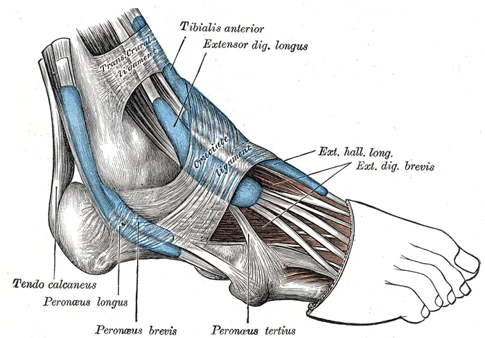
The outer surface of the calcaneus (heel bone) carries a ridge called the peroneal tubercle6. A metatarsal is one of the long bones in the middle of the foot. The two tendons take different paths past these landmarks2:
| Where | Peroneus brevis tendon | Peroneus longus tendon |
|---|---|---|
| Behind the lower end of the fibula | Fairly flat, right against the bone2 | Rounder, behind the brevis tendon2 |
| On the heel bone | Above the peroneal tubercle2 | Below the peroneal tubercle2 |
| Where it ends | Base of the fifth metatarsal7 | Base of the first metatarsal and a bone called the medial cuneiform7 |
Around the ankle, the two tendons share one synovial sheath, a fluid-lined sleeve that a tendon glides in2. It runs from about 4 cm above the tip of the lateral malleolus to about 1 cm below it2. Lower down, the tendons part into two sheaths and cross the outer wall of the heel bone2.
The longus tendon then passes under the cuboid bone and crosses the sole at a slant2. Its tunnel there is formed by a groove in the cuboid and the long plantar ligament2. A small extra bone, the os peroneum, can sit inside the far end of the longus tendon where it enters this tunnel1. Estimates of how often it is present range from 4 to 30%1.
What holds the tendons in place
At the tip of the fibula, both tendons run through a tunnel of bone and fibrous tissue1. A retinaculum is a band of tissue that holds tendons in place where they cross a joint. The peroneal retinaculum has an upper and a lower band2.
Both typically start on the lowest 2 cm of the fibula2. The upper band reaches the Achilles tendon and the lower band the outer side of the heel bone2. The upper band is the main restraint that keeps the tendons from slipping out of place2.
The shape of the groove behind the fibula is set mainly by a thick cushion of fibrocartilage, a tough, fibrous kind of cartilage8. The bone itself matters less8. Two studies sorted the groove by shape, and their figures differ. One used cadavers (bodies donated for study) and one MRI (magnetic resonance imaging)2,9:
| Groove shape | 178 cadaver fibulae | MRI of 65 volunteers with symptom-free ankles |
|---|---|---|
| Concave | 82%2 | 28%9 |
| Flat | 11%2 | 43%9 |
| Convex | 7%2 | 18%9 |
The MRI study classed the remaining 11% of grooves as irregular9.
What the tendons do
The peroneal tendons are the foot’s primary evertors, and they steady the outer side of the ankle2. Sources differ on which tendon does more2,10:
- A narrative review, a summary of research written without a set search method, credits the peroneus brevis with 63% of total eversion power2.
- Six cadaver feet were tested in a simulated early heel rise11. Loading the brevis tendon tilted the heel bone 0.9 degrees further outward than the same load on the longus tendon11.
- A review of the subtalar joint, the joint just below the ankle joint, names the peroneus longus as the strongest evertor crossing it10.
The peroneus longus also steadies the inner column of the foot in stance, when the foot is on the ground2. In ten cadaver feet, a force pushed the first metatarsal inward12. The bone shifted significantly only after the longus tendon was cut12.
How blood reaches the tendons
In ten fresh-frozen cadavers, the tendons got their blood mainly from the peroneal artery13. It arrived through a band of tissue on the back-outer side that connects both tendons13. Whether the tendons have avascular zones, meaning areas without blood vessels, at the sites where they most often tear is disputed13. Three studies that injected the vessels reached different answers13,14,15:
- One reported that the brevis tendon is almost avascular in the fibular groove, where it is squeezed between the longus tendon and bone14.
- One, of ten cadaver specimens, found no avascular zones in the brevis tendon and zones with a poor blood supply in eight longus tendons13.
- One, of 22 cadaver lower limbs, found avascular areas in the brevis tendon in 22.7% and in no longus tendon15.
Breakdown
Three kinds of problem
A consensus panel, a group of experts who meet to agree a shared view, sorted peroneal tendon problems into three categories1:
- tendinopathy, meaning local pain in a tendon that comes with activities that load it1;
- tear or rupture1;
- instability or dislocation1.
In the panel’s terms, a “tear” usually means a lengthwise tear or an incomplete rupture1. A “rupture” means the tendon is completely separated1. A narrative review states that tears may be acute (recent), but most are chronic (long-lasting)16. In peroneal tendinosis the tendon thickens and swells, with patches of degeneration, more often below the outer ankle bone2.
How common tears are
How common peroneal tendon tears are in the general population is unknown1. Cadaver studies have found tears in 11 to 38% of ankles1. In one dissection of 124 cadaver ankles, the brevis tendon was worn in 14 (11.3%) and the longus tendon in none17.
Scans can show changes without symptoms. In 170 people with no peroneal symptoms, ultrasound showed abnormalities in 20% of longus tendons and 12% of brevis tendons18.
Which tendon tears, and where
One series covered 234 patients in a row who had surgery for long-lasting, symptomatic tears3:
| Tendon torn | Share of tears |
|---|---|
| Peroneus brevis only | 73%3 |
| Peroneus longus only | 8%3 |
| Both tendons | 19%3 |
A narrative review describes the usual brevis tear as lengthwise and centred at the tip of the fibula16. There the tendon turns sharply and is compressed between the longus tendon and the fibula16. In a cadaver dissection study, brevis splits averaged 1.9 cm long, and none was a complete rupture17. The same review states that longus tears are much less common16. Most sit between the peroneal tubercle and the cuboid tunnel16.
What goes with tears
Studies and a consensus statement link tears with several factors1,3:
- Foot shape. The 234 patients were compared with matched controls3. A high-arched, inward-tilted foot shape, called cavovarus, had a statistically significant association with tears3.
- A crowded tunnel. A consensus statement names overfilling of the tunnel as an important factor in peroneal tendon problems, more so than the groove’s shape1.
- Ankle instability. Sixty-one patients had a first ligament reconstruction for chronic outer-ankle instability, and 25% had a brevis tear19.
- An earlier sprain. Eighteen patients had surgery for brevis splits with chronic ankle instability20. On average, six years passed between the ankle sprain and pain behind the outer ankle20.
In Swedish register data on 719 patients having first-time peroneal tendon surgery, patients averaged 49 years of age at surgery23. Brevis rupture and tendinosis were more prevalent in women than in men23.
How the tendons slip out of place
Peroneal tendon dislocation occurs in 0.3 to 0.5% of all traumatic ankle events4. An acute dislocation typically follows a forced eccentric contraction, with the ankle in dorsiflexion and eversion1. Eccentric means the peroneal muscles work while they lengthen, and dorsiflexion means pulling the foot up toward the shin. The upper retinaculum can then rupture, letting the tendons dislocate forward over the outer ankle bone4. Dislocation is most prevalent in athletes, mainly in sports with cutting movements such as skiing, soccer, basketball, ice skating and gymnastics4.
One study compared scans of 30 patients with repeated dislocation and 30 matched controls24. A low-lying brevis muscle belly, the thick middle part of the muscle, was significantly associated with dislocation24. The bony shape of the groove was not24.
Recovery
How well tears heal
Peroneal tendon tears are described as seeming to have a poor tendency to heal13. The consensus panel agreed that acute injuries have a better healing tendency than chronic ones1. Its statement notes that evidence on outcomes without surgery in acute peroneal tendinopathy is limited and varied1.
What studies of tear surgery found
- A systematic review of nine studies, with 336 patients, found an average overall complication rate of 38.7%25. Ankle pain was the most commonly reported minor complication25.
- Forty-one patients were followed for at least 1 year after surgery for 49 tears26. Their average return to activity took 3.49 months, and 14 of the 16 athletes got back to full sporting level26.
- The consensus statement notes that elite athletes may not get back to their earlier level of sport after surgery for a rupture1.
What studies of dislocation found
A consensus statement reports that evidence on dislocation managed without surgery is limited to case reports and small series1. These suggest a risk of repeated peroneal instability of about 50%1.
A systematic review of 14 studies of surgery found return-to-sport rates of 83 to 100% in the two surgical groups it analysed4. The redislocation rate in those groups was below 1.5%4. All 14 studies were scored as low quality, and none had a control group, randomisation or masked assessment4.
Connections
The muscles and bones around them
The peroneal muscles share the lateral compartment of the leg with the common fibular nerve and the superficial fibular nerve27. Below the fibula, on the calcaneus, the peroneal tubercle separates an upper groove for the brevis tendon from a lower one for the longus6. The brevis tendon ends on the fifth of the metatarsals and is consistently present28.
The ligaments beside them
The tendons and their sheaths cross over the calcaneofibular ligament, one of the lateral ankle ligaments29. Only about 1 cm of that ligament is left uncovered29. Slipping of the peroneal tendons is commonly associated with lengthwise brevis splits and with outer-ankle instability8. Disrupted lateral ankle ligaments put considerable strain on the upper retinaculum8.
Running
Related exercise pages: running. One study recorded muscle activity with fine-wire electrodes, thin wire sensors placed into a muscle, in 15 injury-free recreational and competitive runners30. Across three paces, peroneus brevis activity rose significantly as pace increased30.
Peroneal tendons is loaded by
Peroneal tendons carries force from
Peroneal tendons runs behind the lower end of
Peroneal tendons crosses the outer side of
Peroneal tendons attaches to
Peroneal tendons crosses over
Peroneal tendons is often injured alongside
Sources
- van Dijk PA, Miller D, Calder J, DiGiovanni CW, Kennedy JG, Kerkhoffs GM, Kynsburg A, Haverkamp D, Guillo S, Oliva XM, Pearce CJ, Pereira H, Spennacchio P, Stephen JM, van Dijk CN (2018). The ESSKA-AFAS international consensus statement on peroneal tendon pathologies. Knee Surgery, Sports Traumatology, Arthroscopy. doi:10.1007/s00167-018-4971-x PMID 29767272
- Davda K, Malhotra K, O'Donnell P, Singh D, Cullen N (2017). Peroneal tendon disorders. EFORT Open Reviews. doi:10.1302/2058-5241.2.160047 PMID 28736620
- Taniguchi A, Alejandro SF, Kane JM, Daoud Y, Tanaka Y, Ford SE, Brodsky JW (2021). Association of Cavovarus Foot Alignment With Peroneal Tendon Tears. Foot & Ankle International. doi:10.1177/1071100721990348 PMID 33847151
- van Dijk PA, Gianakos AL, Kerkhoffs GM, Kennedy JG (2016). Return to sports and clinical outcomes in patients treated for peroneal tendon dislocation: a systematic review. Knee Surgery, Sports Traumatology, Arthroscopy. doi:10.1007/s00167-015-3833-z PMID 26519186
- Brockett CL, Chapman GJ (2016). Biomechanics of the ankle.. Orthopaedics and Trauma. doi:10.1016/j.mporth.2016.04.015 PMID 27594929
- Gray H; Lewis WH (editor) (1918). Anatomy of the Human Body. Lea & Febiger, Philadelphia (20th edition). Link
- Walt J, Massey P (2023). Peroneal Tendon Syndromes. StatPearls. PMID 31335074
- Ferran NA, Oliva F, Maffulli N (2006). Recurrent subluxation of the peroneal tendons. Sports Medicine. doi:10.2165/00007256-200636100-00003 PMID 17004847
- Saupe N, Mengiardi B, Pfirrmann CW, Vienne P, Seifert B, Zanetti M (2007). Anatomic variants associated with peroneal tendon disorders: MR imaging findings in volunteers with asymptomatic ankles. Radiology. doi:10.1148/radiol.2422051993 PMID 17255421
- Krähenbühl N, Horn-Lang T, Hintermann B, Knupp M (2017). The subtalar joint: A complex mechanism. EFORT open reviews. doi:10.1302/2058-5241.2.160050 PMID 28828179
- Otis JC, Deland JT, Lee S, Gordon J (2004). Peroneus brevis is a more effective evertor than peroneus longus. Foot & Ankle International. doi:10.1177/107110070402500408 PMID 15132932
- Bohne WH, Lee KT, Peterson MG (1997). Action of the peroneus longus tendon on the first metatarsal against metatarsus primus varus force. Foot & Ankle International. doi:10.1177/107110079701800810 PMID 9278747
- van Dijk PA, Madirolas FX, Carrera A, Kerkhoffs GM, Reina F (2016). Peroneal tendons well vascularized: results from a cadaveric study. Knee Surgery, Sports Traumatology, Arthroscopy. doi:10.1007/s00167-015-3946-4 PMID 26740089
- Petersen W, Bobka T, Stein V, Tillmann B (2000). Blood supply of the peroneal tendons: injection and immunohistochemical studies of cadaver tendons. Acta Orthopaedica Scandinavica. doi:10.1080/000164700317413148 PMID 10852323
- Mota Gomes T, Guerra-Pinto F, Soares S, Andrade R, Pereira B, Espregueira-Mendes J, Oliva XM (2021). The vascularization of the peroneal tendons: An anatomic study. Foot and Ankle Surgery. doi:10.1016/j.fas.2020.06.004 PMID 32600968
- Danna NR, Brodsky JW (2020). Diagnosis and Operative Treatment of Peroneal Tendon Tears. Foot & Ankle Orthopaedics. doi:10.1177/2473011420910407 PMID 35097372
- Sobel M, Bohne WH, Levy ME (1990). Longitudinal attrition of the peroneus brevis tendon in the fibular groove: an anatomic study. Foot & Ankle. doi:10.1177/107110079001100302 PMID 2131804
- Smoker BA, Giakas AM, Mills FB 4th, Williams KA, Bornemann PH, Jackson JB 3rd (2023). Prevalence of Abnormal Ultrasonographic Findings in Asymptomatic Peroneal Tendons. Foot & Ankle Orthopaedics. doi:10.1177/24730114231164146 PMID 37009419
- DiGiovanni BF, Fraga CJ, Cohen BE, Shereff MJ (2000). Associated injuries found in chronic lateral ankle instability. Foot & Ankle International. doi:10.1177/107110070002101003 PMID 11128010
- Bonnin M, Tavernier T, Bouysset M (1997). Split lesions of the peroneus brevis tendon in chronic ankle laxity. The American Journal of Sports Medicine. doi:10.1177/036354659702500520 PMID 9302480
- Ziai P, Benca E, Wenzel F, Schuh R, Krall C, Auffahrt A, Hofstetter M, Windhager R, Buchhorn T (2016). Peroneal tendinosis as a predisposing factor for the acute lateral ankle sprain in runners. Knee Surgery, Sports Traumatology, Arthroscopy. doi:10.1007/s00167-015-3562-3 PMID 25786820
- Ritter S, Moore M (2008). The relationship between lateral ankle sprain and ankle tendinitis in ballet dancers. Journal of Dance Medicine & Science. PMID 19618575
- André F, Cöster M, Carling M, Pellnor J, Gunnarsson B, Helander KN (2026). A demographic and descriptive analysis of patients who underwent peroneal tendon surgery: Data from the Swedish Foot and Ankle Register (Swefoot). Foot (Edinburgh). doi:10.1016/j.foot.2026.102249 PMID 41887048
- Nishimura A, Nakazora S, Senga Y, Fukuda A, Sudo A (2023). Anatomic Features of Patients With Recurrent Peroneal Tendon Dislocation. The American Journal of Sports Medicine. doi:10.1177/03635465231158100 PMID 36876737
- Mercer NP, Gianakos AL, Mercurio AM, Kennedy JG (2021). Clinical Outcomes of Peroneal Tendon Tears: A Systematic Review. The Journal of Foot and Ankle Surgery. doi:10.1053/j.jfas.2021.03.003 PMID 33785239
- Saxena A, Cassidy A (2003). Peroneal tendon injuries: an evaluation of 49 tears in 41 patients. The Journal of Foot and Ankle Surgery. doi:10.1016/s1067-2516(03)70031-3 PMID 12907932
- Khan IA, Mahabadi N, D'Abarno A, Varacallo MA (2023). Anatomy, Bony Pelvis and Lower Limb: Leg Lateral Compartment.. StatPearls. PMID 30137811
- Liu X, Hong CC, Reina F, Carrera A, Alcolea-Madera E, Del Vecchio JJ, Dalmau-Pastor M (2026). Navicular and fifth metatarsal base stress fractures: An illustrated anatomical review. Knee surgery, sports traumatology, arthroscopy : official journal of the ESSKA. doi:10.1002/ksa.70413 PMID 42171011
- Golanó P, Vega J, de Leeuw PA, Malagelada F, Manzanares MC, Götzens V, van Dijk CN (2010). Anatomy of the ankle ligaments: a pictorial essay.. Knee Surgery, Sports Traumatology, Arthroscopy. doi:10.1007/s00167-010-1100-x PMID 20309522
- Reber L, Perry J, Pink M (1993). Muscular control of the ankle in running.. The American journal of sports medicine. doi:10.1177/036354659302100608 PMID 8291630
Changes
- First published.