Neck extensors
Also called neck extensor muscles, cervical extensors, splenius, semispinalis, splenius cervicis, semispinalis capitis, splenius colli, complexus
The splenius and semispinalis muscles at the back of the neck, which tip the head back. Where they run, what they do, what studies of whiplash and neck pain found in them, and how they change with training.
- Region
- Neck
- Starts on (splenius capitis)
- the neck ligament's lower half and spinous processes from the lowest neck vertebra (C7) to the third or fourth thoracic (Cervical vertebrae)1,3
- Starts on (splenius cervicis)
- the spinous processes of the third to sixth thoracic vertebrae, by a narrow band of tendon3
- Starts on (semispinalis capitis)
- side spurs of the seventh neck to sixth or seventh thoracic vertebrae, and joint processes of neck vertebrae 4–61,3
- Starts on (semispinalis cervicis)
- the side spurs (transverse processes) of the upper five or six thoracic vertebrae3
- Ends on (splenius capitis)
- the mastoid process and the back of the skull just below the outer third of the superior nuchal line1,3
- Ends on (splenius cervicis)
- the back bumps (posterior tubercles) on the side spurs (transverse processes) of the top two or three neck vertebrae3
- Ends on (semispinalis capitis)
- the back of the skull (occipital bone), between the superior and inferior nuchal lines1,3
- Ends on (semispinalis cervicis)
- the spinous processes of the second to fifth neck vertebrae, with its largest bundle on the second (the axis)3
- Muscles
- splenius capitis, splenius cervicis, semispinalis capitis, semispinalis cervicis
- Updated
- October 28, 2026
- Reviewed
- October 28, 2026 · how entries are checked
Function
Two pairs at the back of the neck
OpenStax describes the back-of-neck muscles as mainly concerned with moving the head, such as extension (bending backward)1. Gray’s Anatomy lists the splenius and semispinalis among the deep muscles of the back3.
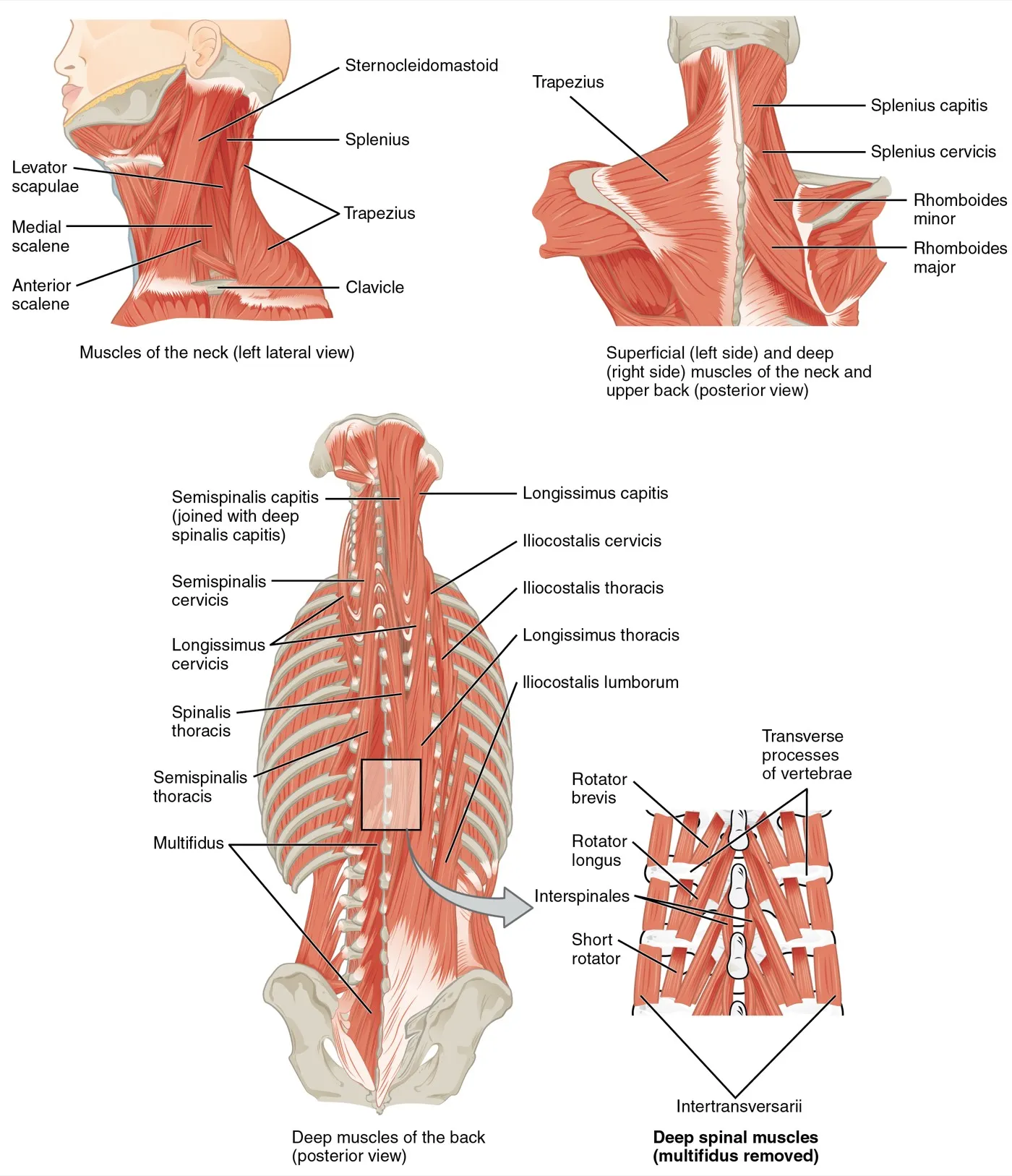
The two splenius muscles start at the midline and run outward and upward1. The splenius capitis reaches the head, and the splenius cervicis reaches the neck vertebrae1. The semispinalis muscles belong to the transversospinales, deep back muscles running from the side spurs of the vertebrae to the bony points behind1. They are named for the region they serve: capitis for the head, cervicis for the neck1. The semispinalis capitis lies beneath the splenius, toward the midline from the longissimus3.
| Muscle | Starts on | Ends on |
|---|---|---|
| Splenius capitis | The lower half of the nuchal ligament (neck ligament), and the spinous processes of the seventh neck vertebra and top three or four thoracic vertebrae3 | The mastoid process (the bony bump behind the ear) and the occipital bone (back of the skull), under the sternocleidomastoid3 |
| Splenius cervicis | The spinous processes of the third to sixth thoracic vertebrae, by a narrow tendon band3 | The transverse processes of the top two or three neck vertebrae3 |
| Semispinalis capitis | Transverse processes of the top six or seven thoracic vertebrae and the seventh neck vertebra; articular processes of the three neck vertebrae above3 | The occipital bone, between two ridges on the back of the skull3 |
| Semispinalis cervicis | The transverse processes of the top five or six thoracic vertebrae3 | Spinous processes from the axis (C2, the second vertebra of the neck) to the fifth neck vertebra. The bundle to the axis is largest3 |
Some of these attachments vary between people3,7. In Gray’s account, the splenius origin often sits one or two vertebrae higher or lower, and extra slips are sometimes found3. In 24 cadavers (bodies donated for study), the semispinalis cervicis insertion on C2 varied considerably in shape7.
What they move
Gray states that the two splenius muscles together draw the head straight back3. One alone draws the head to its side and turns the face slightly to the same side3. They also help hold the head upright3. Acting on both sides, the semispinalis muscles help extend the spine3.
An electromyography (EMG) study, which picks up a muscle’s electrical signals, put wires into the muscles of 19 healthy people8. The splenius capitis had two main jobs: extending the neck and turning the head to the same side8. The semispinalis capitis acted mainly as an extensor, with a minor part in tilting the head sideways8. The authors concluded that head motion comes from muscle forces, ligament forces and gravity working together8.
Which way the semispinalis capitis turns the head
Sources disagree on which way one semispinalis capitis turns the head1,3:
- Gray’s Anatomy states that one semispinalis capitis draws the head to its side and turns the face to the opposite side3.
- An OpenStax table states that one side bends and rotates the head to the same side1.
- In the EMG study of 19 healthy people, most people’s semispinalis capitis was gradually recruited when turning the head to the same side8.
Leverage and strength
A three-dimensional model of the neck was built from measured muscle data2. With the head upright, the semispinalis capitis and splenius capitis had the largest moment arms (leverage) and capacity to produce turning force for extension2. The splenius capitis’s force capacity varied substantially with head posture in the model2.
In 11 men and 5 women, the largest maximal neck moment (turning force) was in extension9. In 149 high school and college athletes, neck strength was on average greatest in extension10. In 16 young healthy people, extension strength rose with the area of the back-of-neck muscles on CT, a type of X-ray scan11.
Autopsy samples came from 13 young men12. Their splenius capitis had more type 1 (slow-contracting) fibres than the vastus lateralis, a thigh muscle: 51.2% against 39.7%12.
Their nerves
The splenius muscles are supplied by branches of the back divisions of the middle and lower neck nerves3. Gray states that the deep back muscles, the semispinalis among them, are supplied by the back divisions of the spinal nerves3. The greater occipital nerve, from the second neck nerve, pierces the semispinalis capitis near the skull and gives it branches3.
Breakdown
In a modelled rear-end impact
A neck model driven by data from people in rear-end car impacts estimated how far each muscle stretched13. The splenius capitis, semispinalis capitis and trapezius lengthened during the rebound phase13. Peak stretch of their fibre bundles was 21% in the splenius capitis and 18% in the semispinalis capitis13. These strains passed a previously reported injury threshold for one stretch of active muscle13. The authors note this fits reports of pain mainly at the back of the neck13.
Fat in the muscles after whiplash
In an MRI study of 79 women with lasting whiplash and 34 controls, the whiplash group had more fat in every extensor measured4. Fat was greatest in the deeper muscles of the upper neck4. It did not depend on age, reported pain, body mass index or how long symptoms had lasted4.
Follow-up studies tracked when and where the fat appears14,15:
- In 44 people followed from 4 weeks after whiplash, fat did not differ between groups at the start14. It rose in those with moderate or severe disability at 6 months14.
- In the same group, early post-traumatic stress symptoms mediated the link between pain and fat at 6 months; early loss of movement did not14.
- At 12 months, in 61 people, fat in the multifidus and semispinalis cervicis sat mostly in the inner parts of the muscles15. It was larger there in those with severe chronic whiplash15.
A meta-analysis found the evidence for changes in neck muscle size or fat after whiplash inconsistent5. A systematic review covered 14 papers on chronic neck pain16. Among the groups studied, fat in the neck extensors seems to occur only after whiplash16.
Size and strength in neck pain
Studies of neck pain report mixed findings in these muscles16:
- In 23 women whose neck pain began without an injury, extensor fat on MRI was not a feature17.
- A systematic review found smaller extensors in most studies of idiopathic (no known cause) neck pain16. After whiplash, they were larger at the top of the neck16.
- In 20 female office workers, those with one-sided neck pain had a smaller semispinalis capitis, smaller on the painful side18.
- In another MRI study, people with idiopathic neck pain had greater volume and fat in the multifidus with semispinalis cervicis than matched controls19.
- A meta-analysis of 15 studies found lower extension strength in chronic neck pain than in healthy controls20.
In 10 women with chronic neck pain and 10 controls, the semispinalis cervicis was less active and less tied to one direction in the patients21. In 32 people with chronic neck pain and 35 without, the pain group had smaller deep extensors and lower endurance22. Endurance was not linked to pain or disability22.
Activity in rugby tackles
A cross-sectional study measured 27 club rugby players making tackles23. Those with a concussion in the past year had the highest head acceleration23. They also had lower splenius capitis activity than players with no concussion history23.
Recovery
Growth with neck-specific training
One study assigned 22 active college students to three groups; its abstract does not say the assignment was random6. One group added three sets of ten head extensions, three days a week, to 12 weeks of lifting6. Their 3 × 10 repetition maximum rose from 17.9 to 23.9 kg6. Their splenius capitis and semispinalis capitis and cervicis grew 24% to 25% in area on MRI6. Students who did the lifts without the neck exercise showed no change in neck muscle area6.
The authors suggest these three muscles are mainly responsible for head extension6. After training, the muscle area used for a submaximal head extension fell by 31%24. The area used for a maximal effort rose by 16%24. The authors suggest such adaptations need a specific neck exercise24.
Other settings
- In six crew members on 4- to 6-month spaceflights with a resistance exercise device, semispinalis capitis area rose 11.5% on average25. The splenius capitis showed no significant change25.
- In a randomised trial of 30 healthy adults, 6 weeks of isometric neck holds was followed by a 25% rise in isokinetic extension strength26. Isometric means the muscle works but the joint does not move; an isokinetic test holds the limb to a fixed speed.
- In a randomised trial of 46 women with chronic neck pain, an 8-week programme made neck muscle activity more tied to one direction27. It was recorded from the sternocleidomastoid and splenius capitis27.
In a randomised trial of 180 female office workers with chronic neck pain, maximal isometric extension strength rose in each group28.
| Group in the office worker trial | Rise in extension strength |
|---|---|
| High-intensity isometric training | 69%28 |
| Endurance training | 16%28 |
| Control group | 7%28 |
A systematic review of 35 randomised trials in adults with neck pain found that neck extensor exercises were mostly combined with other approaches29. Only two of the studies tested them on their own29.
Connections
Neighbours at the back of the neck
The splenius capitis ends under the sternocleidomastoid3. The semispinalis capitis lies toward the midline from the longissimus capitis, part of the erector spinae1,3. An overview article describes the deep neck extensors as able to control single neck segments together with the deep neck flexors30. Their attachments sit on the cervical vertebrae and upper thoracic vertebrae3.
Do they act as one group?
Two studies with wires in the muscles looked at this31,32. In nine seated people doing isometric neck efforts, the working muscles from all layers shared a common neural drive (nerve signal)31. Those recorded included the splenius capitis, semispinalis, multifidus and levator scapulae31. In a study of three men, the back-of-neck muscles other than splenius capitis acted together in voluntary movement, but not when jolted on a sled32. The authors conclude that assuming all back-of-neck muscles act together as extensors is not reasonable32.
Exercises that load them
Related exercise pages: neck isometric hold33,34. Where the push sat changed how hard the semispinalis cervicis worked relative to the splenius capitis33,34:
| Who was studied | What studies found |
|---|---|
| 10 women with chronic neck pain | Resistance at C2 gave more semispinalis cervicis activity, relative to splenius capitis, than resistance at the back of the head or at C533 |
| 10 healthy volunteers | Resistance over C1 or C4 made the semispinalis cervicis about twice as active as the splenius capitis at the level just below34 |
Neck extensors is loaded by
Neck extensors attaches to
Neck extensors ends under, in part, the
Neck extensors lies beside the longissimus of the
Neck extensors shares a common drive in neck efforts with
Neck extensors lengthens in a whiplash model with
Neck extensors controls neck segments with
Neck extensors has been measured in studies of
Sources
- Betts JG, Young KA, Wise JA, Johnson E, Poe B, Kruse DH, Korol O, Johnson JE, Womble M, DeSaix P (2022). Anatomy and Physiology 2e. OpenStax. Link
- Vasavada AN, Li S, Delp SL (1998). Influence of muscle morphometry and moment arms on the moment-generating capacity of human neck muscles.. Spine. doi:10.1097/00007632-199802150-00002 PMID 9516695
- Gray H; Lewis WH (editor) (1918). Anatomy of the Human Body. Lea & Febiger, Philadelphia (20th edition). Link
- Elliott J, Jull G, Noteboom JT, Darnell R, Galloway G, Gibbon WW (2006). Fatty infiltration in the cervical extensor muscles in persistent whiplash-associated disorders: a magnetic resonance imaging analysis.. Spine. doi:10.1097/01.brs.0000240841.07050.34 PMID 17047533
- Owers DS, Perriman DM, Smith PN, Neeman T, Webb AL (2018). Evidence for cervical muscle morphometric changes on magnetic resonance images after whiplash: A systematic review and meta-analysis.. Injury. doi:10.1016/j.injury.2017.12.001 PMID 29269107
- Conley MS, Stone MH, Nimmons M, Dudley GA (1997). Specificity of resistance training responses in neck muscle size and strength.. European journal of applied physiology and occupational physiology. doi:10.1007/s004210050186 PMID 9189733
- Takeuchi K, Yokoyama T, Aburakawa S, Itabashi T, Toh S (2005). Anatomic study of the semispinalis cervicis for reattachment during laminoplasty.. Clinical orthopaedics and related research. doi:10.1097/01.blo.0000160710.11991.33 PMID 15995430
- Mayoux-Benhamou MA, Revel M, Vallee C (1997). Selective electromyography of dorsal neck muscles in humans.. Experimental brain research. doi:10.1007/BF02450333 PMID 9063721
- Vasavada AN, Li S, Delp SL (2001). Three-dimensional isometric strength of neck muscles in humans.. Spine. doi:10.1097/00007632-200109010-00018 PMID 11568704
- Hildenbrand KJ, Vasavada AN (2013). Collegiate and high school athlete neck strength in neutral and rotated postures.. Journal of strength and conditioning research. doi:10.1519/JSC.0b013e31828a1fe2 PMID 23439331
- Mayoux-Benhamou MA, Wybier M, Revel M (1989). Strength and cross-sectional area of the dorsal neck muscles.. Ergonomics. doi:10.1080/00140138908966121 PMID 2767044
- Pollak N, Ugwoke CK, Umek N, Alibegović A, Janáček J, Radochová B, et al. (2025). Fiber-type composition and 3D capillary analysis of the human splenius capitis muscle.. Biomolecules & biomedicine. doi:10.17305/bb.2024.10787 PMID 39340391
- Vasavada AN, Brault JR, Siegmund GP (2007). Musculotendon and fascicle strains in anterior and posterior neck muscles during whiplash injury.. Spine. doi:10.1097/01.brs.0000259058.00460.69 PMID 17414909
- Elliott J, Pedler A, Kenardy J, Galloway G, Jull G, Sterling M (2011). The temporal development of fatty infiltrates in the neck muscles following whiplash injury: an association with pain and posttraumatic stress.. PloS one. doi:10.1371/journal.pone.0021194 PMID 21698170
- Smith AC, Albin SR, Abbott R, Crawford RJ, Hoggarth MA, Wasielewski M, et al. (2020). Confirming the geography of fatty infiltration in the deep cervical extensor muscles in whiplash recovery.. Scientific reports. doi:10.1038/s41598-020-68452-x PMID 32651447
- De Pauw R, Coppieters I, Kregel J, De Meulemeester K, Danneels L, Cagnie B (2016). Does muscle morphology change in chronic neck pain patients? - A systematic review.. Manual therapy. doi:10.1016/j.math.2015.11.006 PMID 26724855
- Elliott J, Sterling M, Noteboom JT, Darnell R, Galloway G, Jull G (2008). Fatty infiltrate in the cervical extensor muscles is not a feature of chronic, insidious-onset neck pain.. Clinical radiology. doi:10.1016/j.crad.2007.11.011 PMID 18455560
- Rezasoltani A, Ahmadipoor A, Khademi-Kalantari K, Javanshir K (2012). The sign of unilateral neck semispinalis capitis muscle atrophy in patients with chronic non-specific neck pain.. Journal of back and musculoskeletal rehabilitation. doi:10.3233/BMR-2012-0303 PMID 22398268
- Snodgrass SJ, Stanwell P, Weber KA, Shepherd S, Kennedy O, Thompson HJ, et al. (2022). Greater muscle volume and muscle fat infiltrate in the deep cervical spine extensor muscles (multifidus with semispinalis cervicis) in individuals with chronic idiopathic neck pain compared to age and sex-matched asymptomatic controls: a cross-sectional study.. BMC musculoskeletal disorders. doi:10.1186/s12891-022-05924-3 PMID 36357864
- Miranda IF, Wagner Neto ES, Dhein W, Brodt GA, Loss JF (2019). Individuals With Chronic Neck Pain Have Lower Neck Strength Than Healthy Controls: A Systematic Review With Meta-Analysis.. Journal of manipulative and physiological therapeutics. doi:10.1016/j.jmpt.2018.12.008 PMID 31771837
- Schomacher J, Farina D, Lindstroem R, Falla D (2012). Chronic trauma-induced neck pain impairs the neural control of the deep semispinalis cervicis muscle.. Clinical neurophysiology : official journal of the International Federation of Clinical Neurophysiology. doi:10.1016/j.clinph.2011.11.033 PMID 22206690
- Ghamkhar L, Kahlaee AH (2019). Is forward head posture relevant to cervical muscles performance and neck pain? A case-control study.. Brazilian journal of physical therapy. doi:10.1016/j.bjpt.2018.08.007 PMID 30145129
- Bussey MD, McLean M, Pinfold J, Anderson N, Kiely R, Romanchuk J, Salmon D (2019). History of concussion is associated with higher head acceleration and reduced cervical muscle activity during simulated rugby tackle: An exploratory study.. Physical therapy in sport. doi:10.1016/j.ptsp.2019.03.012 PMID 30928840
- Conley MS, Stone MH, Nimmons M, Dudley GA (1997). Resistance training and human cervical muscle recruitment plasticity.. Journal of applied physiology (Bethesda, Md. : 1985). doi:10.1152/jappl.1997.83.6.2105 PMID 9390988
- McNamara KP, Greene KA, Tooze JA, Dang J, Khattab K, Lenchik L, Weaver AA (2019). Neck Muscle Changes Following Long-Duration Spaceflight.. Frontiers in physiology. doi:10.3389/fphys.2019.01115 PMID 31572205
- Tsang SM, Chan KT, Ho PL, Kwok JC, Tse DH, Tsoi HH (2021). Comparison between velocity-specific exercise and isometric exercise on neck muscle functions and performance: a randomised clinical trial.. BMC musculoskeletal disorders. doi:10.1186/s12891-021-03943-0 PMID 33446159
- Falla D, Lindstrøm R, Rechter L, Boudreau S, Petzke F (2013). Effectiveness of an 8-week exercise programme on pain and specificity of neck muscle activity in patients with chronic neck pain: a randomized controlled study.. European journal of pain (London, England). doi:10.1002/j.1532-2149.2013.00321.x PMID 23649799
- Ylinen J, Takala EP, Nykänen M, Häkkinen A, Mälkiä E, Pohjolainen T, Karppi SL, Kautiainen H, Airaksinen O (2003). Active Neck Muscle Training in the Treatment of Chronic Neck Pain in Women. JAMA. doi:10.1001/jama.289.19.2509 PMID 12759322
- Colman D, Demoulin C, Vanderthommen M, Saive O, Durieux N, Cagnie B, et al. (2023). Exercise therapy including the cervical extensor muscles in individuals with neck pain: A systematic review.. Clinical rehabilitation. doi:10.1177/02692155231184973 PMID 37424506
- Schomacher J, Falla D (2013). Function and structure of the deep cervical extensor muscles in patients with neck pain.. Manual therapy. doi:10.1016/j.math.2013.05.009 PMID 23849933
- Blouin JS, Siegmund GP, Carpenter MG, Inglis JT (2007). Neural control of superficial and deep neck muscles in humans.. Journal of Neurophysiology. doi:10.1152/jn.00183.2007 PMID 17537909
- Siegmund GP, Blouin JS, Brault JR, Hedenstierna S, Inglis JT (2007). Electromyography of superficial and deep neck muscles during isometric, voluntary, and reflex contractions.. Journal of biomechanical engineering. doi:10.1115/1.2401185 PMID 17227100
- Schomacher J, Petzke F, Falla D (2012). Localised resistance selectively activates the semispinalis cervicis muscle in patients with neck pain.. Manual therapy. doi:10.1016/j.math.2012.05.012 PMID 22728210
- Schomacher J, Erlenwein J, Dieterich A, Petzke F, Falla D (2015). Can neck exercises enhance the activation of the semispinalis cervicis relative to the splenius capitis at specific spinal levels?. Manual therapy. doi:10.1016/j.math.2015.04.010 PMID 25935795
Changes
- First published.