Medial patellofemoral ligament
Also called MPFL
The thin band on the inner side of the knee that holds the kneecap against outward pull. Where it attaches, how much it restrains, how it tears in a dislocation, and what studies report after a tear.
- Region
- Knee
- Updated
- October 28, 2026
- Reviewed
- October 28, 2026 · how entries are checked
Function
Where it runs
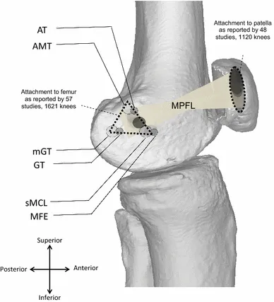
The medial patellofemoral ligament (MPFL) is a ligament, a tough band of tissue that holds bones together at a joint1. It runs from the inner side of the lower femur (thigh bone) to the inner edge of the patella (kneecap)1.
A systematic review (a study that searches for the research on a question in a set, checkable way) of 67 anatomy studies found these features1:
- Thigh-bone end: a triangle between three bony bumps, the adductor tubercle, the medial epicondyle and the gastrocnemius tubercle1.
- Width there: an area rather than a single point, 9 to 17 mm wide1.
- Kneecap end: 20 to 30 mm wide, mostly on the upper part of the inner border1.
- Shape: an hourglass that fans out at both ends, 56 mm long on average1.
- Thickness: 1.0 mm in men, 0.9 mm in women, in one cited MRI (a scan that shows soft tissue) study1.
How the reports differ
Studies disagree on the ligament’s size and even its presence1. Cadaver studies, using bodies donated for study, have found it in 35% to 100% of specimens1. Yet one dissection of 50 knees found it in each of them4. Lengths ran from 45 to 50 mm in eight cadaver knees to about 60 mm in that dissection4,5.
In the 50-knee dissection, the ligament had two origins4. One was a 10.6 mm strip from a bony groove lying between two of the bumps, the medial epicondyle and adductor tubercle4. The other was fibres crossing over from the superficial medial collateral ligament4.
In 36 child cadavers aged 2 to 11, the thigh-bone end lay below the growth plate in 23 and at or above it in 136. The growth plate is a layer of cartilage near the end of a growing bone. On average, the attachment sat 3.0 mm below it6.
What it holds back
In nine cadaver knees bent to 20 degrees, the MPFL gave 60% of the force holding the kneecap back2. Other cadaver studies also found it the main inner restraint, with shares that varied5,7,8:
| Study | Knee bend | MPFL share of the inner restraint |
|---|---|---|
| 9 cadaver knees | 20 degrees | 60%2 |
| 25 knee specimens | Not given | 53% on average7 |
| 8 cadaver knees | 30 degrees | More than 50%5 |
| One cadaver study | As the kneecap enters the groove | 50% to 60%8 |
In 12 cadaver knees bent from 0 to 90 degrees, it did more of this work than the medial quadriceps tendon-femoral ligament9.
Cadaver studies that cut the ligament found these changes10,11:
- In 17 knee specimens, cutting it alone raised outward movement of the kneecap by 50%10.
- In ten specimens, cutting it alone greatly raised outward movement between 20 and 90 degrees of bend12.
- In eight knees, cutting it shifted and tilted the kneecap outward early in the bend11. Pressure fell on the inner side of the kneecap joint and peaked higher on the outer side11.
From a study of six specimens, the authors concluded it restrains mainly when the kneecap is under high outward stress13. They judged that it adds little during unloaded knee bending13. A systematic review of 55 articles found data suggesting the kneecap sits further outward early in the bend without an MPFL14. Its authors suggest this plausibly raises the risk of dislocation14.
How its length and strength change
The 55-article review found the MPFL keeps close to the same length over the first 50 to 60 degrees of bend14. It then shortens step by step into deep bending14. In 15 healthy people imaged during a lunge, it was tightest with the knee straight15. It shortened until about 30 degrees, then stayed near one length15.
The review found reported failure loads from 72 to 208 newtons (a unit of force)14. Stiffness, the force needed to stretch it by a given amount, ranged from 8.0 to 42.5 newtons per mm14. In ten cadaver knees, it failed at 208 newtons on average, after stretching 26 mm16.
Breakdown
How it tears
Most kneecap instability comes from a bending and twisting injury without contact, which pushes the kneecap outward17. Such an injury often also tears the soft tissues holding the kneecap from the inner side17. The 67-study review cites evidence that the MPFL ruptures once stretched beyond 12 to 18 mm1. On that evidence, even a first dislocation would injure it1.
A meta-analysis of 35 articles (2,558 patients) found MPFL injury in 94.7% of sudden outward dislocations3. It reported where the ligament tore3:
| Where it tore | Share of injuries |
|---|---|
| Kneecap end | 37.1%3 |
| Thigh-bone end | 36.8%3 |
| More than one site | 25.1%3 |
| Middle of the ligament | 15.6%3 |
It tore most often at the kneecap end in people aged 16 or under, and at the thigh-bone end in those over 163.
Complete and partial tears
Imaging and surgery studies report how badly it tears18,19:
- In 73 patients scanned within 7 weeks of a dislocation, 72 had an MPFL injury18. Of these, 51.4% were complete tears, most often at the thigh bone, and 48.6% partial18.
- A systematic review of patients under 18 after a first dislocation found, in four studies of 248 knees, 47% complete tears and 41% partial19. No tear showed on MRI in 13%19.
- A case series, a report with no comparison group, followed 74 children aged 9 to 14 after a first dislocation20. Of these, 99% had an MPFL injury20. Keyhole surgery found injury at the kneecap end alone in 81%, and a total rupture there in 66%20.
How the shape of the knee plays a part
In the 73-patient study, tears at the kneecap end went with a larger TT-TG distance18. That distance measures how far outward the bony bump on the shin sits from the thigh-bone groove. The authors concluded that tear patterns depend on trochlear dysplasia, kneecap height and that distance18. Trochlear dysplasia is a shallow or badly shaped groove at the lower end of the thigh bone.
The groove’s shape also mattered in one cadaver study. In eight knees, a flat outer condyle (rounded knob) of the thigh bone cut the kneecap’s outward stability by 70% at 30 degrees21. The patellar dislocation page covers how the injury is defined, who it affects and what trials found.
Recovery
What happens to the torn ligament
The background section of one systematic review states that a damaged MPFL may heal if the knee is held still22. It adds that the ligament can end up longer and looser, leaving the kneecap unstable outward22.
The 67-study review cites one study where the MPFL was longer in unstable kneecaps (54 mm) than in normal ones (49 mm)1. It was 64 mm in kneecaps that dislocated again and again1. Other studies reported 47 to 72 mm in normal knees1. The review notes it is not known which comes first1. People may dislocate because the ligament was longer, or it may be longer because it healed that way1.
A cohort study, which follows a group of people over time, took 53 patients managed without surgery after a first dislocation23. Of these, 35 had torn it at the thigh-bone end23. After 7 years on average, 13 of the 15 with an unstable kneecap had that type of tear23. So did 8 of the 9 who had dislocated again23.
When the ligament is rebuilt
MPFL reconstruction replaces the damaged ligament with a tendon graft (a piece of tissue moved to rebuild a damaged part)22. The graft is fixed in a tunnel drilled in the thigh bone and held with a screw22.
A systematic review of seven studies (380 knees) found that tunnel had widened in 38.7% to 77.1% of knees22. Evidence conflicted on how the width changes over time22. Five low-quality studies in that review found no worse knee scores with a wider tunnel22. The review noted the low level of that evidence22.
In ten cadaver knees, the intact MPFL held 208 newtons16. Stitched repairs and tendon grafts fixed in those knees held between 37 and 195 newtons, depending on the method16.
What ACL grafts show
The studies in this part are of grafts replacing the anterior cruciate ligament (ACL), not the MPFL. They do not show whether MPFL grafts change the same way. A systematic review of tissue-sample studies in people found an ACL tendon graft survives in the joint24. It goes through ligamentisation, the slow change of a tendon graft into tissue like a ligament24. The mature ACL graft may then look like a normal ACL under the microscope24.
Mature ACL grafts still differ in how their finest strands of collagen, a strong rope-like protein, are laid out24. There is no agreement on how long each stage takes24. A review of 23 studies (706 patients) found ACL grafts still changing a year after surgery25.
Connections
The joint and bones it joins
The MPFL steadies the kneecap within the knee joint, where the kneecap slides up and down in a groove on the femur1,26. The MPFL holds the inner side, joining the femur to the patella1.
A dissection found it also attaches under the lower end of the quadriceps mechanism7. In 12 cadaver knees, these upper inner restraints attached half on the quadriceps tendon and half on the kneecap9. A review notes this has led to the name “medial patellofemoral complex”27.
Muscles and ligaments that share its work
The vastus medialis is the inner muscle of the quadriceps26. Its lowest fibres are called the vastus medialis obliquus. In 50 knees, the MPFL merged with this part’s tendon before attaching to the kneecap4. In eight cadaver knees, the two sets of fibres meshed near the kneecap5. Relaxing this muscle in another eight knees cut the kneecap’s outward stability by 30%21.
Other bands share the job of holding the kneecap2,21:
- In nine cadaver knees at 20 degrees, the medial patellomeniscal ligament gave 13% of the restraining force2.
- In eight knees under quadriceps tension, tearing the inner retinacula cut outward stability most with the knee straight, by 49%21. The retinacula are bands of tough tissue at each side of the kneecap.
In the 50-knee dissection, the MPFL took fibres from the medial collateral ligament, the ligament on the inner side of the knee4. Related exercise pages: lunge, during which one study imaged its length in 15 healthy people15.
Medial patellofemoral ligament steadies
Medial patellofemoral ligament attaches to
Medial patellofemoral ligament joins
Medial patellofemoral ligament shares fibres with
Medial patellofemoral ligament is injured in
Medial patellofemoral ligament has been measured during
Sources
- Aframian A, Smith TO, Tennent TD, Cobb JP, Hing CB (2017). Origin and insertion of the medial patellofemoral ligament: a systematic review of anatomy.. Knee surgery, sports traumatology, arthroscopy : official journal of the ESSKA. doi:10.1007/s00167-016-4272-1 PMID 27631645
- Desio SM, Burks RT, Bachus KN (1998). Soft tissue restraints to lateral patellar translation in the human knee.. The American journal of sports medicine. doi:10.1177/03635465980260012701 PMID 9474403
- Kluczynski MA, Miranda L, Marzo JM (2020). Prevalence and Site of Medial Patellofemoral Ligament Injuries in Patients With Acute Lateral Patellar Dislocations: A Systematic Review and Meta-analysis.. Orthopaedic journal of sports medicine. doi:10.1177/2325967120967338 PMID 33403210
- Baldwin JL (2009). The anatomy of the medial patellofemoral ligament.. The American journal of sports medicine. doi:10.1177/0363546509339909 PMID 19729366
- Panagiotopoulos E, Strzelczyk P, Herrmann M, Scuderi G (2006). Cadaveric study on static medial patellar stabilizers: the dynamizing role of the vastus medialis obliquus on medial patellofemoral ligament.. Knee surgery, sports traumatology, arthroscopy : official journal of the ESSKA. doi:10.1007/s00167-005-0631-z PMID 16001289
- Shea KG, Martinson WD, Cannamela PC, Richmond CG, Fabricant PD, Anderson AF, Polousky JD, Ganley TJ (2018). Variation in the Medial Patellofemoral Ligament Origin in the Skeletally Immature Knee: An Anatomic Study.. The American journal of sports medicine. doi:10.1177/0363546517738002 PMID 29083227
- Conlan T, Garth WP Jr, Lemons JE (1993). Evaluation of the medial soft-tissue restraints of the extensor mechanism of the knee.. The Journal of bone and joint surgery. American volume. doi:10.2106/00004623-199305000-00007 PMID 8501083
- Philippot R, Boyer B, Testa R, Farizon F, Moyen B (2012). The role of the medial ligamentous structures on patellar tracking during knee flexion.. Knee surgery, sports traumatology, arthroscopy : official journal of the ESSKA. doi:10.1007/s00167-011-1598-6 PMID 21748394
- Christian DR, Redondo ML, Cancienne JM, Shewman EF, Farr J, Cole BJ, Yanke AB (2020). Differential Contributions of the Quadriceps and Patellar Attachments of the Proximal Medial Patellar Restraints to Resisting Lateral Patellar Translation.. Arthroscopy : the journal of arthroscopic & related surgery : official publication of the Arthroscopy Association of North America and the International Arthroscopy Association. doi:10.1016/j.arthro.2020.01.058 PMID 32061974
- Hautamaa PV, Fithian DC, Kaufman KR, Daniel DM, Pohlmeyer AM (1998). Medial soft tissue restraints in lateral patellar instability and repair.. Clinical orthopaedics and related research. doi:10.1097/00003086-199804000-00021 PMID 9584380
- Stephen JM, Kader D, Lumpaopong P, Deehan DJ, Amis AA (2013). Sectioning the medial patellofemoral ligament alters patellofemoral joint kinematics and contact mechanics.. Journal of orthopaedic research : official publication of the Orthopaedic Research Society. doi:10.1002/jor.22371 PMID 23629829
- Nomura E, Horiuchi Y, Kihara M (2000). Medial patellofemoral ligament restraint in lateral patellar translation and reconstruction.. The Knee. doi:10.1016/s0968-0160(00)00038-7 PMID 10788776
- Zaffagnini S, Colle F, Lopomo N, Sharma B, Bignozzi S, Dejour D, Marcacci M (2013). The influence of medial patellofemoral ligament on patellofemoral joint kinematics and patellar stability.. Knee surgery, sports traumatology, arthroscopy : official journal of the ESSKA. doi:10.1007/s00167-012-2307-9 PMID 23179455
- Huber C, Zhang Q, Taylor WR, Amis AA, Smith C, Hosseini Nasab SH (2020). Properties and Function of the Medial Patellofemoral Ligament: A Systematic Review.. The American journal of sports medicine. doi:10.1177/0363546519841304 PMID 31091114
- Kernkamp WA, Wang C, Li C, Hu H, van Arkel ERA, Nelissen RGHH, LaPrade RF, van de Velde SK, Tsai TY (2019). The Medial Patellofemoral Ligament Is a Dynamic and Anisometric Structure: An In Vivo Study on Length Changes and Isometry.. The American journal of sports medicine. doi:10.1177/0363546519840278 PMID 31070936
- Mountney J, Senavongse W, Amis AA, Thomas NP (2005). Tensile strength of the medial patellofemoral ligament before and after repair or reconstruction.. The Journal of bone and joint surgery. British volume. doi:10.1302/0301-620x.87b1.14924 PMID 15686235
- Sanders TL, Pareek A, Hewett TE, Stuart MJ, Dahm DL, Krych AJ (2018). Incidence of First-Time Lateral Patellar Dislocation: A 21-Year Population-Based Study.. Sports Health. doi:10.1177/1941738117725055 PMID 28795924
- Balcarek P, Ammon J, Frosch S, Walde TA, Schüttrumpf JP, Ferlemann KG, Lill H, Stürmer KM, Frosch KH (2010). Magnetic resonance imaging characteristics of the medial patellofemoral ligament lesion in acute lateral patellar dislocations considering trochlear dysplasia, patella alta, and tibial tuberosity-trochlear groove distance.. Arthroscopy : the journal of arthroscopic & related surgery : official publication of the Arthroscopy Association of North America and the International Arthroscopy Association. doi:10.1016/j.arthro.2009.11.004 PMID 20620792
- Reed JM, Wollenman CC, Tisano BK, Dale KM, LeClere LE (2026). Frequency of Adjunctive MRI Findings on First-Time Patellar Dislocations in Pediatric Patients: A Systematic Review.. The American journal of sports medicine. doi:10.1177/03635465251383851 PMID 41508815
- Askenberger M, Arendt EA, Ekström W, Voss U, Finnbogason T, Janarv PM (2016). Medial Patellofemoral Ligament Injuries in Children With First-Time Lateral Patellar Dislocations: A Magnetic Resonance Imaging and Arthroscopic Study.. The American journal of sports medicine. doi:10.1177/0363546515611661 PMID 26602152
- Senavongse W, Amis AA (2005). The effects of articular, retinacular, or muscular deficiencies on patellofemoral joint stability: a biomechanical study in vitro.. The Journal of bone and joint surgery. British volume. doi:10.1302/0301-620X.87B4.14768 PMID 15795215
- Abelleyra Lastoria DA, Gopinath V, Divekar O, Smith T, Roberts TRW, Hing CB (2023). Does medial patellofemoral ligament reconstruction result in femoral tunnel enlargement? A systematic review.. Knee surgery & related research. doi:10.1186/s43019-023-00187-1 PMID 37131234
- Sillanpää PJ, Peltola E, Mattila VM, Kiuru M, Visuri T, Pihlajamäki H (2009). Femoral avulsion of the medial patellofemoral ligament after primary traumatic patellar dislocation predicts subsequent instability in men: a mean 7-year nonoperative follow-up study.. The American journal of sports medicine. doi:10.1177/0363546509333010 PMID 19376939
- Claes S, Verdonk P, Forsyth R, Bellemans J (2011). The “Ligamentization” Process in Anterior Cruciate Ligament Reconstruction. The American journal of sports medicine. doi:10.1177/0363546511402662 PMID 21515806
- van Groningen B, van der Steen MC, Janssen DM, van Rhijn LW, van der Linden AN, Janssen RPA (2020). Assessment of Graft Maturity After Anterior Cruciate Ligament Reconstruction Using Autografts: A Systematic Review of Biopsy and Magnetic Resonance Imaging studies.. Arthroscopy, sports medicine, and rehabilitation. doi:10.1016/j.asmr.2020.02.008 PMID 32875303
- Betts JG, Young KA, Wise JA, Johnson E, Poe B, Kruse DH, Korol O, Johnson JE, Womble M, DeSaix P (2022). Anatomy and Physiology 2e. OpenStax. Link
- Loeb AE, Tanaka MJ (2018). The medial patellofemoral complex.. Current reviews in musculoskeletal medicine. doi:10.1007/s12178-018-9475-2 PMID 29728862
Changes
- First published.